Loading...
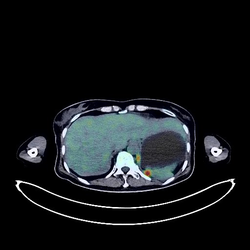
Cervical Cancer PET/CT (case 983827-000142 from PETWB-REP)
0 views9 days agoWhole-body 18F-FDG PET/CT scan in a patient with Cervical Cancer taken from the PETWB-REP dataset. The following English report (translated from original Chinese) is taken verbatim from the public dataset and has not been modified or otherwise checked for accuracy (see the end for citation). Impression a. Post-cervical cancer surgery, multiple space-occupying lesions in the surgical area, vaginal stump, abdominal wall, bilateral adnexa, pelvic floor peritoneum, and pelvic wall, with significantly increased FDG metabolism, suggestive of tumor recurrence with multiple implantation metastases, and involvement of the adjacent sigmoid colon. b. Metastasis to the right iliac fossa, bilateral inguinal regions, bilateral posterior diaphragmatic crura, and right mid-lower abdominal mesenteric lymph nodes. Metastatic tumors in the right psoas major muscle and left rectus abdominis muscle. c. Multiple metastatic tumors in the bilateral pleura. Possible metastasis to the right hilar lymph nodes. d. Scattered multiple patchy and nodular lesions in both lungs, with significantly increased FDG metabolism, suggesting a high probability of inflammatory lesions mixed with metastases. e. Nodular FDG metabolism increase near the right atrium, suggesting a high probability of pericardial metastasis. Dense glandular tissue in both breasts. Manifestations of chronic gastritis. Right renal cyst. Percutaneous external drainage tubes in both renal pelvises. Post-radiotherapy changes in the lumbosacral spine. Osteophyte formation in some vertebral bodies. No obvious abnormalities seen on cranial scintigraphy. Minor chronic inflammation of both maxillary sinuses. This case is from PETWB-REP, a curated dataset of whole-body 18F-FDG PET/CT scans and corresponding radiology reports from 490 patients with a broad spectrum of malignancies. The data were retrospectively collected from patients who underwent clinically indicated whole-body 18F-FDG PET/CT scans at the Shanghai Universal Medical Imaging Diagnostic Center between 2021 and 2024. License: Creative Commons Attribution 4.0 International (CC BY 4.0) Citation: Xue, L., Feng, G., Wenbo, Z., Zhang, Y., Li, L., Wang, S., Peng, L., Peng, S., & Gao, X. (2026). PETWB-REP: A Multi-Cancer Whole-Body FDG PET/CT Dataset with Corresponding Radiology Reports [Data set]. Zenodo. https://doi.org/10.5281/zenodo.18670487
Whole BodyPET/CT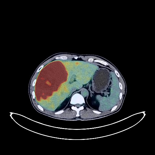
Liver Cancer PET/CT (case 983827-000079 from PETWB-REP)
0 views9 days agoWhole-body 18F-FDG PET/CT scan in a patient with Liver Cancer taken from the PETWB-REP dataset. The following English report (translated from original Chinese) is taken verbatim from the public dataset and has not been modified or otherwise checked for accuracy (see the end for citation). Impression Multiple space-occupying lesions in the liver with increased FDG metabolism; focal increased FDG metabolism in the lower pole of the right kidney; enlarged lymph nodes in the porta hepatis and right supradiaphragmatic region with increased FDG metabolism. All of these suggest malignancy. Primary malignant liver tumors (such as cholangiocarcinoma) with multiple metastases are highly probable; please correlate with clinicopathology. Small amount of pelvic effusion. Several small solid nodules in both lungs with clear borders; FDG metabolism is normal. Metastases need to be ruled out; close CT observation is recommended. A few post-inflammatory lesions in both lungs. Concentrated bile in the gallbladder. Calcification in the prostate. Chronic inflammatory changes in the cardia and antrum of the stomach; hemorrhoidal changes. Slight bulging of the L4/5 and L5/S1 intervertebral discs. Calcification in the left lobe of the thyroid gland with focal increased FDG metabolism; further ultrasound examination is recommended. No obvious abnormalities were found on cranial scintigraphy. A few chronic inflammations in the right maxillary sinus. Reactive hyperplasia of bilateral cervical lymph nodes. This case is from PETWB-REP, a curated dataset of whole-body 18F-FDG PET/CT scans and corresponding radiology reports from 490 patients with a broad spectrum of malignancies. The data were retrospectively collected from patients who underwent clinically indicated whole-body 18F-FDG PET/CT scans at the Shanghai Universal Medical Imaging Diagnostic Center between 2021 and 2024. License: Creative Commons Attribution 4.0 International (CC BY 4.0) Citation: Xue, L., Feng, G., Wenbo, Z., Zhang, Y., Li, L., Wang, S., Peng, L., Peng, S., & Gao, X. (2026). PETWB-REP: A Multi-Cancer Whole-Body FDG PET/CT Dataset with Corresponding Radiology Reports [Data set]. Zenodo. https://doi.org/10.5281/zenodo.18670487
Whole BodyPET/CT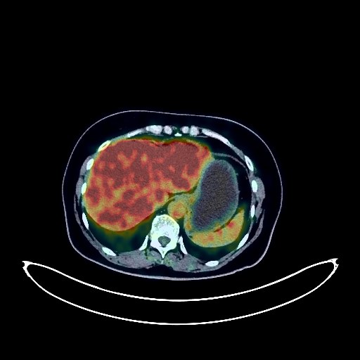
Cervical Cancer PET/CT (case 983827-000088 from PETWB-REP)
0 views9 days agoWhole-body 18F-FDG PET/CT scan in a patient with Cervical Cancer taken from the PETWB-REP dataset. The following English report (translated from original Chinese) is taken verbatim from the public dataset and has not been modified or otherwise checked for accuracy (see the end for citation). Impression a. Heterogeneous density in the cervix and cervical canal with increased FDG metabolism suggests a high probability of neoplastic lesions. b. Possible effusion or cystic mass in the uterine body. c. Cystic mass in the right adnexa, with increased FDG uptake in part of the cyst wall and septa, suggests a possible cystadenoma, with local malignancy to be ruled out. Further enhanced MRI is recommended, and hysteroscopic biopsy may be necessary. a. Mixed ground-glass nodule in the apical-posterior segment of the left upper lobe, with low FDG metabolism, suggests a possible chronic inflammatory nodule, but lung cancer cannot be ruled out. Close observation with HRCT is recommended. b. Multiple solid chronic inflammatory nodules in both lungs. Scattered chronic inflammation and remnants in both lungs. Punctate calcification in the left breast. Calcification of some arterial walls. Mild fatty liver. Partial vertebral osteophyte formation. No obvious abnormalities seen on cranial scintigraphy. Chronic inflammation of the left maxillary sinus. This case is from PETWB-REP, a curated dataset of whole-body 18F-FDG PET/CT scans and corresponding radiology reports from 490 patients with a broad spectrum of malignancies. The data were retrospectively collected from patients who underwent clinically indicated whole-body 18F-FDG PET/CT scans at the Shanghai Universal Medical Imaging Diagnostic Center between 2021 and 2024. License: Creative Commons Attribution 4.0 International (CC BY 4.0) Citation: Xue, L., Feng, G., Wenbo, Z., Zhang, Y., Li, L., Wang, S., Peng, L., Peng, S., & Gao, X. (2026). PETWB-REP: A Multi-Cancer Whole-Body FDG PET/CT Dataset with Corresponding Radiology Reports [Data set]. Zenodo. https://doi.org/10.5281/zenodo.18670487
Whole BodyPET/CT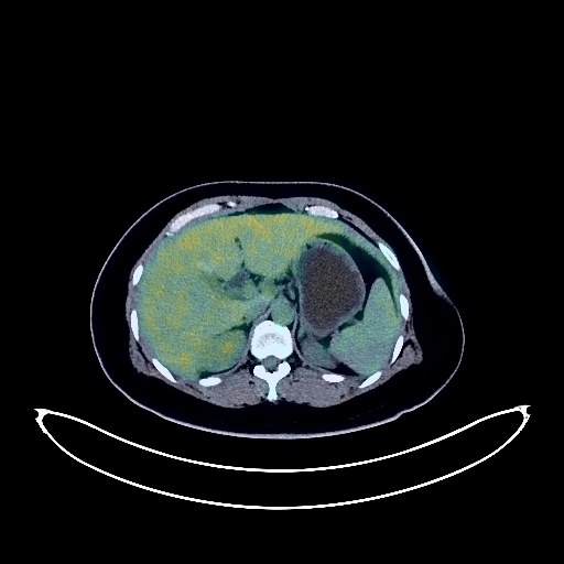
Cervical Cancer PET/CT (case 983827-000155 from PETWB-REP)
0 views9 days agoWhole-body 18F-FDG PET/CT scan in a patient with Cervical Cancer taken from the PETWB-REP dataset. The following English report (translated from original Chinese) is taken verbatim from the public dataset and has not been modified or otherwise checked for accuracy (see the end for citation). Impression a. Cervical mass, elevated FDG metabolism, involving the lower part of the uterine body, consistent with cervical cancer based on pathology. b. Reactive hyperplasia of bilateral pelvic walls, bilateral inguinal lymph nodes, and para-aortic lymph nodes is highly probable; follow-up is recommended to rule out other possible complications. c. Fatty liver. a. Ground-glass nodule in the posterior segment of the right lower lobe, FDG metabolism normal, suggestive of inflammatory nodule or atypical adenomatous hyperplasia; annual HRCT follow-up is recommended. b. Chronic inflammatory micronodules (solid) in the apical segment of the right upper lobe and the apical-posterior segment of the left upper lobe. A few chronic inflammations and old lesions in both lungs. Mild degenerative changes in the spine. L4/5 and L5/S1 intervertebral disc bulges. No abnormalities found on cranial scintigraphy. Bilateral maxillary sinusitis. Left sclerotic mastoid. This case is from PETWB-REP, a curated dataset of whole-body 18F-FDG PET/CT scans and corresponding radiology reports from 490 patients with a broad spectrum of malignancies. The data were retrospectively collected from patients who underwent clinically indicated whole-body 18F-FDG PET/CT scans at the Shanghai Universal Medical Imaging Diagnostic Center between 2021 and 2024. License: Creative Commons Attribution 4.0 International (CC BY 4.0) Citation: Xue, L., Feng, G., Wenbo, Z., Zhang, Y., Li, L., Wang, S., Peng, L., Peng, S., & Gao, X. (2026). PETWB-REP: A Multi-Cancer Whole-Body FDG PET/CT Dataset with Corresponding Radiology Reports [Data set]. Zenodo. https://doi.org/10.5281/zenodo.18670487
Whole BodyPET/CT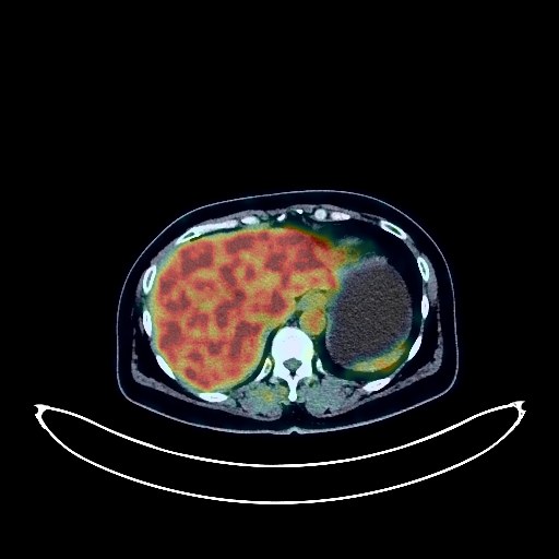
Cervical Cancer PET/CT (case 983827-000076 from PETWB-REP)
0 views9 days agoWhole-body 18F-FDG PET/CT scan in a patient with Cervical Cancer taken from the PETWB-REP dataset. The following English report (translated from original Chinese) is taken verbatim from the public dataset and has not been modified or otherwise checked for accuracy (see the end for citation). Impression Cervical mass with elevated FDG metabolism, consistent with cervical cancer; multiple lymph node metastases near the left iliac vessels. A few fibrotic lesions in both lungs. A pneumocystic cavity in the right upper lobe. Calcification of some arterial walls (including the coronary arteries). Bilateral breast hyperplasia, with calcification in the left breast. Post-cholecystectomy changes. Mild left adrenal hyperplasia. Mild vertebral osteophyte formation. No obvious abnormalities on cranial scintigraphy. Right superior alveolar ulceration. Chronic inflammation of the right lateral nasopharyngeal wall. This case is from PETWB-REP, a curated dataset of whole-body 18F-FDG PET/CT scans and corresponding radiology reports from 490 patients with a broad spectrum of malignancies. The data were retrospectively collected from patients who underwent clinically indicated whole-body 18F-FDG PET/CT scans at the Shanghai Universal Medical Imaging Diagnostic Center between 2021 and 2024. License: Creative Commons Attribution 4.0 International (CC BY 4.0) Citation: Xue, L., Feng, G., Wenbo, Z., Zhang, Y., Li, L., Wang, S., Peng, L., Peng, S., & Gao, X. (2026). PETWB-REP: A Multi-Cancer Whole-Body FDG PET/CT Dataset with Corresponding Radiology Reports [Data set]. Zenodo. https://doi.org/10.5281/zenodo.18670487
Whole BodyPET/CT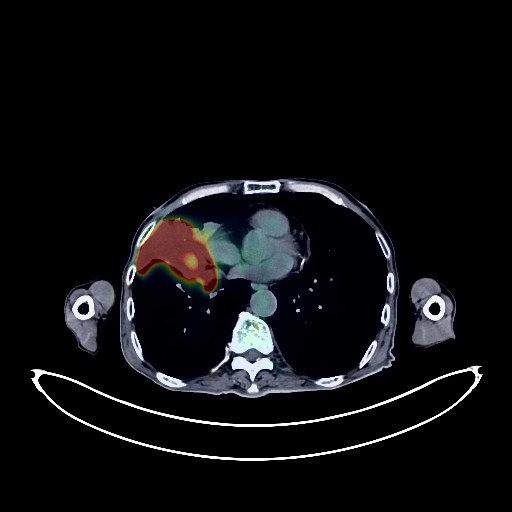
Lung Cancer PET/CT (case 983827-000203 from PETWB-REP)
0 views9 days agoWhole-body 18F-FDG PET/CT scan in a patient with Lung Cancer taken from the PETWB-REP dataset. The following English report (translated from original Chinese) is taken verbatim from the public dataset and has not been modified or otherwise checked for accuracy (see the end for citation). Impression a. Soft tissue mass in the upper and middle lobes of the right lung with increased FDG metabolism; solid nodules in the anterior segment of the left upper lobe and the lateral basal segment of the right lower lobe with increased FDG metabolism; slightly low-density nodules in the body and tail of the pancreas with increased FDG metabolism; enlarged lymph nodes in the retroperitoneum and bilateral common iliac arteries with increased FDG metabolism; multiple bone destruction throughout the body with increased FDG metabolism; nodular FDG hypermetabolic foci in the left gluteus maximus muscle. Considering all of the above, this is likely a malignant tumor, possibly lymphoma with multi-systemic infiltration, and lung cancer with multiple metastases should be ruled out. Please confirm the diagnosis with pathological examination. b. Small solid nodules in the remaining two lungs, with normal FDG metabolism, suggestive of chronic inflammatory nodules. Please follow up with CT scans to rule out other possibilities. Slight inflammation in the left lower lobe, and paraseptal emphysema in the left upper lobe. Possible reactive hyperplasia of mediastinal lymph nodes. Calcification of some arterial walls (including coronary arteries). Anemia. Nodular FDG hypermetabolic lesion in the lower pole of the left kidney. No abnormal density shadows were seen on the same CT scan. Physiological change is considered possible. Occult space-occupying lesion needs to be ruled out. Enhanced MRI is recommended for follow-up. After comprehensive treatment for left tongue cancer: No obvious signs of tumor recurrence were seen in the surgical area. No enlarged lymph nodes were seen in both sides of the neck. Please follow up with clinical and enhanced MRI findings. Increased FDG metabolism in the left lateral pterygoid muscle is considered likely to be a physiological change. Localized high-density lesion in the left parietal lobe. No abnormal FDG metabolism was seen. Benign lesion is considered possible. Enhanced MRI is recommended to rule out other possibilities. A few ischemic lesions in the deep brain, age-related brain changes. Gallstones. Prostatic calcification. Partial vertebral osteophyte formation. L4/5 and L5/S1 intervertebral disc bulge. Chronic inflammation of the right maxillary sinus. Inflammatory changes in the right maxillary gingival region are likely significant; please consult a specialist. This case is from PETWB-REP, a curated dataset of whole-body 18F-FDG PET/CT scans and corresponding radiology reports from 490 patients with a broad spectrum of malignancies. The data were retrospectively collected from patients who underwent clinically indicated whole-body 18F-FDG PET/CT scans at the Shanghai Universal Medical Imaging Diagnostic Center between 2021 and 2024. License: Creative Commons Attribution 4.0 International (CC BY 4.0) Citation: Xue, L., Feng, G., Wenbo, Z., Zhang, Y., Li, L., Wang, S., Peng, L., Peng, S., & Gao, X. (2026). PETWB-REP: A Multi-Cancer Whole-Body FDG PET/CT Dataset with Corresponding Radiology Reports [Data set]. Zenodo. https://doi.org/10.5281/zenodo.18670487
Whole BodyPET/CT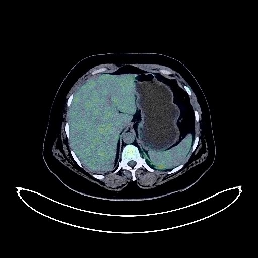
Lung Cancer PET/CT (case 983827-000159 from PETWB-REP)
0 views9 days agoWhole-body 18F-FDG PET/CT scan in a patient with Lung Cancer taken from the PETWB-REP dataset. The following English report (translated from original Chinese) is taken verbatim from the public dataset and has not been modified or otherwise checked for accuracy (see the end for citation). Impression a. A mass in the lower lobe of the left lung with increased FDG metabolism, suggestive of lung cancer with surrounding obstructive inflammation and mediastinal lymph node metastasis. Please correlate with clinicopathology. b. Multiple chronic inflammatory nodules in the left lung, some with metastatic involvement to be ruled out. Follow-up CT scan recommended. A few remnants of chronic inflammation in both lungs. Slight thickening of the left pleura. Increased FDG metabolism in the nasopharyngeal wall, suggestive of inflammation. Specialist examination recommended if necessary. a. Increased FDG uptake in the gastric cardia and lower thoracic esophagus, suggestive of inflammation or physiological uptake. Endoscopic follow-up recommended. b. Diverticulum in the horizontal part of the duodenum. Multiple small diverticula in the sigmoid colon. Increased FDG metabolism in the terminal rectum, suggestive of hemorrhoids or physiological uptake. Please correlate with clinical findings. Fatty liver, small cyst beside the gallbladder in the right anterior lobe of the liver. Gallstones. Left adrenal hyperplasia. Multiple reactive hyperplasia of lymph nodes in both groins. Spinal osteophyte formation, with mild anterior slippage of the L4 vertebral body. L4/5 intervertebral disc bulge. Multiple ischemic lesions deep in the brain parenchyma; MRI examination may be necessary. Multiple reactive hyperplasia of lymph nodes in the bilateral deep cervical spaces, submandibular region, and submandibular region. This case is from PETWB-REP, a curated dataset of whole-body 18F-FDG PET/CT scans and corresponding radiology reports from 490 patients with a broad spectrum of malignancies. The data were retrospectively collected from patients who underwent clinically indicated whole-body 18F-FDG PET/CT scans at the Shanghai Universal Medical Imaging Diagnostic Center between 2021 and 2024. License: Creative Commons Attribution 4.0 International (CC BY 4.0) Citation: Xue, L., Feng, G., Wenbo, Z., Zhang, Y., Li, L., Wang, S., Peng, L., Peng, S., & Gao, X. (2026). PETWB-REP: A Multi-Cancer Whole-Body FDG PET/CT Dataset with Corresponding Radiology Reports [Data set]. Zenodo. https://doi.org/10.5281/zenodo.18670487
Whole BodyPET/CT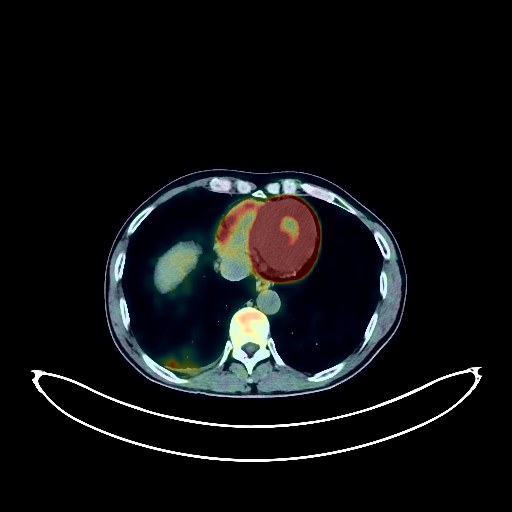
Lung Cancer PET/CT (case 983827-000170 from PETWB-REP)
0 views9 days agoWhole-body 18F-FDG PET/CT scan in a patient with Lung Cancer taken from the PETWB-REP dataset. The following English report (translated from original Chinese) is taken verbatim from the public dataset and has not been modified or otherwise checked for accuracy (see the end for citation). Impression a. A mass in the posterior segment of the right lower lobe with increased FDG metabolism, consistent with lung cancer with obstructive inflammation. Multiple lymph node metastases in the right hilum and mediastinum. Reactive hyperplasia of the left hilar lymph nodes. b. Chronic inflammatory micronodules in the remaining two lungs are likely; follow-up CT is recommended. Mild emphysema in both lungs. A few post-inflammatory lesions in both lungs. Small amount of pleural effusion with pleural thickening on the right side. Partial arteriosclerosis. Cyst in the left lobe of the liver. Cyst in the right kidney. Residual contrast agent in the urinary tract. Calcifications in the prostate. Bilateral isthmic discontinuity at L5. Degenerative changes in the spine, bulging of the L3/4, L4/5, and L5/S1 intervertebral discs. No obvious abnormalities were found on cranial scintigraphy. Chronic inflammation of both ethmoid sinuses. This case is from PETWB-REP, a curated dataset of whole-body 18F-FDG PET/CT scans and corresponding radiology reports from 490 patients with a broad spectrum of malignancies. The data were retrospectively collected from patients who underwent clinically indicated whole-body 18F-FDG PET/CT scans at the Shanghai Universal Medical Imaging Diagnostic Center between 2021 and 2024. License: Creative Commons Attribution 4.0 International (CC BY 4.0) Citation: Xue, L., Feng, G., Wenbo, Z., Zhang, Y., Li, L., Wang, S., Peng, L., Peng, S., & Gao, X. (2026). PETWB-REP: A Multi-Cancer Whole-Body FDG PET/CT Dataset with Corresponding Radiology Reports [Data set]. Zenodo. https://doi.org/10.5281/zenodo.18670487
Whole BodyPET/CT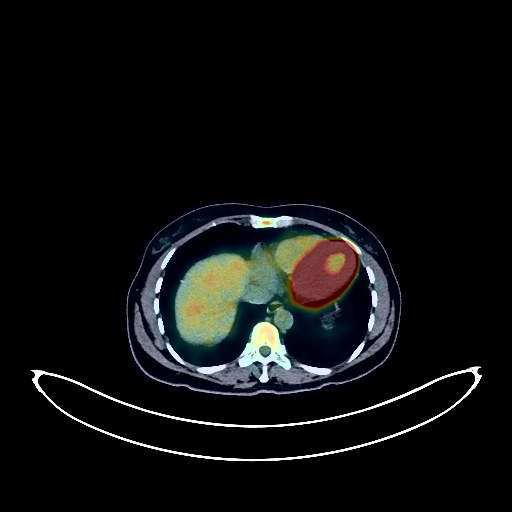
Cervical Cancer PET/CT (case 983827-000006 from PETWB-REP)
0 views9 days agoWhole-body 18F-FDG PET/CT scan in a patient with Cervical Cancer taken from the PETWB-REP dataset. The following English report (translated from original Chinese) is taken verbatim from the public dataset and has not been modified or otherwise checked for accuracy (see the end for citation). Impression a. Soft tissue mass on the left cervical wall with increased FDG metabolism, consistent with cervical cancer. The boundary between the lesion and the upper vagina and uterine body is unclear; please correlate with clinical findings. Metastasis to the left uterine body and the left external iliac lymph nodes. b. Possible uterine fibroid. Left adnexal ovarian cyst. Chronic inflammatory nodule in the lateral basal segment of the left lower lobe of the lung; please follow up with CT. Scattered chronic inflammation and remnants in both lungs. Cyst in the right posterior lobe of the liver. Possible hemangioma in the left medial lobe of the liver; MRI follow-up is recommended. Horseshoe kidney. Partial vertebral osteophyte formation. L4/5 intervertebral disc bulge. No obvious abnormalities seen on cranial scintigraphy. Right maxillary sinusitis. Reactive hyperplasia of bilateral deep cervical interspace and axillary lymph nodes. This case is from PETWB-REP, a curated dataset of whole-body 18F-FDG PET/CT scans and corresponding radiology reports from 490 patients with a broad spectrum of malignancies. The data were retrospectively collected from patients who underwent clinically indicated whole-body 18F-FDG PET/CT scans at the Shanghai Universal Medical Imaging Diagnostic Center between 2021 and 2024. License: Creative Commons Attribution 4.0 International (CC BY 4.0) Citation: Xue, L., Feng, G., Wenbo, Z., Zhang, Y., Li, L., Wang, S., Peng, L., Peng, S., & Gao, X. (2026). PETWB-REP: A Multi-Cancer Whole-Body FDG PET/CT Dataset with Corresponding Radiology Reports [Data set]. Zenodo. https://doi.org/10.5281/zenodo.18670487
Whole BodyPET/CT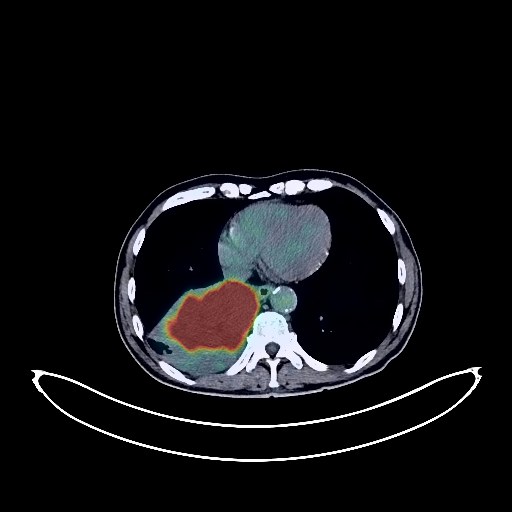
Lung Cancer PET/CT (case 983827-000173 from PETWB-REP)
0 views9 days agoWhole-body 18F-FDG PET/CT scan in a patient with Lung Cancer taken from the PETWB-REP dataset. The following English report (translated from original Chinese) is taken verbatim from the public dataset and has not been modified or otherwise checked for accuracy (see the end for citation). Impression a. A mass in the lower lobe of the right lung, with increased FDG metabolism, suggestive of central lung cancer with obstructive atelectasis. Multiple lymph node metastases in the right hilum and mediastinum. b. Metastasis to the right parasternal pleura and right lower pleura. Small amount of pleural effusion on the right side. c. Heterogeneous bone density in the right iliac bone, upper femur of the left side, and left 6th rib, with increased FDG metabolism, suggesting a high probability of bone metastasis; further specialist examination is recommended. d. Several small, solid, chronic inflammatory nodules in both lungs. Mild emphysema in the upper lobes of both lungs. Scattered chronic inflammation and old lesions in both lungs. A few ischemic lesions in the deep bilateral brain regions; senile cerebral encephalopathy; MRI is recommended to rule out other possibilities. A low-density nodule in the right lobe of the thyroid gland, with normal FDG uptake, suggestive of benignity; follow-up ultrasound is recommended. Calcification of some arterial walls (including coronary arteries). Multiple liver cysts. Hemorrhoids. Benign prostatic hyperplasia with calcification. Degenerative changes in the spine. Lumbar instability, L4/5 and L5/S1 disc bulging, L5/S1 disc pneumothorax. This case is from PETWB-REP, a curated dataset of whole-body 18F-FDG PET/CT scans and corresponding radiology reports from 490 patients with a broad spectrum of malignancies. The data were retrospectively collected from patients who underwent clinically indicated whole-body 18F-FDG PET/CT scans at the Shanghai Universal Medical Imaging Diagnostic Center between 2021 and 2024. License: Creative Commons Attribution 4.0 International (CC BY 4.0) Citation: Xue, L., Feng, G., Wenbo, Z., Zhang, Y., Li, L., Wang, S., Peng, L., Peng, S., & Gao, X. (2026). PETWB-REP: A Multi-Cancer Whole-Body FDG PET/CT Dataset with Corresponding Radiology Reports [Data set]. Zenodo. https://doi.org/10.5281/zenodo.18670487
Whole BodyPET/CT