Loading...
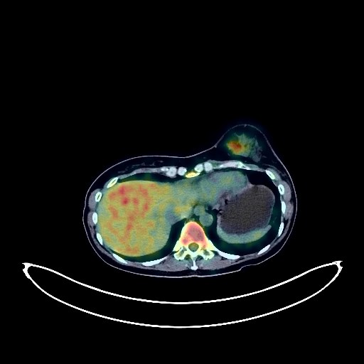
Breast Cancer PET/CT (case 983827-000248 from PETWB-REP)
2 views9 days agoWhole-body 18F-FDG PET/CT scan in a patient with Breast Cancer taken from the PETWB-REP dataset. The following English report (translated from original Chinese) is taken verbatim from the public dataset and has not been modified or otherwise checked for accuracy (see the end for citation). Impression a. Left breast mass with elevated FDG metabolism, consistent with breast cancer; left axillary lymph node metastasis. b. Right breast hyperplasia, possibly with hyperplastic nodules or fibroadenoma; ultrasound follow-up recommended. a. Right lower lobe anterior basal segment mixed ground-glass nodule, FDG metabolism normal, suggestive of atypical adenomatous hyperplasia; HRCT follow-up every six months recommended to rule out early lung cancer. b. Right upper lobe chronic inflammatory micronodule (solid); CT follow-up recommended. A few post-inflammatory lesions in both lungs. Mild anemia. Right frontal arachnoid cyst is the primary consideration; MRI follow-up recommended. Partial vertebral osteophyte formation. This case is from PETWB-REP, a curated dataset of whole-body 18F-FDG PET/CT scans and corresponding radiology reports from 490 patients with a broad spectrum of malignancies. The data were retrospectively collected from patients who underwent clinically indicated whole-body 18F-FDG PET/CT scans at the Shanghai Universal Medical Imaging Diagnostic Center between 2021 and 2024. License: Creative Commons Attribution 4.0 International (CC BY 4.0) Citation: Xue, L., Feng, G., Wenbo, Z., Zhang, Y., Li, L., Wang, S., Peng, L., Peng, S., & Gao, X. (2026). PETWB-REP: A Multi-Cancer Whole-Body FDG PET/CT Dataset with Corresponding Radiology Reports [Data set]. Zenodo. https://doi.org/10.5281/zenodo.18670487
Whole BodyPET/CT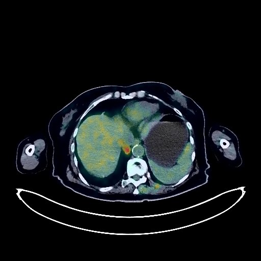
Renal Cancer PET/CT (case 983827-000219 from PETWB-REP)
0 views9 days agoWhole-body 18F-FDG PET/CT scan in a patient with Renal Cancer taken from the PETWB-REP dataset. The following English report (translated from original Chinese) is taken verbatim from the public dataset and has not been modified or otherwise checked for accuracy (see the end for citation). Impression a. Right renal mass with increased FDG metabolism, suggestive of renal cell carcinoma with tumor thrombus formation in the right renal vein and inferior vena cava. Please correlate with clinicopathology. Retroperitoneal and mesenteric lymph node metastasis to be ruled out. b. Left renal soft tissue nodule with increased FDG metabolism, renal cell carcinoma to be ruled out. Please correlate with enhanced MRI for comprehensive analysis. Left renal angiomyolipoma. c. Multiple metastatic tumors in both lungs. Multiple lymph node metastases in both hilar and mediastinal regions. d. Metastatic tumors in the left humerus and left scapula. Scattered post-inflammatory lesions in both lungs. Anemic changes, partial arterial wall calcification (including coronary arteries). Left lobe hepatic cyst. Chronic cholecystitis, gallstones. Bilateral adrenal hyperplasia. Post-hysterectomy changes. Chronic inflammatory changes or physiological uptake in parts of the stomach wall and intestines. Osteoporosis, scoliosis, degenerative changes in the spine, anterior slippage of the L4 and L5 vertebrae, multiple intervertebral disc bulges with pneumoconiosis and degeneration. Bilateral subcutaneous calcifications in the buttocks. Age-related brain conditions, deep lacunar infarcts. This case is from PETWB-REP, a curated dataset of whole-body 18F-FDG PET/CT scans and corresponding radiology reports from 490 patients with a broad spectrum of malignancies. The data were retrospectively collected from patients who underwent clinically indicated whole-body 18F-FDG PET/CT scans at the Shanghai Universal Medical Imaging Diagnostic Center between 2021 and 2024. License: Creative Commons Attribution 4.0 International (CC BY 4.0) Citation: Xue, L., Feng, G., Wenbo, Z., Zhang, Y., Li, L., Wang, S., Peng, L., Peng, S., & Gao, X. (2026). PETWB-REP: A Multi-Cancer Whole-Body FDG PET/CT Dataset with Corresponding Radiology Reports [Data set]. Zenodo. https://doi.org/10.5281/zenodo.18670487
Whole BodyPET/CT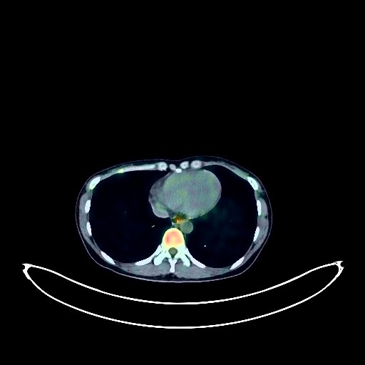
Lung Cancer PET/CT (case 983827-000085 from PETWB-REP)
0 views9 days agoWhole-body 18F-FDG PET/CT scan in a patient with Lung Cancer taken from the PETWB-REP dataset. The following English report (translated from original Chinese) is taken verbatim from the public dataset and has not been modified or otherwise checked for accuracy (see the end for citation). Impression a. Irregular nodule in the anterior segment of the right upper lobe, with increased FDG metabolism, strongly suggestive of lung cancer; further clinical and pathological examination is recommended. b. Ground-glass nodule in the apical segment of the right upper lobe, with normal FDG metabolism, suggestive of inflammatory nodule or atypical adenomatous hyperplasia; annual HRCT follow-up is recommended. Chronic inflammatory nodule in the apical segment of the right upper lobe. Cystic mass in the head of the pancreas, with absent FDG uptake, suggestive of a cyst; neoplastic lesion to be ruled out; further enhanced MRI is recommended. Increased FDG metabolism in some intestinal segments, suggestive of inflammatory or physiological uptake. Full-shaped uterus with uneven density and unevenly increased FDG metabolism, strongly suggestive of adenomyosis; bilateral adnexal ovarian physiological cysts are also highly likely. Ultrasound follow-up is recommended for all of the above. Reactive hyperplasia of lymph nodes adjacent to the left iliac vessels and bilateral pelvic walls. Small amount of pelvic effusion. No abnormalities found on cranial scintigraphy. This case is from PETWB-REP, a curated dataset of whole-body 18F-FDG PET/CT scans and corresponding radiology reports from 490 patients with a broad spectrum of malignancies. The data were retrospectively collected from patients who underwent clinically indicated whole-body 18F-FDG PET/CT scans at the Shanghai Universal Medical Imaging Diagnostic Center between 2021 and 2024. License: Creative Commons Attribution 4.0 International (CC BY 4.0) Citation: Xue, L., Feng, G., Wenbo, Z., Zhang, Y., Li, L., Wang, S., Peng, L., Peng, S., & Gao, X. (2026). PETWB-REP: A Multi-Cancer Whole-Body FDG PET/CT Dataset with Corresponding Radiology Reports [Data set]. Zenodo. https://doi.org/10.5281/zenodo.18670487
Whole BodyPET/CT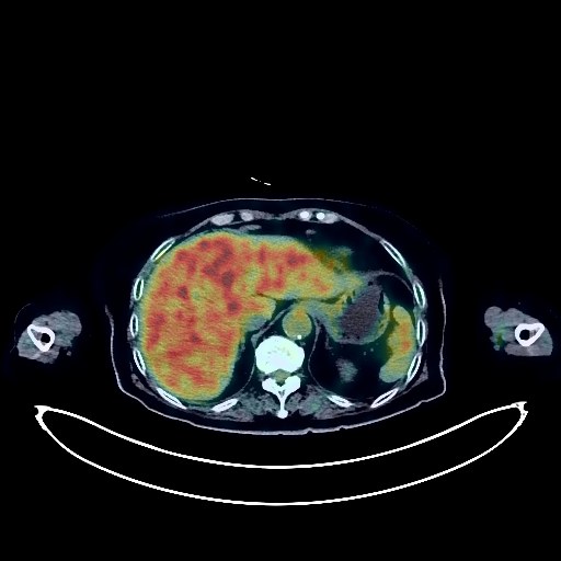
Renal Cancer PET/CT (case 983827-000221 from PETWB-REP)
0 views9 days agoWhole-body 18F-FDG PET/CT scan in a patient with Renal Cancer taken from the PETWB-REP dataset. The following English report (translated from original Chinese) is taken verbatim from the public dataset and has not been modified or otherwise checked for accuracy (see the end for citation). Impression Right renal pelvis mass with increased FDG uptake, suggestive of malignancy, most likely renal pelvis carcinoma; please correlate with clinicopathology. Multiple lymph node metastases in the right renal hilum and retroperitoneum. Residual contrast agent in the right renal parenchyma. a. Bronchiectasis with infection in the apical-posterior segment of the left upper lobe and the apical segment of the right upper lobe; multiple chronic inflammatory micronodules with calcifications in both lungs; small air-filled cysts in the right lower lobe; a few post-inflammatory lesions in both lungs. b. Slight pleural thickening with right-sided calcification. Slight pericardial thickening; partial calcification of the aorta and coronary artery walls, post-pacemaker implantation changes; mild anemia. Duodenal diverticulum in the descending part. Splenic calcifications. Left ovarian cyst; ultrasound follow-up recommended. Low-density nodule in the left lobe of the thyroid gland; no abnormal uptake was observed on FDG, suggestive of nodular goiter; ultrasound and thyroid function tests are recommended. Osteoporosis. Scoliosis with bone hyperplasia. L2 vertebral body wedge-shaped deformity with slight posterior slippage. L3/4, L4/5, and L5/S1 intervertebral disc bulges. Bilateral frozen shoulder. Senile cerebral atrophy. This case is from PETWB-REP, a curated dataset of whole-body 18F-FDG PET/CT scans and corresponding radiology reports from 490 patients with a broad spectrum of malignancies. The data were retrospectively collected from patients who underwent clinically indicated whole-body 18F-FDG PET/CT scans at the Shanghai Universal Medical Imaging Diagnostic Center between 2021 and 2024. License: Creative Commons Attribution 4.0 International (CC BY 4.0) Citation: Xue, L., Feng, G., Wenbo, Z., Zhang, Y., Li, L., Wang, S., Peng, L., Peng, S., & Gao, X. (2026). PETWB-REP: A Multi-Cancer Whole-Body FDG PET/CT Dataset with Corresponding Radiology Reports [Data set]. Zenodo. https://doi.org/10.5281/zenodo.18670487
Whole BodyPET/CT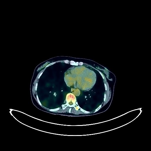
Pancreatic Cancer PET/CT (case 983827-000257 from PETWB-REP)
0 views9 days agoWhole-body 18F-FDG PET/CT scan in a patient with Pancreatic Cancer taken from the PETWB-REP dataset. The following English report (translated from original Chinese) is taken verbatim from the public dataset and has not been modified or otherwise checked for accuracy (see the end for citation). Impression a. Massive FDG-promoted lesions in the tail of the pancreas, suggestive of malignancy, pancreatic cancer is the primary consideration. b. Metastasis to the para-tailed, retroperitoneal, and para-aortic lymph nodes. Bone metastases to the C5, T4 vertebral bodies and appendages, and L2 vertebral body. c. Multiple liver metastases and liver cysts. Abdominal and pelvic peritoneal implantation metastases to be ruled out. Multiple solid nodules in both lungs, some with increased FDG metabolism, some metastases are the primary consideration; close CT observation is recommended. Interstitial changes in both lungs with scattered inflammation and remnants (including calcifications). Reactive hyperplasia of the hilar and mediastinal lymph nodes in both lungs. Bilateral breast hyperplasia. Calcification in the middle of the left kidney, left renal calculus, no obvious space-occupying lesions; please combine with contrast-enhanced MRI images from other hospitals for comprehensive analysis. Increased FDG metabolism in parts of the stomach wall and intestines, possibly due to physiological uptake or chronic inflammation; please follow up with endoscopy. Lumbar sacralization. Spinal degenerative changes. L3/4 and L4/5 intervertebral disc bulge. No obvious abnormalities seen on cranial scintigraphy. Uneven thyroid density; ultrasound follow-up recommended. Reactive hyperplasia of cervical lymph nodes. This case is from PETWB-REP, a curated dataset of whole-body 18F-FDG PET/CT scans and corresponding radiology reports from 490 patients with a broad spectrum of malignancies. The data were retrospectively collected from patients who underwent clinically indicated whole-body 18F-FDG PET/CT scans at the Shanghai Universal Medical Imaging Diagnostic Center between 2021 and 2024. License: Creative Commons Attribution 4.0 International (CC BY 4.0) Citation: Xue, L., Feng, G., Wenbo, Z., Zhang, Y., Li, L., Wang, S., Peng, L., Peng, S., & Gao, X. (2026). PETWB-REP: A Multi-Cancer Whole-Body FDG PET/CT Dataset with Corresponding Radiology Reports [Data set]. Zenodo. https://doi.org/10.5281/zenodo.18670487
Whole BodyPET/CT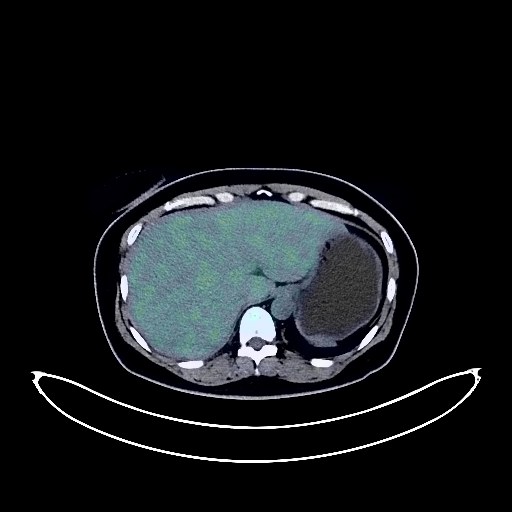
Cervical Cancer PET/CT (case 983827-000228 from PETWB-REP)
0 views9 days agoWhole-body 18F-FDG PET/CT scan in a patient with Cervical Cancer taken from the PETWB-REP dataset. The following English report (translated from original Chinese) is taken verbatim from the public dataset and has not been modified or otherwise checked for accuracy (see the end for citation). Impression a. Cervical mass with elevated FDG metabolism, highly suggestive of cervical cancer; please correlate with clinicopathology. Bilateral iliac lymph node metastasis to be ruled out; reactive hyperplasia of retroperitoneal and bilateral inguinal lymph nodes. b. Cystic mass in the left adnexal region, with uneven thickening of the cyst wall and slightly elevated FDG metabolism, suggestive of cyst or cystadenoma; please correlate with enhanced MRI. c. Possible adenomyosis with fibroids; Nabothian cyst of the cervix. Ground-glass nodules in the apical-posterior segment of the left upper lobe, the lateral segment of the right middle lobe, and the posterior segment of the left lower lobe, suggestive of inflammation or atypical adenomatous hyperplasia; follow-up CT is recommended. A few post-inflammatory lesions in both lungs. Partial arteriosclerosis. Bilateral breast hyperplasia. Reactive hyperplasia of bilateral axillary lymph nodes. Mild fatty liver, cyst in the right lobe of the liver. Partial chronic inflammatory changes in the gastric wall; please follow up with endoscopy. Degenerative changes in the spine, L5/S1 intervertebral disc bulge with posterior calcification. No obvious abnormalities were found on cranial scintigraphy. This case is from PETWB-REP, a curated dataset of whole-body 18F-FDG PET/CT scans and corresponding radiology reports from 490 patients with a broad spectrum of malignancies. The data were retrospectively collected from patients who underwent clinically indicated whole-body 18F-FDG PET/CT scans at the Shanghai Universal Medical Imaging Diagnostic Center between 2021 and 2024. License: Creative Commons Attribution 4.0 International (CC BY 4.0) Citation: Xue, L., Feng, G., Wenbo, Z., Zhang, Y., Li, L., Wang, S., Peng, L., Peng, S., & Gao, X. (2026). PETWB-REP: A Multi-Cancer Whole-Body FDG PET/CT Dataset with Corresponding Radiology Reports [Data set]. Zenodo. https://doi.org/10.5281/zenodo.18670487
Whole BodyPET/CT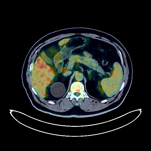
Nasopharyngeal Cancer PET/CT (case 983827-000188 from PETWB-REP)
0 views9 days agoWhole-body 18F-FDG PET/CT scan in a patient with Nasopharyngeal Cancer taken from the PETWB-REP dataset. The following English report (translated from original Chinese) is taken verbatim from the public dataset and has not been modified or otherwise checked for accuracy (see the end for citation). Impression Nasopharyngeal mass with increased FDG metabolism, consistent with nasopharyngeal carcinoma. Right deep cervical lymph node metastasis to be ruled out; reactive hyperplasia of other cervical lymph nodes is possible. Follow-up is recommended. Right parotid gland soft tissue nodule with calcification and increased FDG metabolism, suggestive of mixed tumor, malignancy to be ruled out; please confirm with pathology. Anterior to it is a soft tissue nodule with normal FDG uptake, considered benign. a. Bilateral chronic inflammatory micronodules in the lungs. Interstitial changes in both lungs, bilateral emphysema, calcification in the right upper lobe, and a few post-inflammatory remnants in both lungs. b. Slight thickening of the pleura bilaterally, with a localized nodular protrusion on the left pleura; normal FDG uptake suggests a high probability of benign nodule, follow-up is recommended. Reactive hyperplasia of mediastinal lymph nodes. c. Localized widening of the aortic arch, aneurysm to be ruled out; enhanced CT scan recommended; partial aortic wall calcification; changes after coronary stent placement. Right renal cyst. Benign prostatic hyperplasia with calcification. Degenerative changes in the spine, L4/5 and L5/S1 disc bulges. Age-related brain, deep lacunar infarcts; follow-up with MRI. Right otitis media/mastoid inflammation. This case is from PETWB-REP, a curated dataset of whole-body 18F-FDG PET/CT scans and corresponding radiology reports from 490 patients with a broad spectrum of malignancies. The data were retrospectively collected from patients who underwent clinically indicated whole-body 18F-FDG PET/CT scans at the Shanghai Universal Medical Imaging Diagnostic Center between 2021 and 2024. License: Creative Commons Attribution 4.0 International (CC BY 4.0) Citation: Xue, L., Feng, G., Wenbo, Z., Zhang, Y., Li, L., Wang, S., Peng, L., Peng, S., & Gao, X. (2026). PETWB-REP: A Multi-Cancer Whole-Body FDG PET/CT Dataset with Corresponding Radiology Reports [Data set]. Zenodo. https://doi.org/10.5281/zenodo.18670487
Whole BodyPET/CT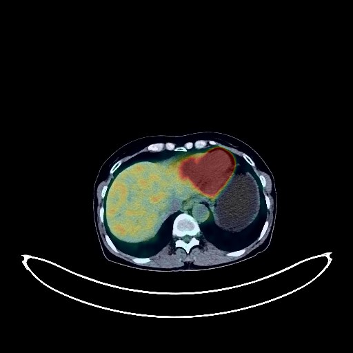
Nasopharyngeal Cancer PET/CT (case 983827-000258 from PETWB-REP)
0 views9 days agoWhole-body 18F-FDG PET/CT scan in a patient with Nasopharyngeal Cancer taken from the PETWB-REP dataset. The following English report (translated from original Chinese) is taken verbatim from the public dataset and has not been modified or otherwise checked for accuracy (see the end for citation). Impression A mass on the right lateral wall and posterior wall of the nasopharynx, with increased FDG metabolism, consistent with nasopharyngeal carcinoma. Reactive hyperplasia of bilateral deep cervical lymph nodes is highly probable; follow-up is recommended. Chronic inflammation of the right mastoid process. Several small, solid, chronic inflammatory nodules in both lungs; follow-up with CT scan is recommended. A few post-inflammatory lesions in both lungs. Partial arteriosclerosis. Slight thickening of the walls of part of the gastric body and antrum, with mildly increased FDG uptake, suggestive of chronic gastritis; increased FDG metabolism in part of the intestinal tract, suggestive of inflammatory or physiological uptake. Follow-up gastroscopy and colonoscopy are recommended for all of the above. Osteophyte formation at the vertebral margins in some areas. L4/5 and L5/S1 intervertebral disc bulges. L5/S1 intervertebral disc pneumatosis and degeneration. No obvious abnormalities were found on cranial parenchymal scintigraphy. A few inflammations in the bilateral ethmoid sinuses and right maxillary sinus. The thyroid gland has uneven density; ultrasound follow-up is recommended. This case is from PETWB-REP, a curated dataset of whole-body 18F-FDG PET/CT scans and corresponding radiology reports from 490 patients with a broad spectrum of malignancies. The data were retrospectively collected from patients who underwent clinically indicated whole-body 18F-FDG PET/CT scans at the Shanghai Universal Medical Imaging Diagnostic Center between 2021 and 2024. License: Creative Commons Attribution 4.0 International (CC BY 4.0) Citation: Xue, L., Feng, G., Wenbo, Z., Zhang, Y., Li, L., Wang, S., Peng, L., Peng, S., & Gao, X. (2026). PETWB-REP: A Multi-Cancer Whole-Body FDG PET/CT Dataset with Corresponding Radiology Reports [Data set]. Zenodo. https://doi.org/10.5281/zenodo.18670487
Whole BodyPET/CT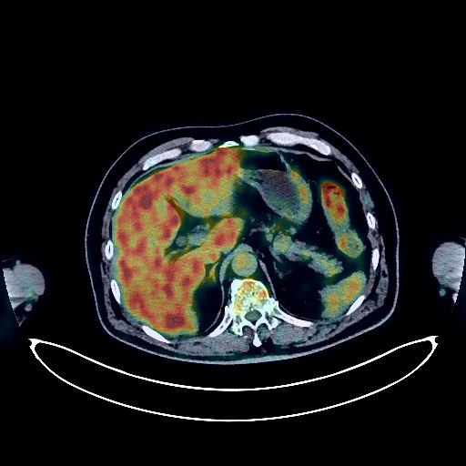
Lung Cancer PET/CT (case 983827-000057 from PETWB-REP)
0 views9 days agoWhole-body 18F-FDG PET/CT scan in a patient with Lung Cancer taken from the PETWB-REP dataset. The following English report (translated from original Chinese) is taken verbatim from the public dataset and has not been modified or otherwise checked for accuracy (see the end for citation). Impression a. Solid plaque lesion with increased FDG metabolism in the posterior segment of the left upper lobe, suggestive of malignancy; please confirm the diagnosis with pathology. b. Metastasis to some lymph nodes in the left hilum and mediastinum. c. Metastasis to the right gluteus medius muscle; metastasis to the deep lobe of the right parotid gland is pending; follow-up is recommended. Multiple bone metastases to the T8 vertebral body, L5 spinous process, and sacrum. d. Interstitial inflammation in both lungs. Chronic inflammatory nodules in both lungs are highly probable; please follow up with CT scans. Slight thickening of the pleura bilaterally. Calcification of some arterial walls. Post-rectal cancer surgery, no obvious signs of tumor recurrence. Physiological or inflammatory uptake of the stomach and part of the colon; endoscopic follow-up is recommended. Lacunar infarcts in the deep brain bilaterally; age-related brain; no obvious abnormalities seen on cranial FDG imaging; no obvious abnormal FDG metabolic lesions seen in either eye. Enhanced MRI is recommended for follow-up to rule out occult lesions. Right maxillary sinusitis. Mild fatty liver. Post-cholecystectomy. Bilateral renal cysts. Left renal pelvis dilation. Right testicular tunica vaginalis calcification. Spinal degenerative changes. L4/5 and L5/S1 intervertebral disc bulges. This case is from PETWB-REP, a curated dataset of whole-body 18F-FDG PET/CT scans and corresponding radiology reports from 490 patients with a broad spectrum of malignancies. The data were retrospectively collected from patients who underwent clinically indicated whole-body 18F-FDG PET/CT scans at the Shanghai Universal Medical Imaging Diagnostic Center between 2021 and 2024. License: Creative Commons Attribution 4.0 International (CC BY 4.0) Citation: Xue, L., Feng, G., Wenbo, Z., Zhang, Y., Li, L., Wang, S., Peng, L., Peng, S., & Gao, X. (2026). PETWB-REP: A Multi-Cancer Whole-Body FDG PET/CT Dataset with Corresponding Radiology Reports [Data set]. Zenodo. https://doi.org/10.5281/zenodo.18670487
Whole BodyPET/CT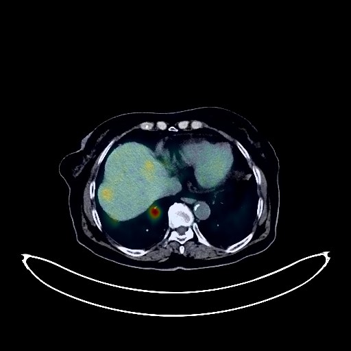
Lung Cancer PET/CT (case 983827-000011 from PETWB-REP)
0 views9 days agoWhole-body 18F-FDG PET/CT scan in a patient with Lung Cancer taken from the PETWB-REP dataset. The following English report (translated from original Chinese) is taken verbatim from the public dataset and has not been modified or otherwise checked for accuracy (see the end for citation). Impression After treatment of the left lung mass, combined with PET/CT images from another hospital: a. The mass in the anterior basal segment of the left lower lobe has shrunk compared to before, and FDG metabolism has not increased, suggesting that tumor activity has been suppressed after treatment. b. Partially shrunk lymph nodes in the bilateral hilar, mediastinal, right supraclavicular, hepatic hilum, and hilar space, with increased FDG metabolism, suggesting that tumor activity has been partially suppressed after treatment, with some still remaining. c. The liver metastases show significant activity; please follow up with enhanced MRI. a. Mixed ground-glass nodules in the anterior basal segment of the right lower lobe, with normal FDG metabolism; malignancy cannot be ruled out. Please compare with the previous images and repeat HRCT after anti-inflammatory treatment. b. Ground-glass nodules in the apical-posterior segment of the left upper lobe and the posterior segment of the right upper lobe, with normal FDG metabolism; chronic inflammatory nodules or atypical adenomatous hyperplasia are possible. Please follow up annually with HRCT. c. Multiple solid nodules in both lungs, with normal FDG metabolism; chronic inflammatory nodules are possible. Please follow up with CT to rule out other possibilities. Chronic inflammation and sequelae in both lungs, with an air sac in the lower lobe of the left lung. Mild bronchial dilatation in both lower lobes. Mild pleural thickening bilaterally. Small amount of pericardial effusion. Partial arteriosclerosis (including coronary arteries). Thyroid gland density is uneven; uterus is slightly full in shape, with uneven density and FDG uptake in the uterine body; please correlate with ultrasound examination. Left renal cyst. Hemorrhoidal changes. Localized increased density in the right 5th rib with increased FDG metabolism; please compare with previous images and follow up to rule out metastasis; large bone islands in the right 3rd and 8th ribs; please follow up. Scoliosis with degenerative changes. Mild anterior slippage of the L5 vertebral body. L3 and L4 vertebral endplate inflammation. L3/4 and L4/5 intervertebral disc bulge with pneumatosis and degeneration. Deep lacunar ischemic foci in both cerebral regions, senile encephalopathy. Chronic inflammation of both palatine tonsils. Unclear visualization of both lenses; please correlate with clinical findings. This case is from PETWB-REP, a curated dataset of whole-body 18F-FDG PET/CT scans and corresponding radiology reports from 490 patients with a broad spectrum of malignancies. The data were retrospectively collected from patients who underwent clinically indicated whole-body 18F-FDG PET/CT scans at the Shanghai Universal Medical Imaging Diagnostic Center between 2021 and 2024. License: Creative Commons Attribution 4.0 International (CC BY 4.0) Citation: Xue, L., Feng, G., Wenbo, Z., Zhang, Y., Li, L., Wang, S., Peng, L., Peng, S., & Gao, X. (2026). PETWB-REP: A Multi-Cancer Whole-Body FDG PET/CT Dataset with Corresponding Radiology Reports [Data set]. Zenodo. https://doi.org/10.5281/zenodo.18670487
Whole BodyPET/CT