Loading...
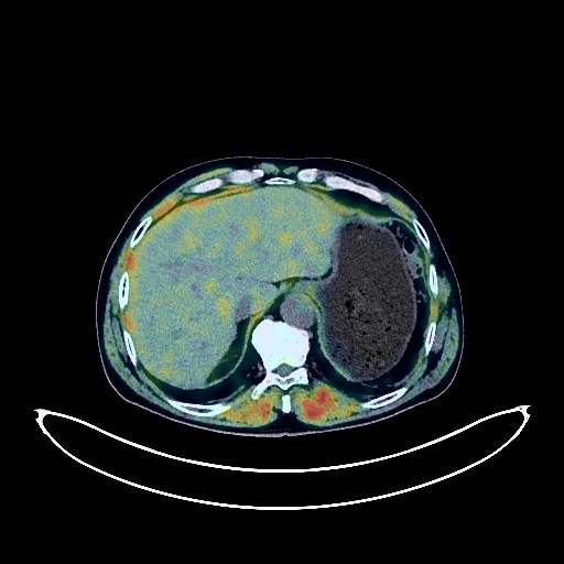
Lung Cancer PET/CT (case 983827-000246 from PETWB-REP)
0 views9 days agoWhole-body 18F-FDG PET/CT scan in a patient with Lung Cancer taken from the PETWB-REP dataset. The following English report (translated from original Chinese) is taken verbatim from the public dataset and has not been modified or otherwise checked for accuracy (see the end for citation). Impression a. A mass in the posterior segment of the right upper lobe, with increased FDG metabolism, consistent with lung cancer, and metastasis to the right hilar and part of the mediastinal lymph nodes. b. Metastasis to the posterior segment of the right lower lobe and the apical segment of the right upper lobe; several solid micronodules in the remaining two lungs, without high FDG metabolism; please follow up with CT scan. A few post-inflammatory lesions in both lungs. Mild pleural thickening bilaterally. Spleen absent post-surgery. Benign prostatic hyperplasia. Highly likely reactive hyperplasia of the bilateral inguinal lymph nodes; ultrasound follow-up is recommended. Degenerative changes in the spine. L4/5 and L5/S1 intervertebral disc bulge. Post-fracture changes of the right 5th/6th rib. No obvious abnormalities seen on cranial scintigraphy; MRI is recommended. Left sclerotic mastoid process; right mastoid air cell hypoplasia. Reactive hyperplasia of bilateral deep cervical spaces and submandibular lymph nodes. This case is from PETWB-REP, a curated dataset of whole-body 18F-FDG PET/CT scans and corresponding radiology reports from 490 patients with a broad spectrum of malignancies. The data were retrospectively collected from patients who underwent clinically indicated whole-body 18F-FDG PET/CT scans at the Shanghai Universal Medical Imaging Diagnostic Center between 2021 and 2024. License: Creative Commons Attribution 4.0 International (CC BY 4.0) Citation: Xue, L., Feng, G., Wenbo, Z., Zhang, Y., Li, L., Wang, S., Peng, L., Peng, S., & Gao, X. (2026). PETWB-REP: A Multi-Cancer Whole-Body FDG PET/CT Dataset with Corresponding Radiology Reports [Data set]. Zenodo. https://doi.org/10.5281/zenodo.18670487
Whole BodyPET/CT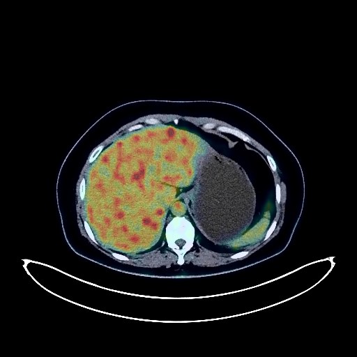
Lung Cancer PET/CT (case 983827-000098 from PETWB-REP)
0 views9 days agoWhole-body 18F-FDG PET/CT scan in a patient with Lung Cancer taken from the PETWB-REP dataset. The following English report (translated from original Chinese) is taken verbatim from the public dataset and has not been modified or otherwise checked for accuracy (see the end for citation). Impression a. Irregular soft tissue nodules in the lower lingular segment of the left upper lobe with mildly increased FDG metabolism, suggestive of lung cancer, but chronic inflammatory lesions need to be ruled out. A biopsy is recommended for definitive diagnosis. b. Multiple enlarged lymph nodes in the left hilum, mediastinum, and bilateral supraclavicular fossae with increased FDG uptake, highly suggestive of metastatic tumors. c. Multiple lesions in the right cerebral hemisphere and cerebellar vermis, highly suggestive of metastatic tumors. a. Chronic inflammatory solid nodules in the right middle lobe. Bilateral lower lobe aspiration effect. b. Bilateral dense glandular tissue, suspicious nodule in the central region of the left breast with mildly increased FDG metabolism, suggestive of fibroadenoma; ultrasound examination is recommended. Bilateral axillary lymph node reactive hyperplasia. Uterine cavity physiological uptake is highly likely, Nabothian cysts of the cervix, cystic lesions in the left ovary; follow-up ultrasound is recommended. Reactive hyperplasia of small retroperitoneal lymph nodes. Gastric antrum contraction with physiological uptake or chronic inflammatory changes; endoscopic re-examination may be necessary. Physiological uptake of some intestinal segments. Fatty liver, small hepatic cysts. Degenerative changes in the spine, L5 vertebral body grade I anterior slippage with pars interarticularis fracture, L1/2 intervertebral disc herniation with calcification. Reactive hyperplasia of bilateral cervical small lymph nodes. This case is from PETWB-REP, a curated dataset of whole-body 18F-FDG PET/CT scans and corresponding radiology reports from 490 patients with a broad spectrum of malignancies. The data were retrospectively collected from patients who underwent clinically indicated whole-body 18F-FDG PET/CT scans at the Shanghai Universal Medical Imaging Diagnostic Center between 2021 and 2024. License: Creative Commons Attribution 4.0 International (CC BY 4.0) Citation: Xue, L., Feng, G., Wenbo, Z., Zhang, Y., Li, L., Wang, S., Peng, L., Peng, S., & Gao, X. (2026). PETWB-REP: A Multi-Cancer Whole-Body FDG PET/CT Dataset with Corresponding Radiology Reports [Data set]. Zenodo. https://doi.org/10.5281/zenodo.18670487
Whole BodyPET/CT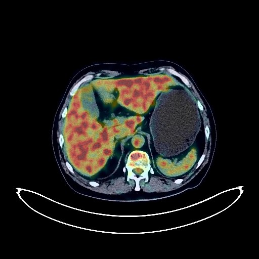
Lung Cancer PET/CT (case 983827-000097 from PETWB-REP)
0 views9 days agoWhole-body 18F-FDG PET/CT scan in a patient with Lung Cancer taken from the PETWB-REP dataset. The following English report (translated from original Chinese) is taken verbatim from the public dataset and has not been modified or otherwise checked for accuracy (see the end for citation). Impression a. Soft tissue lesions in the right middle lobe of the lung with increased FDG metabolism, highly suggestive of lung cancer with right hilar lymph node metastasis; please confirm with pathology. b. Reactive hyperplasia of mediastinal lymph nodes. c. Old lesion in the right apex of the lung. Multiple chronic inflammatory nodules, chronic inflammation, and sequelae in both lungs. Chronic bronchitis in both lungs, more pronounced in the right lung. Calcification of some arterial walls (including coronary arteries). a. Space-occupying lesion in the right cerebellopontine angle with decreased FDG metabolism, suggestive of a schwannoma; please provide a comprehensive analysis with enhanced MRI. b. Multiple ischemic and softening lesions in the deep brain. Bilateral ethmoid sinusitis and right maxillary sinusitis. Postoperative changes after thyroid cancer surgery; no signs of tumor recurrence were observed in the surgical area. Cyst in the right lobe of the liver. Right kidney stone. Physiological or inflammatory uptake of some intestinal segments. Degenerative changes in the spine. Left frozen shoulder. This case is from PETWB-REP, a curated dataset of whole-body 18F-FDG PET/CT scans and corresponding radiology reports from 490 patients with a broad spectrum of malignancies. The data were retrospectively collected from patients who underwent clinically indicated whole-body 18F-FDG PET/CT scans at the Shanghai Universal Medical Imaging Diagnostic Center between 2021 and 2024. License: Creative Commons Attribution 4.0 International (CC BY 4.0) Citation: Xue, L., Feng, G., Wenbo, Z., Zhang, Y., Li, L., Wang, S., Peng, L., Peng, S., & Gao, X. (2026). PETWB-REP: A Multi-Cancer Whole-Body FDG PET/CT Dataset with Corresponding Radiology Reports [Data set]. Zenodo. https://doi.org/10.5281/zenodo.18670487
Whole BodyPET/CT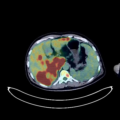
Liver Cancer PET/CT (case 983827-000003 from PETWB-REP)
0 views9 days agoWhole-body 18F-FDG PET/CT scan in a patient with Liver Cancer taken from the PETWB-REP dataset. The following English report (translated from original Chinese) is taken verbatim from the public dataset and has not been modified or otherwise checked for accuracy (see the end for citation). Impression a. Multiple intrahepatic lesions with increased FDG metabolism, suggestive of hepatocellular carcinoma, most likely hepatocellular carcinoma with multiple intrahepatic lesions; please correlate with clinicopathology. Right adrenal metastasis. Involvement of the inferior vena cava and main portal vein. b. Multiple lymph node metastases near the porta hepatis, in the hepatogastric space, retroperitoneum, and bilateral iliac vessels. Diffuse bone metastases throughout the body (see description), pathological fracture of the L5 vertebra. Metastases in both lungs and bilateral pleura. Localized increased FDG metabolism in the left temporal lobe, metastasis to be ruled out; enhanced MRI is recommended. A few ischemic lesions in the deep brain. Age-related brain changes. Gallbladder enlargement with cholestasis. Gallbladder neck stones. Splenomegaly. Accessory spleen. Small kidney stones in the left kidney. Chronic inflammation and sequelae in both lungs. Emphysema in the right upper lobe. Calcification of some arterial walls. Continuous increased FDG metabolism in the colon, suggestive of inflammatory uptake; please correlate with colonoscopy. Calcification of the tunica vaginalis in the left testis. Partial vertebral osteophyte formation. Right shoulder arthritis. Bilateral maxillary sinusitis. Reactive hyperplasia of the bilateral deep cervical space and submandibular lymph nodes. This case is from PETWB-REP, a curated dataset of whole-body 18F-FDG PET/CT scans and corresponding radiology reports from 490 patients with a broad spectrum of malignancies. The data were retrospectively collected from patients who underwent clinically indicated whole-body 18F-FDG PET/CT scans at the Shanghai Universal Medical Imaging Diagnostic Center between 2021 and 2024. License: Creative Commons Attribution 4.0 International (CC BY 4.0) Citation: Xue, L., Feng, G., Wenbo, Z., Zhang, Y., Li, L., Wang, S., Peng, L., Peng, S., & Gao, X. (2026). PETWB-REP: A Multi-Cancer Whole-Body FDG PET/CT Dataset with Corresponding Radiology Reports [Data set]. Zenodo. https://doi.org/10.5281/zenodo.18670487
Whole BodyPET/CT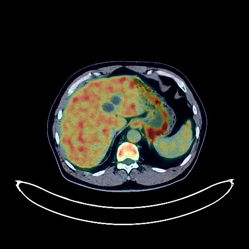
Lung Cancer PET/CT (case 983827-000245 from PETWB-REP)
0 views9 days agoWhole-body 18F-FDG PET/CT scan in a patient with Lung Cancer taken from the PETWB-REP dataset. The following English report (translated from original Chinese) is taken verbatim from the public dataset and has not been modified or otherwise checked for accuracy (see the end for citation). Impression a. A mass in the posterior segment of the right upper lobe with elevated FDG metabolism, suggestive of lung cancer; please confirm with pathology. Multiple lymph node metastases in the right hilum, mediastinum, and left deep cervical space. b. Multiple bone metastases throughout the body (see description for details). Small patchy low-density lesions in the right basal ganglia region, with normal FDG metabolism, suggestive of ischemic lesions; further MRI is recommended to rule out metastases. Chronic inflammatory micronodules in both lungs; follow-up with CT is recommended. A few post-inflammatory remnants in the right lung. Right pleural thickening. Calcification of some arterial walls (including coronary arteries). Multiple cysts in the liver. Chronic inflammatory changes or physiological changes in part of the gastric wall, resembling hemorrhoids; please confirm with endoscopy. Degenerative changes in the spine, L5/S1 disc bulge with pneumoconiosis and degeneration. This case is from PETWB-REP, a curated dataset of whole-body 18F-FDG PET/CT scans and corresponding radiology reports from 490 patients with a broad spectrum of malignancies. The data were retrospectively collected from patients who underwent clinically indicated whole-body 18F-FDG PET/CT scans at the Shanghai Universal Medical Imaging Diagnostic Center between 2021 and 2024. License: Creative Commons Attribution 4.0 International (CC BY 4.0) Citation: Xue, L., Feng, G., Wenbo, Z., Zhang, Y., Li, L., Wang, S., Peng, L., Peng, S., & Gao, X. (2026). PETWB-REP: A Multi-Cancer Whole-Body FDG PET/CT Dataset with Corresponding Radiology Reports [Data set]. Zenodo. https://doi.org/10.5281/zenodo.18670487
Whole BodyPET/CT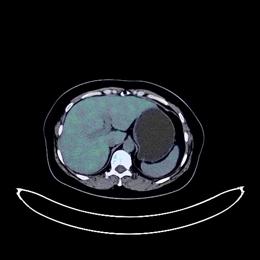
Lung Cancer PET/CT (case 983827-000237 from PETWB-REP)
0 views9 days agoWhole-body 18F-FDG PET/CT scan in a patient with Lung Cancer taken from the PETWB-REP dataset. The following English report (translated from original Chinese) is taken verbatim from the public dataset and has not been modified or otherwise checked for accuracy (see the end for citation). Impression a. A mass in the posterior segment of the left upper lobe, with elevated FDG metabolism, suggestive of peripheral lung cancer with metastatic nodules. b. A nodule in the lateral basal segment of the left lower lobe, closely attached to the diaphragm and pleura, with elevated FDG metabolism, suggesting a high probability of pleural invasion by a metastatic tumor; possible left hilar lymph node metastasis; please correlate with clinicopathology. c. Pure ground-glass nodules in the apical segment of the right upper lobe and the posterior segments of both lower lobes, with normal FDG metabolism, suggestive of chronic inflammatory nodules or atypical adenomatous hyperplasia; annual HRCT follow-up is recommended. d. Scattered chronic inflammatory nodules (solid and calcified) in the posterior segment of the left lower lobe and the right lung. Scattered chronic inflammation and remnants in both lungs. Enlarged cardiac silhouette, partial arteriosclerosis. Mild bilateral breast hyperplasia. A teratoma is highly probable in the left pelvis. Small uterine fibroids; Nabothian cysts of the cervix. Specialist and ultrasound follow-up is recommended for the above. Left renal cyst; bilateral renal microhamartomas are highly probable. Right renal stones or calcifications. Calcifications at the left adrenal junction. Splenic microcysts. Fatty liver. Gallstones. Chronic antral gastritis. Degenerative changes in the spine. Schmorl's nodes at the lower margin of the L4 vertebral body. L3/4, L4/5, and L5/S1 intervertebral disc bulges. Mild age-related brain changes, deep lacunar infarcts in the brain; MRI is recommended. Minor inflammation of the right maxillary sinus. Reactive hyperplasia of bilateral deep cervical interspace, submandibular, and submental lymph nodes. This case is from PETWB-REP, a curated dataset of whole-body 18F-FDG PET/CT scans and corresponding radiology reports from 490 patients with a broad spectrum of malignancies. The data were retrospectively collected from patients who underwent clinically indicated whole-body 18F-FDG PET/CT scans at the Shanghai Universal Medical Imaging Diagnostic Center between 2021 and 2024. License: Creative Commons Attribution 4.0 International (CC BY 4.0) Citation: Xue, L., Feng, G., Wenbo, Z., Zhang, Y., Li, L., Wang, S., Peng, L., Peng, S., & Gao, X. (2026). PETWB-REP: A Multi-Cancer Whole-Body FDG PET/CT Dataset with Corresponding Radiology Reports [Data set]. Zenodo. https://doi.org/10.5281/zenodo.18670487
Whole BodyPET/CT
Prostate Cancer PET/CT (case 983827-000227 from PETWB-REP)
1 views9 days agoWhole-body 18F-FDG PET/CT scan in a patient with Prostate Cancer taken from the PETWB-REP dataset. The following English report (translated from original Chinese) is taken verbatim from the public dataset and has not been modified or otherwise checked for accuracy (see the end for citation). Impression a. Benign prostatic hyperplasia with calcification; prostatic mass with increased FDG metabolism, suggestive of prostate cancer. Left pelvic wall lymph node metastasis, right pelvic wall lymph node metastasis to be ruled out. b. Focal increased FDG metabolism in the left part of the T12 vertebral body, please combine with MRI to rule out metastasis. Chronic inflammatory micronodules in the right upper lobe of the lung. Calcification in the left lower lobe of the lung, a few post-inflammatory remnants in both lungs. Slight thickening of the pleura bilaterally. Calcification of some arterial walls (including coronary arteries). Chronic cholecystitis. Calcification in the left testicular tunica vaginalis. Chronic inflammatory changes in the antrum of the stomach, please combine with endoscopic follow-up. Degenerative changes in the spine, L4/5 and L5/S1 intervertebral disc bulge. Osteitis condensans of the right sacroiliac joint. A low-density nodule in the left lobe of the thyroid gland with elevated FDG metabolism, suggestive of an adenomatous nodule; please confirm with ultrasound examination. Elderly patient with deep lacunar infarcts and chronic inflammation of both maxillary sinuses. This case is from PETWB-REP, a curated dataset of whole-body 18F-FDG PET/CT scans and corresponding radiology reports from 490 patients with a broad spectrum of malignancies. The data were retrospectively collected from patients who underwent clinically indicated whole-body 18F-FDG PET/CT scans at the Shanghai Universal Medical Imaging Diagnostic Center between 2021 and 2024. License: Creative Commons Attribution 4.0 International (CC BY 4.0) Citation: Xue, L., Feng, G., Wenbo, Z., Zhang, Y., Li, L., Wang, S., Peng, L., Peng, S., & Gao, X. (2026). PETWB-REP: A Multi-Cancer Whole-Body FDG PET/CT Dataset with Corresponding Radiology Reports [Data set]. Zenodo. https://doi.org/10.5281/zenodo.18670487
Whole BodyPET/CT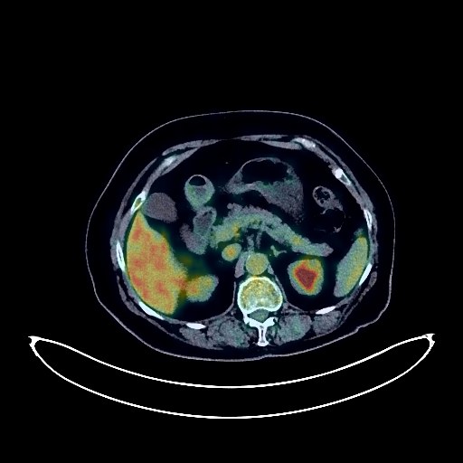
Breast Cancer PET/CT (case 983827-000099 from PETWB-REP)
2 views9 days agoWhole-body 18F-FDG PET/CT scan in a patient with Breast Cancer taken from the PETWB-REP dataset. The following English report (translated from original Chinese) is taken verbatim from the public dataset and has not been modified or otherwise checked for accuracy (see the end for citation). Impression Left breast mass with elevated FDG metabolism, suggestive of breast cancer; please confirm clinicopathological findings. Multiple lymph node metastases in the left axilla, left interpectoral space, left supraclavicular fossa, and para-aortic arch. Chronic inflammatory nodules in both lungs. Scattered post-inflammatory lesions in both lungs. Partial arteriosclerosis. Possible uterine fibroids; please confirm with ultrasound examination. Postoperative changes in the left groin. Chronic inflammatory changes in the gastric antrum; please confirm with endoscopic follow-up. Degenerative changes in the spine; L4/5 disc herniation. Uneven thyroid density; bilateral lobular calcifications; ultrasound follow-up recommended. Deep lacunar infarcts in the brain. Chronic inflammation of the right maxillary sinus. Reactive hyperplasia of bilateral cervical lymph nodes. This case is from PETWB-REP, a curated dataset of whole-body 18F-FDG PET/CT scans and corresponding radiology reports from 490 patients with a broad spectrum of malignancies. The data were retrospectively collected from patients who underwent clinically indicated whole-body 18F-FDG PET/CT scans at the Shanghai Universal Medical Imaging Diagnostic Center between 2021 and 2024. License: Creative Commons Attribution 4.0 International (CC BY 4.0) Citation: Xue, L., Feng, G., Wenbo, Z., Zhang, Y., Li, L., Wang, S., Peng, L., Peng, S., & Gao, X. (2026). PETWB-REP: A Multi-Cancer Whole-Body FDG PET/CT Dataset with Corresponding Radiology Reports [Data set]. Zenodo. https://doi.org/10.5281/zenodo.18670487
Whole BodyPET/CT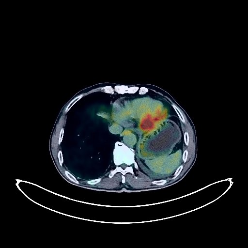
Lung Cancer PET/CT (case 983827-000056 from PETWB-REP)
2 views9 days agoWhole-body 18F-FDG PET/CT scan in a patient with Lung Cancer taken from the PETWB-REP dataset. The following English report (translated from original Chinese) is taken verbatim from the public dataset and has not been modified or otherwise checked for accuracy (see the end for citation). Impression a. A space-occupying lesion in the left upper lobe, adjacent to the mediastinum, with increased FDG metabolism, consistent with lung cancer with obstructive changes, invading adjacent major blood vessels and the pericardium. b. Multiple soft tissue nodules in the left lower 6th-10th intercostal spaces, with normal FDG metabolism, suggestive of metastatic tumors. c. Moderate pleural effusion in the left side. Slight pericardial thickening. Reactive hyperplasia of the hilar and mediastinal lymph nodes is highly probable, with partial metastasis to be ruled out. d. Interstitial changes in both lungs with multiple scattered chronic inflammations and old lesions. Emphysema with bullae in both lungs. Calcification of some arterial walls (including coronary arteries). Duodenal diverticulum in the descending part. Degenerative changes in the spine. L4/5 and L5/S1 intervertebral disc bulges. A few ischemic lesions in the deep bilateral brain, senile cerebral changes. This case is from PETWB-REP, a curated dataset of whole-body 18F-FDG PET/CT scans and corresponding radiology reports from 490 patients with a broad spectrum of malignancies. The data were retrospectively collected from patients who underwent clinically indicated whole-body 18F-FDG PET/CT scans at the Shanghai Universal Medical Imaging Diagnostic Center between 2021 and 2024. License: Creative Commons Attribution 4.0 International (CC BY 4.0) Citation: Xue, L., Feng, G., Wenbo, Z., Zhang, Y., Li, L., Wang, S., Peng, L., Peng, S., & Gao, X. (2026). PETWB-REP: A Multi-Cancer Whole-Body FDG PET/CT Dataset with Corresponding Radiology Reports [Data set]. Zenodo. https://doi.org/10.5281/zenodo.18670487
Whole BodyPET/CT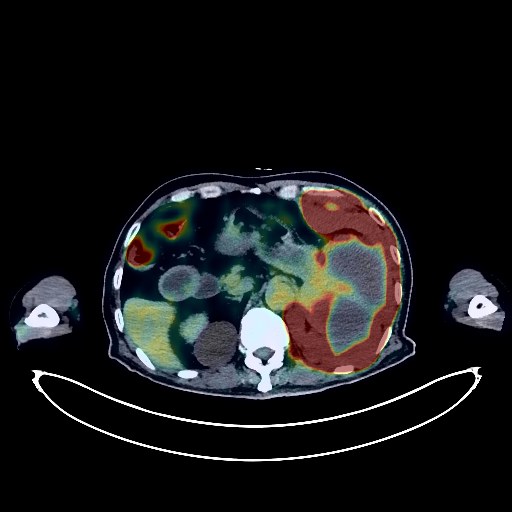
Pancreatic Cancer PET/CT (case 983827-000075 from PETWB-REP)
1 views9 days agoWhole-body 18F-FDG PET/CT scan in a patient with Pancreatic Cancer taken from the PETWB-REP dataset. The following English report (translated from original Chinese) is taken verbatim from the public dataset and has not been modified or otherwise checked for accuracy (see the end for citation). Impression A mass in the tail of the pancreas with increased FDG metabolism; a mass in the spleen with increased FDG metabolism; thickening of the left upper quadrant peritoneum and adjacent diaphragm with increased FDG metabolism; multiple enlarged lymph nodes throughout the body with increased FDG metabolism. These findings suggest malignancy, possibly pancreatic cancer with multiple metastases, or multi-systemic lymphoma. A comprehensive analysis combining tumor markers is recommended, and a biopsy may be necessary. Bronchiectasis in both lungs with chronic inflammation and remnants. Reactive hyperplasia of the hilar and mediastinal lymph nodes in both lungs. Mild pleural thickening bilaterally, with left-sided pleural effusion. Post-coronary stent placement, with partial arteriosclerosis (including coronary arteries). Liver cirrhosis. Multiple renal cysts. Residual contrast agent in the urinary system. Prostatic calcifications. Continuous increased FDG metabolism in parts of the intestine, considered to be physiological uptake or chronic inflammation. Spinal degeneration. L5 vertebral instability. L2/3, L3/4, L4/5 intervertebral disc bulge; L4/5, L5/S1 intervertebral disc pneumoconiosis and degeneration. Benign osteopathy of the right humeral head. Bilateral deep lacunar infarcts, age-related brain encephalopathy. This case is from PETWB-REP, a curated dataset of whole-body 18F-FDG PET/CT scans and corresponding radiology reports from 490 patients with a broad spectrum of malignancies. The data were retrospectively collected from patients who underwent clinically indicated whole-body 18F-FDG PET/CT scans at the Shanghai Universal Medical Imaging Diagnostic Center between 2021 and 2024. License: Creative Commons Attribution 4.0 International (CC BY 4.0) Citation: Xue, L., Feng, G., Wenbo, Z., Zhang, Y., Li, L., Wang, S., Peng, L., Peng, S., & Gao, X. (2026). PETWB-REP: A Multi-Cancer Whole-Body FDG PET/CT Dataset with Corresponding Radiology Reports [Data set]. Zenodo. https://doi.org/10.5281/zenodo.18670487
Whole BodyPET/CT