Loading...
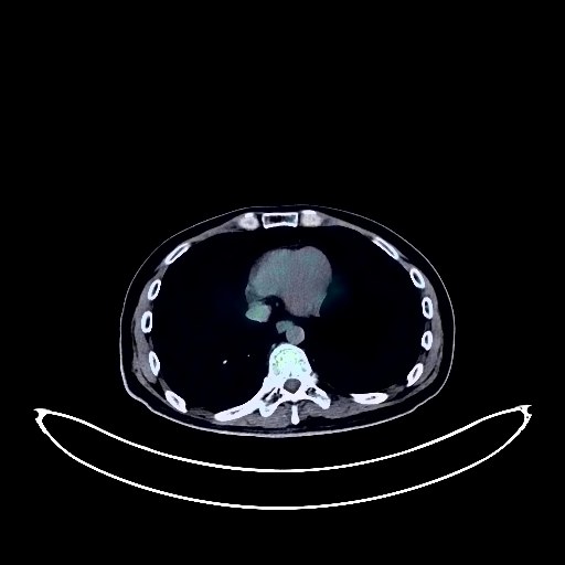
Colon Cancer PET/CT (case 983827-000116 from PETWB-REP)
0 views9 days agoWhole-body 18F-FDG PET/CT scan in a patient with Colon Cancer taken from the PETWB-REP dataset. The following English report (translated from original Chinese) is taken verbatim from the public dataset and has not been modified or otherwise checked for accuracy (see the end for citation). Impression a. Changes from "sigmoid colon cancer resection + partial small bowel resection + ileocecal resection + terminal ileostomy", multiple soft tissue lesions around the anastomoses of the intestines and in the abdominopelvic cavity with significantly increased FDG metabolism, considered to be metastatic tumors, with involvement of the adjacent bladder floor and intestines. b. Metastatic tumors in the left lateral lobe of the liver and the lower segment of the right posterior lobe of the liver. Metastasis to the bilateral iliac vessels and retroperitoneal para-aortic lymph nodes. Linearly increased FDG metabolism in the remaining intestinal tract, considered to be physiological uptake. Poor ventricular relaxation, no increased FDG metabolism observed. Chronic inflammatory ground-glass nodules or atypical adenomatous hyperplasia in the middle and lower lobes of the right lung, chronic inflammatory solid micronodules in the upper lobe of the right lung and the lower lobe of the left lung. A few fibrotic lesions in both lungs. Cyst in the lower segment of the right anterior lobe of the liver; subcapsular calcification in the lower segment of the right posterior lobe of the liver. Gallstones; chronic cholecystitis. Accessory spleen. Bilateral renal cysts. Mild osteophyte formation in some lumbar vertebrae. Physiological uptake of the erector spinae muscles in the cervical spine. No obvious abnormalities were found on cranial scintigraphy. Possible periodontitis in the left lower molar region; please consult a specialist. This case is from PETWB-REP, a curated dataset of whole-body 18F-FDG PET/CT scans and corresponding radiology reports from 490 patients with a broad spectrum of malignancies. The data were retrospectively collected from patients who underwent clinically indicated whole-body 18F-FDG PET/CT scans at the Shanghai Universal Medical Imaging Diagnostic Center between 2021 and 2024. License: Creative Commons Attribution 4.0 International (CC BY 4.0) Citation: Xue, L., Feng, G., Wenbo, Z., Zhang, Y., Li, L., Wang, S., Peng, L., Peng, S., & Gao, X. (2026). PETWB-REP: A Multi-Cancer Whole-Body FDG PET/CT Dataset with Corresponding Radiology Reports [Data set]. Zenodo. https://doi.org/10.5281/zenodo.18670487
Whole BodyPET/CT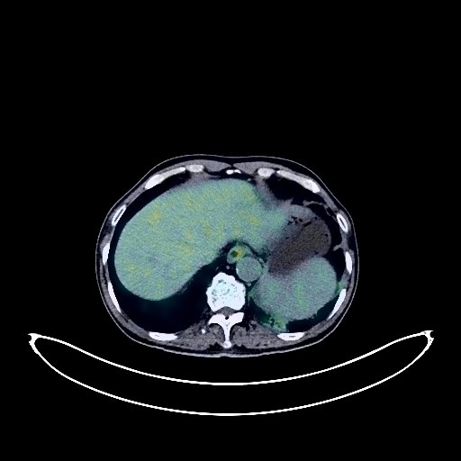
Lung Cancer PET/CT (case 983827-000095 from PETWB-REP)
0 views9 days agoWhole-body 18F-FDG PET/CT scan in a patient with Lung Cancer taken from the PETWB-REP dataset. The following English report (translated from original Chinese) is taken verbatim from the public dataset and has not been modified or otherwise checked for accuracy (see the end for citation). Impression a. Irregular mass in the left lower lobe with increased FDG metabolism, suggestive of lung cancer with obstructive inflammation; please correlate with clinicopathology. b. Multiple metastatic tumors in the left hilum, mediastinum, retroperitoneum, and right pelvic wall. Bilateral emphysema, bilateral chronic inflammatory micronodules. Scattered post-inflammatory lesions in both lungs. Tracheal diverticulum. Calcification of some arterial walls (including coronary arteries). Chronic cholecystitis. Splenic calcification. Benign prostatic hyperplasia with calcification. Schistosomiasis intestinal manifestations, chronic inflammatory changes in the gastric antrum. Osteoporosis, degenerative changes in the spine, multiple intervertebral disc bulges, L4/5 intervertebral disc pneumoconiosis, L5/S1 vertebral endplate inflammation. Inflammation around the left ischial tuberosity. Senile brain, deep lacunar infarcts. Subcutaneous calcified nodules in the occipital region. This case is from PETWB-REP, a curated dataset of whole-body 18F-FDG PET/CT scans and corresponding radiology reports from 490 patients with a broad spectrum of malignancies. The data were retrospectively collected from patients who underwent clinically indicated whole-body 18F-FDG PET/CT scans at the Shanghai Universal Medical Imaging Diagnostic Center between 2021 and 2024. License: Creative Commons Attribution 4.0 International (CC BY 4.0) Citation: Xue, L., Feng, G., Wenbo, Z., Zhang, Y., Li, L., Wang, S., Peng, L., Peng, S., & Gao, X. (2026). PETWB-REP: A Multi-Cancer Whole-Body FDG PET/CT Dataset with Corresponding Radiology Reports [Data set]. Zenodo. https://doi.org/10.5281/zenodo.18670487
Whole BodyPET/CT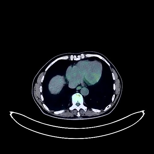
Colon Cancer PET/CT (case 983827-000201 from PETWB-REP)
0 views9 days agoWhole-body 18F-FDG PET/CT scan in a patient with Colon Cancer taken from the PETWB-REP dataset. The following English report (translated from original Chinese) is taken verbatim from the public dataset and has not been modified or otherwise checked for accuracy (see the end for citation). Impression a. Slight thickening of the intestinal wall at the junction of the descending colon and sigmoid colon with increased FDG metabolism, consistent with pathological findings of colorectal cancer, with a high probability of involvement of the serosa. Please correlate with clinical findings. b. Localized symmetrical narrowing of the sigmoid colon near the rectum with increased FDG metabolism, likely due to intestinal contraction and physiological uptake; slight localized thickening of the ileocecal wall with increased FDG metabolism, suggesting a possible space-occupying lesion, but physiological changes need to be ruled out. Please analyze the above in conjunction with enhanced CT scans. c. Reactive hyperplasia of lymph nodes in the bilateral pelvic walls, parailiac vessels, retroperitoneum, hilar space, and hepatic hilum. d. Continuous increased FDG metabolism in the remaining colon, suggesting physiological uptake or inflammatory changes. Please follow up with colonoscopy. Chronic inflammatory micronodules in both lungs. Emphysema in both lungs. Chronic inflammatory lymph nodes in the bilateral hilar and mediastinal regions. Calcification of some arterial walls (including coronary arteries). Liver calcifications. Small gallstones, chronic cholecystitis. Right renal cyst. Prostatic calcifications. Spinal degeneration, L2-4 vertebral instability, L4/5 and L5/S1 vertebral endplate inflammation. L3/4 intervertebral disc bulge, L4/5 intervertebral disc herniation, partial calcification. Benign bone disease of the proximal right femur. Right occipital lobe softening lesion, bilateral deep lacunar infarcts, mild age-related brain changes; please correlate with MRI. Minor inflammation of the right maxillary sinus and bilateral ethmoid sinuses. This case is from PETWB-REP, a curated dataset of whole-body 18F-FDG PET/CT scans and corresponding radiology reports from 490 patients with a broad spectrum of malignancies. The data were retrospectively collected from patients who underwent clinically indicated whole-body 18F-FDG PET/CT scans at the Shanghai Universal Medical Imaging Diagnostic Center between 2021 and 2024. License: Creative Commons Attribution 4.0 International (CC BY 4.0) Citation: Xue, L., Feng, G., Wenbo, Z., Zhang, Y., Li, L., Wang, S., Peng, L., Peng, S., & Gao, X. (2026). PETWB-REP: A Multi-Cancer Whole-Body FDG PET/CT Dataset with Corresponding Radiology Reports [Data set]. Zenodo. https://doi.org/10.5281/zenodo.18670487
Whole BodyPET/CT
Lung Cancer PET/CT (case 983827-000046 from PETWB-REP)
0 views9 days agoWhole-body 18F-FDG PET/CT scan in a patient with Lung Cancer taken from the PETWB-REP dataset. The following English report (translated from original Chinese) is taken verbatim from the public dataset and has not been modified or otherwise checked for accuracy (see the end for citation). Impression a. Space-occupying lesion in the upper lobe of the right lung, with increased FDG metabolism, consistent with lung cancer with obstructive atelectasis. b. Bilateral lung metastases are highly probable; regular CT scans are recommended. c. Right hilar and pretracheal lymph node metastases are highly probable. d. A few ischemic lesions in the deep bilateral brain regions; MRI scans are recommended. Partial arterial wall calcification. Bile duct stones or calcifications in the right lobe of the liver; ultrasound follow-up is recommended. Partial vertebral osteophyte formation. Right-sided frozen shoulder. This case is from PETWB-REP, a curated dataset of whole-body 18F-FDG PET/CT scans and corresponding radiology reports from 490 patients with a broad spectrum of malignancies. The data were retrospectively collected from patients who underwent clinically indicated whole-body 18F-FDG PET/CT scans at the Shanghai Universal Medical Imaging Diagnostic Center between 2021 and 2024. License: Creative Commons Attribution 4.0 International (CC BY 4.0) Citation: Xue, L., Feng, G., Wenbo, Z., Zhang, Y., Li, L., Wang, S., Peng, L., Peng, S., & Gao, X. (2026). PETWB-REP: A Multi-Cancer Whole-Body FDG PET/CT Dataset with Corresponding Radiology Reports [Data set]. Zenodo. https://doi.org/10.5281/zenodo.18670487
Whole BodyPET/CT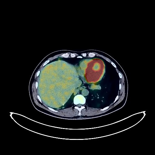
Lung Cancer PET/CT (case 983827-000239 from PETWB-REP)
0 views9 days agoWhole-body 18F-FDG PET/CT scan in a patient with Lung Cancer taken from the PETWB-REP dataset. The following English report (translated from original Chinese) is taken verbatim from the public dataset and has not been modified or otherwise checked for accuracy (see the end for citation). Impression a. Irregular plaque-like lesions with increased FDG metabolism in the anterior segment of the left upper lobe, adjacent to the mediastinum, lung cancer is the primary consideration; suspected space-occupying lesions in both lower lobes; signs of lung cancer-related lymphangitis in both lungs. The above should be compared with older scans and closely monitored. b. Multiple lymph nodes in the bilateral hilar, mediastinal, right axilla, right supraclavicular fossa, right deep cervical space, hepatogastric space, and retroperitoneal region showing increased FDG metabolism, some suggestive of metastases. c. Metastases in the C7 vertebral body and right sacral region. T5 vertebral metastasis to be ruled out. Chronic inflammatory nodules in both lungs; CT follow-up is recommended to rule out partial metastases. Emphysema in both upper lobes; calcification in the left upper lobe. Micropleural effusion in both pleural cavities. Partial arteriosclerosis. Benign prostatic hyperplasia with calcification and uneven FDG metabolism; PSA testing is recommended. Gallstones. Right kidney stones with hydronephrosis. Chronic inflammatory changes in the gastric antrum; please follow up with endoscopy. Degenerative changes in the spine; L4/5 and L5/S1 intervertebral disc bulges. No obvious abnormalities were found on cranial scintigraphy. This case is from PETWB-REP, a curated dataset of whole-body 18F-FDG PET/CT scans and corresponding radiology reports from 490 patients with a broad spectrum of malignancies. The data were retrospectively collected from patients who underwent clinically indicated whole-body 18F-FDG PET/CT scans at the Shanghai Universal Medical Imaging Diagnostic Center between 2021 and 2024. License: Creative Commons Attribution 4.0 International (CC BY 4.0) Citation: Xue, L., Feng, G., Wenbo, Z., Zhang, Y., Li, L., Wang, S., Peng, L., Peng, S., & Gao, X. (2026). PETWB-REP: A Multi-Cancer Whole-Body FDG PET/CT Dataset with Corresponding Radiology Reports [Data set]. Zenodo. https://doi.org/10.5281/zenodo.18670487
Whole BodyPET/CT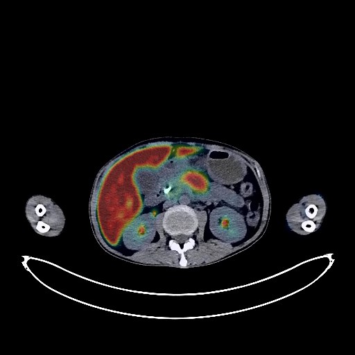
Lymphoma PET/CT (case 983827-000086 from PETWB-REP)
1 views9 days agoWhole-body 18F-FDG PET/CT scan in a patient with Lymphoma taken from the PETWB-REP dataset. The following English report (translated from original Chinese) is taken verbatim from the public dataset and has not been modified or otherwise checked for accuracy (see the end for citation). Impression a. After treatment for nasal lymphoma, no obvious FDG metabolism elevation foci were observed in the nose, suggesting suppressed tumor activity. b. Diffuse FDG metabolism elevation in the liver; pancreatic swelling with surrounding effusion and FDG metabolism elevation; bilateral adrenal gland lesions with FDG metabolism elevation; multiple lymph nodes in the bilateral posterior diaphragmatic crura, hepatogastric space, and retroperitoneum showing FDG metabolism elevation. All of the above suggest lymphoma infiltration, and follow-up examination after treatment is recommended. A few post-inflammatory lesions in both lungs. Anemia. Right anterior chest wall port-a-cath placement. Fatty liver. Post-cholecystectomy changes, intrahepatic bile duct pneumothorax, chronic cholecystitis. Slightly enlarged spleen. Bilateral renal stones. Pelvic effusion. Possible physiological uptake or chronic inflammatory changes in the right lower abdomen, lymphoma infiltration to be ruled out, please follow up. No obvious abnormalities were seen on cranial scintigraphy. Chronic inflammation of bilateral ethmoid and maxillary sinuses. This case is from PETWB-REP, a curated dataset of whole-body 18F-FDG PET/CT scans and corresponding radiology reports from 490 patients with a broad spectrum of malignancies. The data were retrospectively collected from patients who underwent clinically indicated whole-body 18F-FDG PET/CT scans at the Shanghai Universal Medical Imaging Diagnostic Center between 2021 and 2024. License: Creative Commons Attribution 4.0 International (CC BY 4.0) Citation: Xue, L., Feng, G., Wenbo, Z., Zhang, Y., Li, L., Wang, S., Peng, L., Peng, S., & Gao, X. (2026). PETWB-REP: A Multi-Cancer Whole-Body FDG PET/CT Dataset with Corresponding Radiology Reports [Data set]. Zenodo. https://doi.org/10.5281/zenodo.18670487
Whole BodyPET/CT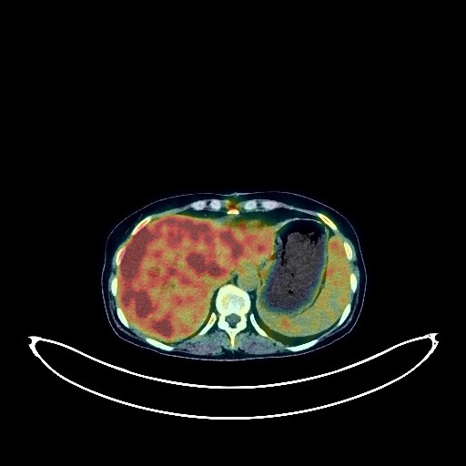
Cholangiocarcinoma PET/CT (case 983827-000205 from PETWB-REP)
0 views9 days agoWhole-body 18F-FDG PET/CT scan in a patient with Cholangiocarcinoma taken from the PETWB-REP dataset. The following English report (translated from original Chinese) is taken verbatim from the public dataset and has not been modified or otherwise checked for accuracy (see the end for citation). Impression a. Postoperative changes following palliative surgery for a space-occupying lesion in the hepatic hilum and bile duct stent placement; dilation of intrahepatic bile ducts, with pneumothorax in some bile ducts; multiple irregular patchy and nodular foci of increased FDG metabolism in the liver, with lesions predominantly accumulating near the distal bile ducts. Considering the medical history, hilar cholangiocarcinoma with intrahepatic metastasis and cholangitis is highly probable. Specialist and MRI follow-up is recommended. b. Splenomegaly; increased FDG metabolism observed in the bone marrow cavity suggests reactive bone marrow hyperplasia. Scattered infectious lesions in the right lung; CT follow-up is recommended. Chronic inflammatory miliary nodules in both lungs. A few chronic inflammations and old lesions in both lungs. Reactive hyperplasia of hilar and mediastinal lymph nodes in both lungs. Small amount of pericardial effusion. Incomplete bilateral mammary regression. Calcification of some arterial walls. Slight thickening of the walls in parts of the gastric body and antrum, with mildly increased FDG uptake, suggestive of chronic gastritis; increased FDG metabolism in parts of the intestines, suggestive of inflammatory or physiological uptake. Follow-up gastroscopy and colonoscopy are recommended. Degenerative changes in the spine. Sacral canal cyst. Irregular morphology of the right iliac bone; please correlate with clinical findings. No obvious abnormalities were found on cranial FDG imaging. This case is from PETWB-REP, a curated dataset of whole-body 18F-FDG PET/CT scans and corresponding radiology reports from 490 patients with a broad spectrum of malignancies. The data were retrospectively collected from patients who underwent clinically indicated whole-body 18F-FDG PET/CT scans at the Shanghai Universal Medical Imaging Diagnostic Center between 2021 and 2024. License: Creative Commons Attribution 4.0 International (CC BY 4.0) Citation: Xue, L., Feng, G., Wenbo, Z., Zhang, Y., Li, L., Wang, S., Peng, L., Peng, S., & Gao, X. (2026). PETWB-REP: A Multi-Cancer Whole-Body FDG PET/CT Dataset with Corresponding Radiology Reports [Data set]. Zenodo. https://doi.org/10.5281/zenodo.18670487
Whole BodyPET/CT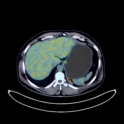
Lung Cancer PET/CT (case 983827-000066 from PETWB-REP)
0 views9 days agoWhole-body 18F-FDG PET/CT scan in a patient with Lung Cancer taken from the PETWB-REP dataset. The following English report (translated from original Chinese) is taken verbatim from the public dataset and has not been modified or otherwise checked for accuracy (see the end for citation). Impression a. Large mass in the lower lobe of the left lung, with increased FDG metabolism, suggestive of lung cancer. b. Multiple lymph node metastases in the bilateral hilar, mediastinal, and bilateral supraclavicular fossa. c. Diffuse metastases in both lungs. L3 and L5 vertebral metastases. Uneven density in the right cerebellopontine angle, no abnormalities in FDG metabolism; please repeat with contrast-enhanced MRI. Scattered chronic inflammation and old lesions in both lungs. Calcification of some arterial walls (including coronary arteries). Stone in the upper segment of the left ureter, mild hydronephrosis and dilation of the proximal ureter and renal pelvis. Degenerative changes in the spine. L4/5 and L5/S1 intervertebral disc bulges. Chronic inflammation of the right maxillary sinus. This case is from PETWB-REP, a curated dataset of whole-body 18F-FDG PET/CT scans and corresponding radiology reports from 490 patients with a broad spectrum of malignancies. The data were retrospectively collected from patients who underwent clinically indicated whole-body 18F-FDG PET/CT scans at the Shanghai Universal Medical Imaging Diagnostic Center between 2021 and 2024. License: Creative Commons Attribution 4.0 International (CC BY 4.0) Citation: Xue, L., Feng, G., Wenbo, Z., Zhang, Y., Li, L., Wang, S., Peng, L., Peng, S., & Gao, X. (2026). PETWB-REP: A Multi-Cancer Whole-Body FDG PET/CT Dataset with Corresponding Radiology Reports [Data set]. Zenodo. https://doi.org/10.5281/zenodo.18670487
Whole BodyPET/CT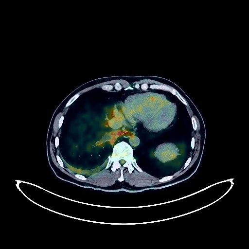
Lung Cancer PET/CT (case 983827-000150 from PETWB-REP)
0 views9 days agoWhole-body 18F-FDG PET/CT scan in a patient with Lung Cancer taken from the PETWB-REP dataset. The following English report (translated from original Chinese) is taken verbatim from the public dataset and has not been modified or otherwise checked for accuracy (see the end for citation). Impression a. A mass in the right hilum with elevated FDG metabolism, stenosis of the posterior segment of the lower lobe bronchus and part of the basal segment bronchus, strongly suggestive of lung cancer, atypical infection to be ruled out; b. High probability of metastasis to the right hilar, mediastinal, and right supraclavicular fossa lymph nodes; inflammatory lymph nodes to be ruled out. Reactive hyperplasia of bilateral deep cervical spaces and bilateral submandibular lymph nodes. c. Obstructive pneumonia in the right lung. Thickening of the right oblique fissure pleura. Small amount of pleural effusion in the right pleural cavity. Fatty liver. Bilateral renal cysts. Pelvic calcifications. Prostatic cysts. Bilateral hydrocele. Spinal degenerative changes. No obvious abnormalities seen on cranial scintigraphy. This case is from PETWB-REP, a curated dataset of whole-body 18F-FDG PET/CT scans and corresponding radiology reports from 490 patients with a broad spectrum of malignancies. The data were retrospectively collected from patients who underwent clinically indicated whole-body 18F-FDG PET/CT scans at the Shanghai Universal Medical Imaging Diagnostic Center between 2021 and 2024. License: Creative Commons Attribution 4.0 International (CC BY 4.0) Citation: Xue, L., Feng, G., Wenbo, Z., Zhang, Y., Li, L., Wang, S., Peng, L., Peng, S., & Gao, X. (2026). PETWB-REP: A Multi-Cancer Whole-Body FDG PET/CT Dataset with Corresponding Radiology Reports [Data set]. Zenodo. https://doi.org/10.5281/zenodo.18670487
Whole BodyPET/CT
Liver Cancer PET/CT (case 983827-000241 from PETWB-REP)
0 views9 days agoWhole-body 18F-FDG PET/CT scan in a patient with Liver Cancer taken from the PETWB-REP dataset. The following English report (translated from original Chinese) is taken verbatim from the public dataset and has not been modified or otherwise checked for accuracy (see the end for citation). Impression A large mass in the liver with unevenly increased FDG uptake suggests a malignant tumor, possibly hepatoblastoma. Please correlate with clinicopathology. Signs of anemia. Inflammatory or physiological uptake in the ileocecal region. No abnormal density shadows or abnormally increased FDG uptake observed in the bones throughout the body. No obvious abnormalities were found on cranial imaging. This case is from PETWB-REP, a curated dataset of whole-body 18F-FDG PET/CT scans and corresponding radiology reports from 490 patients with a broad spectrum of malignancies. The data were retrospectively collected from patients who underwent clinically indicated whole-body 18F-FDG PET/CT scans at the Shanghai Universal Medical Imaging Diagnostic Center between 2021 and 2024. License: Creative Commons Attribution 4.0 International (CC BY 4.0) Citation: Xue, L., Feng, G., Wenbo, Z., Zhang, Y., Li, L., Wang, S., Peng, L., Peng, S., & Gao, X. (2026). PETWB-REP: A Multi-Cancer Whole-Body FDG PET/CT Dataset with Corresponding Radiology Reports [Data set]. Zenodo. https://doi.org/10.5281/zenodo.18670487
Whole BodyPET/CT