Loading...
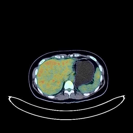
Ovarian Cancer PET/CT (case 983827-000129 from PETWB-REP)
0 views9 days agoWhole-body 18F-FDG PET/CT scan in a patient with Ovarian Cancer taken from the PETWB-REP dataset. The following English report (translated from original Chinese) is taken verbatim from the public dataset and has not been modified or otherwise checked for accuracy (see the end for citation). Impression A large, multilocular cystic-solid lesion in the abdominopelvic cavity, with increased FDG metabolism in the solid portion, is highly suggestive of a right ovarian cystadenoma with local malignant transformation; please confirm with pathology. Physiological uptake in the uterine cavity; physiological cyst in the left adnexal region; please confirm with ultrasound follow-up. Small amount of pelvic effusion. No obvious abnormalities seen on cranial scintigraphy. Reactive hyperplasia of bilateral cervical lymph nodes. This case is from PETWB-REP, a curated dataset of whole-body 18F-FDG PET/CT scans and corresponding radiology reports from 490 patients with a broad spectrum of malignancies. The data were retrospectively collected from patients who underwent clinically indicated whole-body 18F-FDG PET/CT scans at the Shanghai Universal Medical Imaging Diagnostic Center between 2021 and 2024. License: Creative Commons Attribution 4.0 International (CC BY 4.0) Citation: Xue, L., Feng, G., Wenbo, Z., Zhang, Y., Li, L., Wang, S., Peng, L., Peng, S., & Gao, X. (2026). PETWB-REP: A Multi-Cancer Whole-Body FDG PET/CT Dataset with Corresponding Radiology Reports [Data set]. Zenodo. https://doi.org/10.5281/zenodo.18670487
Whole BodyPET/CT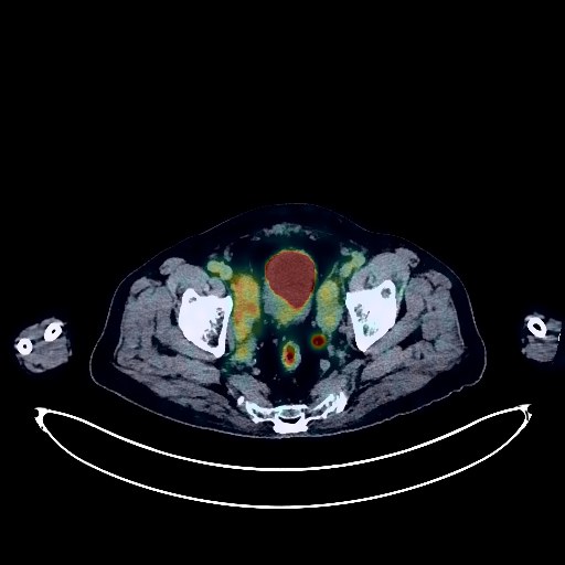
Prostate Cancer PET/CT (case 983827-000054 from PETWB-REP)
0 views9 days agoWhole-body 18F-FDG PET/CT scan in a patient with Prostate Cancer taken from the PETWB-REP dataset. The following English report (translated from original Chinese) is taken verbatim from the public dataset and has not been modified or otherwise checked for accuracy (see the end for citation). Impression a. Irregular prostatic lesion with elevated FDG metabolism, highly suggestive of prostate cancer; please combine PSA and MRI for comprehensive analysis; benign prostatic hyperplasia with calcification. b. Multiple lymph node metastases in bilateral pelvic walls, bilateral iliac vessels, and retroperitoneum are highly probable; lymphoma to be ruled out. a. Chronic inflammatory micronodules in both lungs; CT follow-up is recommended to rule out other complications. Mild emphysema in the upper lobes of both lungs; chronic inflammation and post-inflammatory remnants in both lungs. b. Chronic inflammatory lymph nodes in the hilar and mediastinal regions of both lungs. Pleural thickening bilaterally. Calcification of some arterial walls (including coronary arteries). Multiple liver cysts. Post-cholecystectomy changes; compensatory dilatation of the common bile duct. Fatty infiltration of the pancreas. Right renal cyst. Chronic inflammatory changes in part of the gastric wall and intestinal tract; please combine endoscopic follow-up. Osteoporosis; degenerative changes in the spine; posterior slippage of L2 and L3 vertebral bodies; multiple bulging lumbar intervertebral discs with pneumothorax and degeneration. Age-related brain abnormalities, deep lacunar infarcts, bilateral basal ganglia softening, and chronic inflammation of the right ethmoid sinus and left maxillary sinus. This case is from PETWB-REP, a curated dataset of whole-body 18F-FDG PET/CT scans and corresponding radiology reports from 490 patients with a broad spectrum of malignancies. The data were retrospectively collected from patients who underwent clinically indicated whole-body 18F-FDG PET/CT scans at the Shanghai Universal Medical Imaging Diagnostic Center between 2021 and 2024. License: Creative Commons Attribution 4.0 International (CC BY 4.0) Citation: Xue, L., Feng, G., Wenbo, Z., Zhang, Y., Li, L., Wang, S., Peng, L., Peng, S., & Gao, X. (2026). PETWB-REP: A Multi-Cancer Whole-Body FDG PET/CT Dataset with Corresponding Radiology Reports [Data set]. Zenodo. https://doi.org/10.5281/zenodo.18670487
Whole BodyPET/CT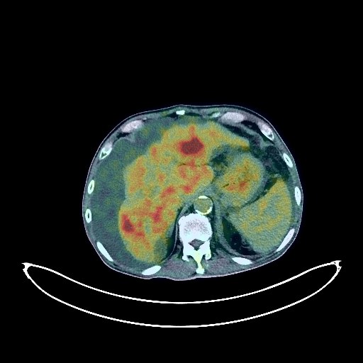
Liver Cancer PET/CT (case 983827-000128 from PETWB-REP)
0 views9 days agoWhole-body 18F-FDG PET/CT scan in a patient with Liver Cancer taken from the PETWB-REP dataset. The following English report (translated from original Chinese) is taken verbatim from the public dataset and has not been modified or otherwise checked for accuracy (see the end for citation). Impression a. Multiple lesions within the liver parenchyma with increased FDG metabolism, highly suggestive of primary liver cancer with right portal vein tumor thrombus formation. Cirrhosis, splenomegaly, and ascites/pelvic effusion. b. Retroperitoneal lymph node metastasis, locally fused and embezzling the abdominal aorta. Peritoneal seeding metastasis and left adrenal metastasis cannot be ruled out; follow-up CT scan is recommended. c. Multiple metastatic tumors in the left upper lobe and both lower lobes. Left pleural metastasis with left pleural effusion is highly probable. a. Irregular ground-glass opacity in the apical segment of the right upper lobe, with slight FDG uptake, early-stage lung cancer to be ruled out; chronic inflammatory ground-glass opacities or atypical adenomatous hyperplasia in the remaining upper lobes of both lungs. b. Soft tissue lesion in the apical segment of the right upper lobe, with slight FDG uptake, highly suggestive of chronic inflammation; a few chronic inflammation lesions and remnants in both lungs. Bilateral emphysema with bullae. Partial arteriosclerosis (including coronary arteries). No obvious abnormal density shadows or increased FDG metabolism shadows seen in the pancreas; please combine with enhanced MRI. Gallbladder wall edema. Benign prostatic hyperplasia with calcification. Poor gastric distension; physiological or inflammatory uptake of the gastric wall. Degenerative changes in the spine. Right occipital subcutaneous lipoma and left posterior cervical intermuscular lipoma. Deep lacunar infarcts in the brain, age-related encephalopathy. Chronic inflammation of the left maxillary sinus. Possible right upper periodontitis; chronic inflammatory changes on the right side of the oropharyngeal wall; please combine with specialist examination. This case is from PETWB-REP, a curated dataset of whole-body 18F-FDG PET/CT scans and corresponding radiology reports from 490 patients with a broad spectrum of malignancies. The data were retrospectively collected from patients who underwent clinically indicated whole-body 18F-FDG PET/CT scans at the Shanghai Universal Medical Imaging Diagnostic Center between 2021 and 2024. License: Creative Commons Attribution 4.0 International (CC BY 4.0) Citation: Xue, L., Feng, G., Wenbo, Z., Zhang, Y., Li, L., Wang, S., Peng, L., Peng, S., & Gao, X. (2026). PETWB-REP: A Multi-Cancer Whole-Body FDG PET/CT Dataset with Corresponding Radiology Reports [Data set]. Zenodo. https://doi.org/10.5281/zenodo.18670487
Whole BodyPET/CT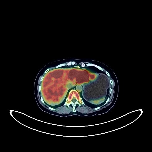
Liver Cancer PET/CT (case 983827-000171 from PETWB-REP)
0 views9 days agoWhole-body 18F-FDG PET/CT scan in a patient with Liver Cancer taken from the PETWB-REP dataset. The following English report (translated from original Chinese) is taken verbatim from the public dataset and has not been modified or otherwise checked for accuracy (see the end for citation). Impression a. Multiple low-density lesions in the liver with increased FDG metabolism, suggestive of malignancy, possibly primary or metastatic. Please combine clinical findings with enhanced MRI for comprehensive analysis. b. Multiple lymph node metastases in the hepatogastric space, hepatic hilum, and pancreatic head region. Two nodules in the left anterior pelvic region with increased FDG metabolism, suggestive of implantation metastasis. Old fibrocalcifications in both lungs; please follow up with CT. Partial calcification of the aorta and coronary artery walls. Bilateral breast hyperplasia, with calcification in the upper outer quadrant of the left breast; ultrasound follow-up is recommended. Calcifications in the spleen. Possible causes include cholestasis or cholesterol crystals in the gallbladder; ultrasound follow-up is recommended. Increased FDG metabolism in parts of the gastric wall and intestinal tract, suggestive of physiological or chronic inflammatory changes; please combine clinical findings with endoscopic follow-up. Scoliosis with degenerative changes, mild anterior slippage of the L4 vertebral body. L3/4, L4/5, and L5/S1 intervertebral disc bulges, with pneumatosis and degeneration of the L5/S1 intervertebral disc. Benign bone disease of the right iliac bone. Bilateral deep lacunar infarcts, age-related brain changes. This case is from PETWB-REP, a curated dataset of whole-body 18F-FDG PET/CT scans and corresponding radiology reports from 490 patients with a broad spectrum of malignancies. The data were retrospectively collected from patients who underwent clinically indicated whole-body 18F-FDG PET/CT scans at the Shanghai Universal Medical Imaging Diagnostic Center between 2021 and 2024. License: Creative Commons Attribution 4.0 International (CC BY 4.0) Citation: Xue, L., Feng, G., Wenbo, Z., Zhang, Y., Li, L., Wang, S., Peng, L., Peng, S., & Gao, X. (2026). PETWB-REP: A Multi-Cancer Whole-Body FDG PET/CT Dataset with Corresponding Radiology Reports [Data set]. Zenodo. https://doi.org/10.5281/zenodo.18670487
Whole BodyPET/CT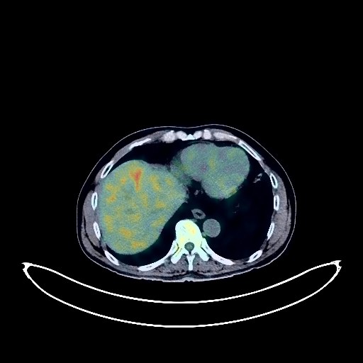
Gallbladder Cancer PET/CT (case 983827-000174 from PETWB-REP)
0 views9 days agoWhole-body 18F-FDG PET/CT scan in a patient with Gallbladder Cancer taken from the PETWB-REP dataset. The following English report (translated from original Chinese) is taken verbatim from the public dataset and has not been modified or otherwise checked for accuracy (see the end for citation). Impression a. Mass at the gallbladder fundus, with increased FDG metabolism, suggestive of gallbladder cancer involving adjacent liver parenchyma. b. Multiple lymph node metastases around the hepatic hilum and pancreatic head. c. Soft tissue shadow within the cystic duct, with increased FDG metabolism, suggestive of tumor embolism; contrast-enhanced MRI is recommended. d. Multiple stones within the left hepatic lobe bile duct. Slight dilation of intrahepatic bile ducts. Several solid, chronic inflammatory micronodules in both lungs. A few chronic inflammations and old lesions in both lungs. Calcification in the lower lobe of the left lung. Small cysts in both kidneys. Osteophyte formation in the cervical, thoracic, and lumbar vertebrae; L4/5 and L5/S1 intervertebral disc bulges. No obvious abnormalities were found on cranial scintigraphy. Cavity of septum pellucidum (normal variation). Chronic inflammation of the right maxillary sinus. This case is from PETWB-REP, a curated dataset of whole-body 18F-FDG PET/CT scans and corresponding radiology reports from 490 patients with a broad spectrum of malignancies. The data were retrospectively collected from patients who underwent clinically indicated whole-body 18F-FDG PET/CT scans at the Shanghai Universal Medical Imaging Diagnostic Center between 2021 and 2024. License: Creative Commons Attribution 4.0 International (CC BY 4.0) Citation: Xue, L., Feng, G., Wenbo, Z., Zhang, Y., Li, L., Wang, S., Peng, L., Peng, S., & Gao, X. (2026). PETWB-REP: A Multi-Cancer Whole-Body FDG PET/CT Dataset with Corresponding Radiology Reports [Data set]. Zenodo. https://doi.org/10.5281/zenodo.18670487
Whole BodyPET/CT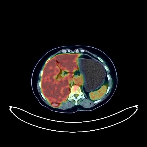
Breast Cancer PET/CT (case 983827-000250 from PETWB-REP)
0 views9 days agoWhole-body 18F-FDG PET/CT scan in a patient with Breast Cancer taken from the PETWB-REP dataset. The following English report (translated from original Chinese) is taken verbatim from the public dataset and has not been modified or otherwise checked for accuracy (see the end for citation). Impression a. A mass in the lateral aspect of the left breast, with increased FDG metabolism, consistent with breast cancer; multiple lymph node metastases in the left internal mammary chain, left axilla, left interpectoral space, and left supraclavicular fossa. b. A slightly dense nodule above the nipple in the left breast, with increased FDG metabolism, breast cancer to be ruled out; please correlate with clinicopathology. c. Multiple bone metastases throughout the body. d. A slightly high-density nodule in the left occipital lobe, with increased FDG metabolism; contrast-enhanced MRI is recommended to rule out metastasis. Age-related brain changes, deep lacunar infarcts. A bulla in the right middle lobe. A few post-inflammatory lesions in both lungs. A very small amount of pleural effusion on the right side. Partial arteriosclerosis. A liver cyst. Degenerative changes in the spine. L4/5 and L5/S1 intervertebral disc bulges. Mild inflammation of the bilateral ethmoid sinuses and right maxillary sinus, and a submucosal cyst of the left maxillary sinus. Reactive hyperplasia of the deep cervical lymph nodes bilaterally. This case is from PETWB-REP, a curated dataset of whole-body 18F-FDG PET/CT scans and corresponding radiology reports from 490 patients with a broad spectrum of malignancies. The data were retrospectively collected from patients who underwent clinically indicated whole-body 18F-FDG PET/CT scans at the Shanghai Universal Medical Imaging Diagnostic Center between 2021 and 2024. License: Creative Commons Attribution 4.0 International (CC BY 4.0) Citation: Xue, L., Feng, G., Wenbo, Z., Zhang, Y., Li, L., Wang, S., Peng, L., Peng, S., & Gao, X. (2026). PETWB-REP: A Multi-Cancer Whole-Body FDG PET/CT Dataset with Corresponding Radiology Reports [Data set]. Zenodo. https://doi.org/10.5281/zenodo.18670487
Whole BodyPET/CT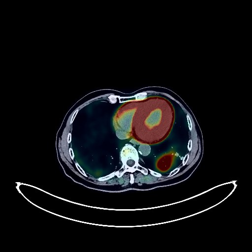
Lung Cancer PET/CT (case 983827-000255 from PETWB-REP)
0 views9 days agoWhole-body 18F-FDG PET/CT scan in a patient with Lung Cancer taken from the PETWB-REP dataset. The following English report (translated from original Chinese) is taken verbatim from the public dataset and has not been modified or otherwise checked for accuracy (see the end for citation). Impression a. A mass in the lower lobe of the left lung with increased FDG metabolism, suggestive of lung cancer. Please confirm the diagnosis with pathology. b. Left hilar lymph node metastasis, likely with reactive hyperplasia of mediastinal and bilateral axillary lymph nodes. c. Bilateral solid nodules with normal FDG metabolism, suggestive of chronic inflammatory nodules. Please follow up with CT scan. Bilateral interstitial lung changes with chronic inflammation and sequelae. Emphysema. Partial arteriosclerosis (including coronary arteries). Liver cyst. Uneven FDG metabolism in the prostate parenchyma, likely physiological or chronic inflammatory. Please follow up with PSA. Increased FDG metabolism in parts of the stomach wall and intestines, suggestive of physiological uptake or chronic inflammation. Please follow up with endoscopy. Reactive hyperplasia of retroperitoneal para-aortic, mesenteric, and bilateral inguinal lymph nodes. Spinal degenerative changes. L3 and L4 vertebral instability. L3 vertebral compression changes. T8 vertebral hemangioma. L3/4 and L4/5 intervertebral disc bulging, the latter with pneumoconiosis and degeneration. Right sternoclavicular arthritis. Bilateral deep lacunar infarcts, age-related brain; MRI recommended. Low-density nodule in the left lobe of the thyroid gland; FDG metabolism normal; nodular goiter suspected; ultrasound follow-up recommended. Reactive hyperplasia of cervical lymph nodes. This case is from PETWB-REP, a curated dataset of whole-body 18F-FDG PET/CT scans and corresponding radiology reports from 490 patients with a broad spectrum of malignancies. The data were retrospectively collected from patients who underwent clinically indicated whole-body 18F-FDG PET/CT scans at the Shanghai Universal Medical Imaging Diagnostic Center between 2021 and 2024. License: Creative Commons Attribution 4.0 International (CC BY 4.0) Citation: Xue, L., Feng, G., Wenbo, Z., Zhang, Y., Li, L., Wang, S., Peng, L., Peng, S., & Gao, X. (2026). PETWB-REP: A Multi-Cancer Whole-Body FDG PET/CT Dataset with Corresponding Radiology Reports [Data set]. Zenodo. https://doi.org/10.5281/zenodo.18670487
Whole BodyPET/CT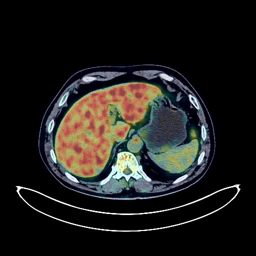
Lung Cancer PET/CT (case 983827-000087 from PETWB-REP)
0 views9 days agoWhole-body 18F-FDG PET/CT scan in a patient with Lung Cancer taken from the PETWB-REP dataset. The following English report (translated from original Chinese) is taken verbatim from the public dataset and has not been modified or otherwise checked for accuracy (see the end for citation). Impression A mass near the hilum in the right upper lobe, with elevated FDG metabolism, suggestive of central lung cancer with obstructive inflammation. Multiple lymph node metastases in the right hilum and mediastinum. Chronic inflammatory miliary nodules in the posterior segment of the right lower lobe. A few old lesions in both lower lobes. Minimal renal cysts in both kidneys. Calcifications in the prostate. Mild osteophyte formation in the cervical, thoracic, and lumbar spine. L4/5 and L5/S1 disc bulges. No abnormalities found on cranial scintigraphy. This case is from PETWB-REP, a curated dataset of whole-body 18F-FDG PET/CT scans and corresponding radiology reports from 490 patients with a broad spectrum of malignancies. The data were retrospectively collected from patients who underwent clinically indicated whole-body 18F-FDG PET/CT scans at the Shanghai Universal Medical Imaging Diagnostic Center between 2021 and 2024. License: Creative Commons Attribution 4.0 International (CC BY 4.0) Citation: Xue, L., Feng, G., Wenbo, Z., Zhang, Y., Li, L., Wang, S., Peng, L., Peng, S., & Gao, X. (2026). PETWB-REP: A Multi-Cancer Whole-Body FDG PET/CT Dataset with Corresponding Radiology Reports [Data set]. Zenodo. https://doi.org/10.5281/zenodo.18670487
Whole BodyPET/CT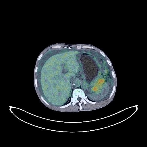
Liver Cancer PET/CT (case 983827-000109 from PETWB-REP)
0 views9 days agoWhole-body 18F-FDG PET/CT scan in a patient with Liver Cancer taken from the PETWB-REP dataset. The following English report (translated from original Chinese) is taken verbatim from the public dataset and has not been modified or otherwise checked for accuracy (see the end for citation). Impression a. Space-occupying lesions in both lobes of the liver with increased FDG uptake, suggestive of malignancy, possible metastasis, hepatocellular carcinoma to be ruled out. b. Extensive peritoneal seeding metastasis in the abdominopelvic region, with a large amount of fluid in the abdominopelvic cavity. Retroperitoneal lymph node metastasis. c. Soft tissue mass in the left upper abdomen with increased FDG metabolism, suggestive of malignancy, highly likely metastasis (peritoneal seeding metastasis), primary tumor to be ruled out. The above suggestions should be combined with enhanced MRI of the upper abdomen for comprehensive analysis. a. Multiple ground-glass nodules in the apical segment of the right upper lobe and the anterior segment of the left upper lobe, with normal FDG metabolism, suggestive of atypical adenomatous hyperplasia or inflammatory nodules, annual HRCT follow-up recommended. b. Multiple chronic inflammatory nodules (solid) in both lungs; calcifications in the middle and upper lobes of the right lung; a few fibrotic lesions in both lungs. Left renal pelvis and calyces stones with dilation and hydronephrosis; small calcifications in the right kidney; bilateral renal cysts. Left adrenal gland not clearly visible. Thickening of the gastric fundus and body mucosa, increased FDG metabolism, suggestive of gastritis. Prostatic calcifications. Calcifications at the lower end of the urachus are likely large. Small amount of hydrocele in the left testis. Spinal degenerative changes, L3/4 and L4/5 intervertebral disc bulge, bilateral pars interarticular isthmic discontinuity of the L5 vertebral body, mild anterior slippage of the L5 vertebral body. Mild age-related brain changes. This case is from PETWB-REP, a curated dataset of whole-body 18F-FDG PET/CT scans and corresponding radiology reports from 490 patients with a broad spectrum of malignancies. The data were retrospectively collected from patients who underwent clinically indicated whole-body 18F-FDG PET/CT scans at the Shanghai Universal Medical Imaging Diagnostic Center between 2021 and 2024. License: Creative Commons Attribution 4.0 International (CC BY 4.0) Citation: Xue, L., Feng, G., Wenbo, Z., Zhang, Y., Li, L., Wang, S., Peng, L., Peng, S., & Gao, X. (2026). PETWB-REP: A Multi-Cancer Whole-Body FDG PET/CT Dataset with Corresponding Radiology Reports [Data set]. Zenodo. https://doi.org/10.5281/zenodo.18670487
Whole BodyPET/CT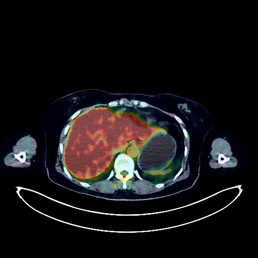
Glioma PET/CT (case 983827-000042 from PETWB-REP)
0 views9 days agoWhole-body 18F-FDG PET/CT scan in a patient with Glioma taken from the PETWB-REP dataset. The following English report (translated from original Chinese) is taken verbatim from the public dataset and has not been modified or otherwise checked for accuracy (see the end for citation). Impression a. A mass in the left temporal lobe with increased FDG metabolism and surrounding extensive edema, suggestive of malignancy, with glioblastoma being more likely than metastasis. b. Increased bone density in the T11 vertebral body, accompanied by increased FDG uptake, metastasis to be ruled out; close observation is recommended. Scattered chronic inflammatory nodules in both lungs; please follow up with CT to rule out other possibilities. Calcifications in the left upper lobe and right lower lobe. Reactive hyperplasia of bilateral hilar and mediastinal lymph nodes. Calcification of some arterial walls. Bilateral breast hyperplasia. Chronic gastritis; some physiological or inflammatory uptake of the intestines; please follow up with endoscopy. Left renal cyst. Partial vertebral osteophyte formation. Nuchal ligament calcification. This case is from PETWB-REP, a curated dataset of whole-body 18F-FDG PET/CT scans and corresponding radiology reports from 490 patients with a broad spectrum of malignancies. The data were retrospectively collected from patients who underwent clinically indicated whole-body 18F-FDG PET/CT scans at the Shanghai Universal Medical Imaging Diagnostic Center between 2021 and 2024. License: Creative Commons Attribution 4.0 International (CC BY 4.0) Citation: Xue, L., Feng, G., Wenbo, Z., Zhang, Y., Li, L., Wang, S., Peng, L., Peng, S., & Gao, X. (2026). PETWB-REP: A Multi-Cancer Whole-Body FDG PET/CT Dataset with Corresponding Radiology Reports [Data set]. Zenodo. https://doi.org/10.5281/zenodo.18670487
Whole BodyPET/CT