Loading...
Lung Cancer PET/CT (case 983827-000244 from PETWB-REP)
0 views9 days agoWhole-body 18F-FDG PET/CT scan in a patient with Lung Cancer taken from the PETWB-REP dataset. The following English report (translated from original Chinese) is taken verbatim from the public dataset and has not been modified or otherwise checked for accuracy (see the end for citation). Impression a. Mass near the hilum of the left upper lobe, with increased FDG metabolism, consistent with central lung cancer, and left hilar lymph node metastasis. b. Chronic inflammatory micronodules in the upper lobes of both lungs. A few chronic inflammations and old lesions (including calcifications) in both lungs. Calcification of some arterial walls. Chronic cholecystitis; gallstones. Accessory spleen. Right renal cyst. Mild bilateral adrenal hyperplasia; follow-up is recommended. Increased FDG metabolism in some intestinal segments, considered inflammatory or physiological uptake; colonoscopy is recommended. Benign prostatic hyperplasia with calcification, increased FDG metabolism in the gland, considered inflammatory or physiological uptake; follow-up PSA and ultrasound are recommended. Degenerative changes in the spine. L3/4 and L4/5 intervertebral disc bulge. Subcutaneous inflammation in the left buttock. A few ischemic lesions in the deep bilateral brain regions; age-related brain condition, MRI recommended. Chronic inflammation of the right maxillary sinus. This case is from PETWB-REP, a curated dataset of whole-body 18F-FDG PET/CT scans and corresponding radiology reports from 490 patients with a broad spectrum of malignancies. The data were retrospectively collected from patients who underwent clinically indicated whole-body 18F-FDG PET/CT scans at the Shanghai Universal Medical Imaging Diagnostic Center between 2021 and 2024. License: Creative Commons Attribution 4.0 International (CC BY 4.0) Citation: Xue, L., Feng, G., Wenbo, Z., Zhang, Y., Li, L., Wang, S., Peng, L., Peng, S., & Gao, X. (2026). PETWB-REP: A Multi-Cancer Whole-Body FDG PET/CT Dataset with Corresponding Radiology Reports [Data set]. Zenodo. https://doi.org/10.5281/zenodo.18670487
Whole BodyPET/CT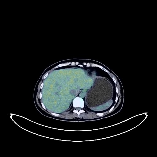
Lung Cancer PET/CT (case 983827-000008 from PETWB-REP)
0 views9 days agoWhole-body 18F-FDG PET/CT scan in a patient with Lung Cancer taken from the PETWB-REP dataset. The following English report (translated from original Chinese) is taken verbatim from the public dataset and has not been modified or otherwise checked for accuracy (see the end for citation). Impression a. Mixed-density lesions in the left basal ganglia and centrum semiovale with increased FDG metabolism, considered a metastatic tumor based on enhanced MRI findings at this center. b. Lacunar infarcts deep in the brain. Submucosal cyst in the left sphenoid sinus. a. Postoperative changes after right lung cancer surgery; no signs of tumor recurrence were observed in the surgical area. b. Multiple ground-glass nodules in the right lower lobe and left lung; FDG metabolism was normal. Early lung cancer is suspected in the apical-posterior segment of the left upper lobe; the remaining nodules are considered inflammatory nodules or atypical adenomatous hyperplasia. Close CT observation is recommended. c. Chronic inflammatory nodule (solid) in the apical-posterior segment of the left upper lobe. A few post-inflammatory lesions in both lungs. Calcifications in the spleen. Left adrenal hyperplasia. Residual contrast agent in the urinary tract. IUD insertion in the endometrial area with physiological uptake. Physiological uptake in the left ovary is possible; further specialist examination is needed. Chronic inflammatory changes in the gastric antrum; endoscopic follow-up is recommended. A nodular low-density lesion was found in the S1 vertebral body. No abnormalities were observed in FDG uptake. Benign bone disease is suspected, and metastasis should be ruled out. This case is from PETWB-REP, a curated dataset of whole-body 18F-FDG PET/CT scans and corresponding radiology reports from 490 patients with a broad spectrum of malignancies. The data were retrospectively collected from patients who underwent clinically indicated whole-body 18F-FDG PET/CT scans at the Shanghai Universal Medical Imaging Diagnostic Center between 2021 and 2024. License: Creative Commons Attribution 4.0 International (CC BY 4.0) Citation: Xue, L., Feng, G., Wenbo, Z., Zhang, Y., Li, L., Wang, S., Peng, L., Peng, S., & Gao, X. (2026). PETWB-REP: A Multi-Cancer Whole-Body FDG PET/CT Dataset with Corresponding Radiology Reports [Data set]. Zenodo. https://doi.org/10.5281/zenodo.18670487
Whole BodyPET/CT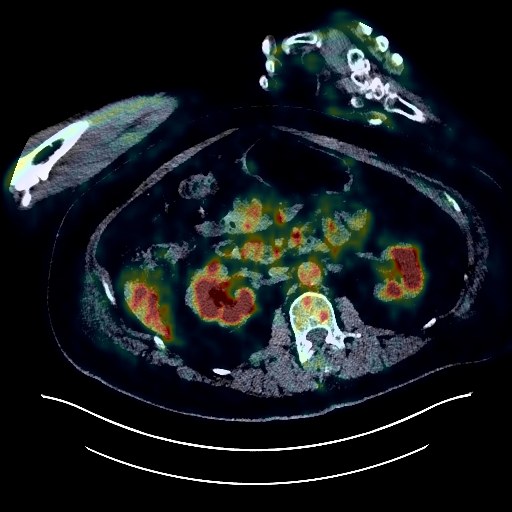
Lung Cancer PET/CT (case 983827-000058 from PETWB-REP)
0 views9 days agoWhole-body 18F-FDG PET/CT scan in a patient with Lung Cancer taken from the PETWB-REP dataset. The following English report (translated from original Chinese) is taken verbatim from the public dataset and has not been modified or otherwise checked for accuracy (see the end for citation). Impression a. Right upper lobe posterior segment lesion, increased FDG metabolism, suggestive of malignancy, please correlate with clinicopathology. b. Multiple metastases to the right pleura, small amount of right pleural effusion. Multiple lymph node metastases to the right hilum and mediastinum. c. Lobe-shaped low-density area in the right temporoparietal lobe, lack of FDG uptake, primarily suggestive of encephalomalacia, metastasis to be ruled out, contrast-enhanced MRI recommended. Age-related brain changes. Soft tissue mass in the gastric cardia region, increased FDG metabolism, suggestive of stromal tumor (malignant), please correlate with clinicopathology. Partial calcification of the aorta and coronary artery walls. Uneven density with calcification in the left and right lobes of the thyroid gland, uneven FDG uptake, suggestive of nodular goiter, ultrasound and related laboratory tests recommended. Left renal cyst. Small amount of hydrocele in both testes. Spinal osteophyte. This case is from PETWB-REP, a curated dataset of whole-body 18F-FDG PET/CT scans and corresponding radiology reports from 490 patients with a broad spectrum of malignancies. The data were retrospectively collected from patients who underwent clinically indicated whole-body 18F-FDG PET/CT scans at the Shanghai Universal Medical Imaging Diagnostic Center between 2021 and 2024. License: Creative Commons Attribution 4.0 International (CC BY 4.0) Citation: Xue, L., Feng, G., Wenbo, Z., Zhang, Y., Li, L., Wang, S., Peng, L., Peng, S., & Gao, X. (2026). PETWB-REP: A Multi-Cancer Whole-Body FDG PET/CT Dataset with Corresponding Radiology Reports [Data set]. Zenodo. https://doi.org/10.5281/zenodo.18670487
Whole BodyPET/CT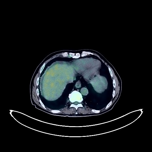
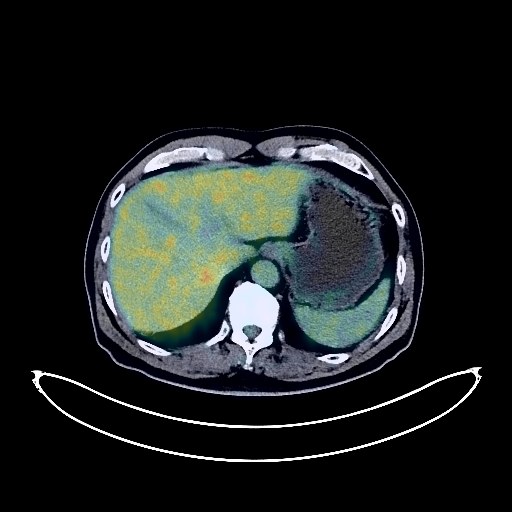
Pancreatic Cancer PET/CT (case 983827-000256 from PETWB-REP)
0 views9 days agoWhole-body 18F-FDG PET/CT scan in a patient with Pancreatic Cancer taken from the PETWB-REP dataset. The following English report (translated from original Chinese) is taken verbatim from the public dataset and has not been modified or otherwise checked for accuracy (see the end for citation). Impression a. Multiple implantation metastases in the abdomen and pelvis, some with unclear boundaries from adjacent stomach walls and intestinal segments. Metastatic tumors in the lower segment of the right posterior lobe of the liver, the left lobe of the liver, and the subcapsular lobe. b. A mass in the tail of the pancreas, with increased FDG metabolism, involving the splenic hilum, suggestive of malignancy; pancreatic cancer is more likely than metastasis. c. Slight thickening of the intestinal wall at the rectosigmoid junction, with increased FDG uptake; combined with colonoscopy pathology, colon cancer cannot be ruled out; periodic colonoscopy follow-up is recommended. Presacral lymph node metastasis is highly probable. d. Reactive hyperplasia of small lymph nodes in the left iliac vessels, mesentery, and retroperitoneum; follow-up is required. Small amount of fluid in the abdomen and pelvis. e. Metastasis to the T11 vertebra and right iliac bone is pending; follow-up is recommended with MRI. a. Ground-glass nodule in the lateral basal segment of the right lower lobe, FDG metabolism normal, suggestive of chronic inflammatory nodule or atypical adenomatous hyperplasia, please combine with annual HRCT follow-up. b. Chronic inflammatory micronodule (solid) in the right lung. A few post-inflammatory remnants and calcifications in both lungs. Partial thickening and calcification of the right pleura. Tracheal diverticulum. Reactive hyperplasia of mediastinal lymph nodes. Calcification of some arterial walls (including coronary arteries). Small liver cyst. Slightly thickened and roughened gallbladder wall. Bilateral renal cysts; likely mild hyperplasia of the left adrenal gland. Prostatic hyperplasia with calcification. Changes characteristic of chronic gastritis. Spinal degenerative changes; mild L4/5 and L5/S1 intervertebral disc protrusion. Right infraspinatus muscle lipoma. Bilateral deep lacunar infarcts, age-related brain changes, and formation of the cavum septum pellucidum. Minor inflammation of the left maxillary and ethmoid sinuses. This case is from PETWB-REP, a curated dataset of whole-body 18F-FDG PET/CT scans and corresponding radiology reports from 490 patients with a broad spectrum of malignancies. The data were retrospectively collected from patients who underwent clinically indicated whole-body 18F-FDG PET/CT scans at the Shanghai Universal Medical Imaging Diagnostic Center between 2021 and 2024. License: Creative Commons Attribution 4.0 International (CC BY 4.0) Citation: Xue, L., Feng, G., Wenbo, Z., Zhang, Y., Li, L., Wang, S., Peng, L., Peng, S., & Gao, X. (2026). PETWB-REP: A Multi-Cancer Whole-Body FDG PET/CT Dataset with Corresponding Radiology Reports [Data set]. Zenodo. https://doi.org/10.5281/zenodo.18670487
Whole BodyPET/CT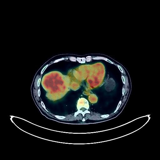
Lung Cancer PET/CT (case 983827-000184 from PETWB-REP)
0 views9 days agoWhole-body 18F-FDG PET/CT scan in a patient with Lung Cancer taken from the PETWB-REP dataset. The following English report (translated from original Chinese) is taken verbatim from the public dataset and has not been modified or otherwise checked for accuracy (see the end for citation). Impression a. Mass in the left upper lobe of the lung, with increased FDG metabolism, suggestive of lung cancer. Multiple lymph node metastases in the left hilum and mediastinum. b. Localized bone destruction in the left 8th rib, with sclerosis at the margins, increased FDG metabolism, metastasis to be ruled out; please correlate with clinical findings and closely monitor. c. Emphysema in both lungs. Minor chronic inflammation and remnants in both lungs. Calcification of some arterial walls. Low-density nodule in the left lobe of the thyroid gland, with increased FDG metabolism, suggestive of an adenomatous nodule; ultrasound examination is recommended, and fine-needle aspiration biopsy may be necessary to rule out malignancy. Liver cyst. Pancreatic calcification. Left renal cyst. Benign prostatic hyperplasia. Segmental increased FDG metabolism in part of the colorectal region, suggestive of inflammatory or physiological uptake. Appendicitis fecalith. Degenerative changes in the spine. L4/5 and L5/S1 intervertebral disc bulge. Left acetabular island. A few ischemic lesions in the deep bilateral brain regions, indicative of age-related brain changes. This case is from PETWB-REP, a curated dataset of whole-body 18F-FDG PET/CT scans and corresponding radiology reports from 490 patients with a broad spectrum of malignancies. The data were retrospectively collected from patients who underwent clinically indicated whole-body 18F-FDG PET/CT scans at the Shanghai Universal Medical Imaging Diagnostic Center between 2021 and 2024. License: Creative Commons Attribution 4.0 International (CC BY 4.0) Citation: Xue, L., Feng, G., Wenbo, Z., Zhang, Y., Li, L., Wang, S., Peng, L., Peng, S., & Gao, X. (2026). PETWB-REP: A Multi-Cancer Whole-Body FDG PET/CT Dataset with Corresponding Radiology Reports [Data set]. Zenodo. https://doi.org/10.5281/zenodo.18670487
Whole BodyPET/CT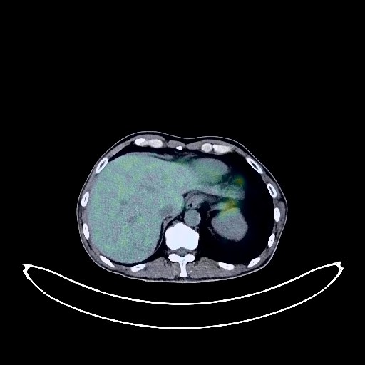
Pancreatic Cancer PET/CT (case 983827-000055 from PETWB-REP)
0 views9 days agoWhole-body 18F-FDG PET/CT scan in a patient with Pancreatic Cancer taken from the PETWB-REP dataset. The following English report (translated from original Chinese) is taken verbatim from the public dataset and has not been modified or otherwise checked for accuracy (see the end for citation). Impression a. A cystic-solid mass in the body and tail of the pancreas, with elevated FDG metabolism in the solid portion, suggestive of malignancy, most likely pancreatic cancer or cystadenocarcinoma, invading the adjacent stomach wall. b. Liver metastasis. Chronic inflammatory micronodules (solid) in the posterior segment of the right upper lobe and the posterior basal segment of the right lower lobe. A few old lesions in both lungs. Small liver cysts. Small kidney stone in the right kidney. Calcifications in the prostate. Degenerative changes in the spine. Bilateral pars intervertebral disc fracture at L5. L4/5 and L5/S1 intervertebral disc herniation. Subcutaneous calcifications in both buttocks. No abnormalities were found on cranial scintigraphy. This case is from PETWB-REP, a curated dataset of whole-body 18F-FDG PET/CT scans and corresponding radiology reports from 490 patients with a broad spectrum of malignancies. The data were retrospectively collected from patients who underwent clinically indicated whole-body 18F-FDG PET/CT scans at the Shanghai Universal Medical Imaging Diagnostic Center between 2021 and 2024. License: Creative Commons Attribution 4.0 International (CC BY 4.0) Citation: Xue, L., Feng, G., Wenbo, Z., Zhang, Y., Li, L., Wang, S., Peng, L., Peng, S., & Gao, X. (2026). PETWB-REP: A Multi-Cancer Whole-Body FDG PET/CT Dataset with Corresponding Radiology Reports [Data set]. Zenodo. https://doi.org/10.5281/zenodo.18670487
Whole BodyPET/CT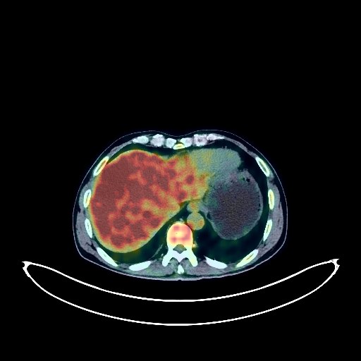
Liver Cancer PET/CT (case 983827-000082 from PETWB-REP)
0 views9 days agoWhole-body 18F-FDG PET/CT scan in a patient with Liver Cancer taken from the PETWB-REP dataset. The following English report (translated from original Chinese) is taken verbatim from the public dataset and has not been modified or otherwise checked for accuracy (see the end for citation). Impression a. A low-density mass with increased FDG metabolism at the top of the diaphragm in the right lobe of the liver, highly suggestive of hepatocellular carcinoma. Please combine clinical findings with enhanced MRI for comprehensive analysis. Liver cyst. b. Possible reactive hyperplasia of the hilar and retroperitoneal lymph nodes. Please follow up to rule out mixed metastases. Benign prostatic hyperplasia with calcification, a space-occupying lesion on the right side of the prostate with increased FDG metabolism, neoplastic lesion to be ruled out. Please combine PSA and enhanced MRI examinations, and perform a biopsy if necessary to confirm the diagnosis. Chronic inflammatory nodules in both lungs, CT follow-up is recommended. Emphysema in the upper lobes of both lungs, a few post-inflammatory remnants in both lungs. Reactive hyperplasia of mediastinal lymph nodes. Gallbladder and upper common bile duct stones, cholecystitis. Accessory spleen. Left adrenal hyperplasia. Bilateral renal cysts. Chronic inflammatory changes in the antrum of the stomach and part of the intestine. Please follow up with endoscopy. Degenerative changes in the spine, L4/5 and L5/S1 intervertebral disc bulges. Cranial scintigraphy showed no obvious abnormalities. Chronic inflammation of the left ethmoid sinus. Reactive hyperplasia of bilateral cervical lymph nodes. This case is from PETWB-REP, a curated dataset of whole-body 18F-FDG PET/CT scans and corresponding radiology reports from 490 patients with a broad spectrum of malignancies. The data were retrospectively collected from patients who underwent clinically indicated whole-body 18F-FDG PET/CT scans at the Shanghai Universal Medical Imaging Diagnostic Center between 2021 and 2024. License: Creative Commons Attribution 4.0 International (CC BY 4.0) Citation: Xue, L., Feng, G., Wenbo, Z., Zhang, Y., Li, L., Wang, S., Peng, L., Peng, S., & Gao, X. (2026). PETWB-REP: A Multi-Cancer Whole-Body FDG PET/CT Dataset with Corresponding Radiology Reports [Data set]. Zenodo. https://doi.org/10.5281/zenodo.18670487
Whole BodyPET/CT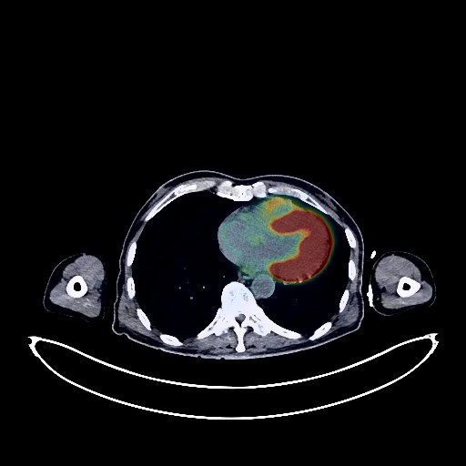
Gallbladder Cancer PET/CT (case 983827-000045 from PETWB-REP)
0 views9 days agoWhole-body 18F-FDG PET/CT scan in a patient with Gallbladder Cancer taken from the PETWB-REP dataset. The following English report (translated from original Chinese) is taken verbatim from the public dataset and has not been modified or otherwise checked for accuracy (see the end for citation). Impression a. Changes after comprehensive treatment for gallbladder cancer; no clear signs of tumor recurrence were observed in the surgical area. Slight local dilation of intrahepatic bile ducts. b. Right quadratus femoris muscle metastasis, enlarged compared to previous scan. Multiple metastases near bilateral external iliac vessels, right inguinal region, and left anterior pubic region (newly added). a. After treatment for brain metastases, no clear space-occupying lesions were observed in the brain; lacunar ischemic lesions in the deep cerebral region and brainstem bilaterally, characteristic of senile brain changes. b. Liver cyst; no obvious abnormal density shadows were observed in the remaining liver parenchyma; no abnormal increase in FDG metabolism was observed. Please follow up with enhanced MRI for the above findings. Benign prostatic hyperplasia; uneven FDG metabolism; please combine PSA and MRI for comprehensive analysis. Chronic inflammatory micronodules in the right lung horizontal fissure. Calcification of some arterial walls. Mild pancreatic atrophy. Multiple renal cysts. Mildly increased FDG metabolism in some gastric walls; slight thickening of the rectal wall with increased FDG metabolism, suggestive of physiological uptake or chronic inflammatory changes; please follow up with endoscopy. Decreased FDG metabolism in some thoracic and lumbar vertebrae, suggestive of post-radiotherapy changes. Spinal degeneration. L5/S1 vertebral endplate inflammation. Minor inflammation of bilateral ethmoid sinuses. This case is from PETWB-REP, a curated dataset of whole-body 18F-FDG PET/CT scans and corresponding radiology reports from 490 patients with a broad spectrum of malignancies. The data were retrospectively collected from patients who underwent clinically indicated whole-body 18F-FDG PET/CT scans at the Shanghai Universal Medical Imaging Diagnostic Center between 2021 and 2024. License: Creative Commons Attribution 4.0 International (CC BY 4.0) Citation: Xue, L., Feng, G., Wenbo, Z., Zhang, Y., Li, L., Wang, S., Peng, L., Peng, S., & Gao, X. (2026). PETWB-REP: A Multi-Cancer Whole-Body FDG PET/CT Dataset with Corresponding Radiology Reports [Data set]. Zenodo. https://doi.org/10.5281/zenodo.18670487
Whole BodyPET/CT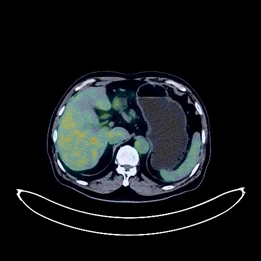
Renal Cancer PET/CT (case 983827-000107 from PETWB-REP)
0 views9 days agoWhole-body 18F-FDG PET/CT scan in a patient with Renal Cancer taken from the PETWB-REP dataset. The following English report (translated from original Chinese) is taken verbatim from the public dataset and has not been modified or otherwise checked for accuracy (see the end for citation). Impression A mass in the upper pole of the left kidney with slightly elevated FDG metabolism, highly suggestive of renal cell carcinoma; please correlate with clinicopathology. Reactive hyperplasia of retroperitoneal lymph nodes. Chronic inflammatory micronodules in the upper lobe of the left lung. Slight bronchial dilatation in the upper lobe of the left lung, a few old lesions in the upper lobe of the left lung. Slight thickening of the left pleura, calcification of some arterial walls (including coronary arteries). Gallstones. Accessory spleen. Bilateral renal cysts (including a complex cyst in the left kidney). Possible left inguinal hernia. Degenerative changes in the spine, L4/5 and L5/S1 intervertebral disc bulge with pneumoconiosis and degeneration. Deep lacunar infarcts in the brain. Physiological uptake of left facial muscles. This case is from PETWB-REP, a curated dataset of whole-body 18F-FDG PET/CT scans and corresponding radiology reports from 490 patients with a broad spectrum of malignancies. The data were retrospectively collected from patients who underwent clinically indicated whole-body 18F-FDG PET/CT scans at the Shanghai Universal Medical Imaging Diagnostic Center between 2021 and 2024. License: Creative Commons Attribution 4.0 International (CC BY 4.0) Citation: Xue, L., Feng, G., Wenbo, Z., Zhang, Y., Li, L., Wang, S., Peng, L., Peng, S., & Gao, X. (2026). PETWB-REP: A Multi-Cancer Whole-Body FDG PET/CT Dataset with Corresponding Radiology Reports [Data set]. Zenodo. https://doi.org/10.5281/zenodo.18670487
Whole BodyPET/CT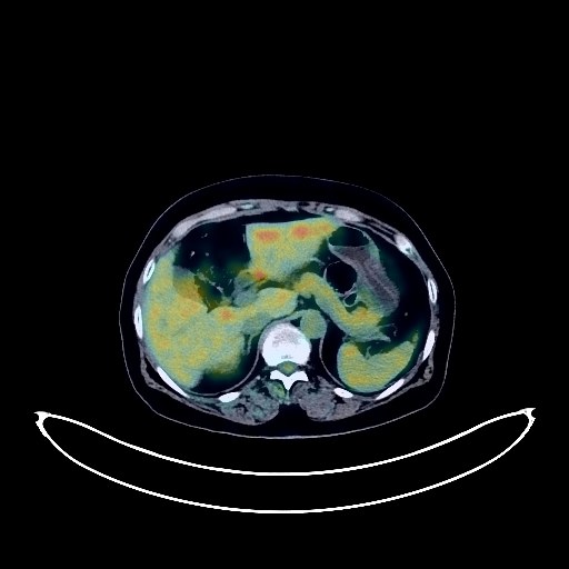
Nasopharyngeal Cancer PET/CT (case 983827-000270 from PETWB-REP)
1 views9 days agoWhole-body 18F-FDG PET/CT scan in a patient with Nasopharyngeal Cancer taken from the PETWB-REP dataset. The following English report (translated from original Chinese) is taken verbatim from the public dataset and has not been modified or otherwise checked for accuracy (see the end for citation). Impression a. After treatment for nasopharyngeal carcinoma, no obvious space-occupying lesions were found in the nasopharynx, and FDG metabolism was normal, suggesting that tumor activity was basically suppressed; bilateral cervical lymph node reactive hyperplasia or post-treatment changes. b. Multiple soft tissue density shadows with increased FDG metabolism in the spinal canal at the C1-2, T2, T7, and T10-L1 levels; T10 vertebral body bone destruction with increased FDG metabolism. These are considered metastatic tumors. Low-density lesion in the left temporal lobe, with normal FDG metabolism, combined with contrast-enhanced MRI images from another hospital, suggests a metastatic tumor. Age-related brain changes. Chronic inflammation of the left sphenoid sinus, bilateral ethmoid sinuses, and bilateral maxillary sinuses. Right otitis media/mastoid inflammation. Chronic inflammatory micronodules in both lungs; CT follow-up is recommended. Right lower lobe bullae. A few post-inflammatory remnants in both lungs. Bilateral pleural thickening. Reactive hyperplasia of mediastinal lymph nodes. Partial arteriosclerosis. Hemangioma of the right lobe of the liver is the primary consideration; enhanced MRI follow-up is recommended. Cyst in the left lobe of the liver. Left renal atrophy. Small cyst in the right kidney. Chronic inflammatory changes or physiological uptake in the entire esophagus, part of the stomach wall, and intestines; hemorrhoidal changes; please follow up with endoscopy. Degenerative changes in the spine; L5/S1 disc pneumothorax; L4/5 and L5/S1 disc bulging. This case is from PETWB-REP, a curated dataset of whole-body 18F-FDG PET/CT scans and corresponding radiology reports from 490 patients with a broad spectrum of malignancies. The data were retrospectively collected from patients who underwent clinically indicated whole-body 18F-FDG PET/CT scans at the Shanghai Universal Medical Imaging Diagnostic Center between 2021 and 2024. License: Creative Commons Attribution 4.0 International (CC BY 4.0) Citation: Xue, L., Feng, G., Wenbo, Z., Zhang, Y., Li, L., Wang, S., Peng, L., Peng, S., & Gao, X. (2026). PETWB-REP: A Multi-Cancer Whole-Body FDG PET/CT Dataset with Corresponding Radiology Reports [Data set]. Zenodo. https://doi.org/10.5281/zenodo.18670487
Whole BodyPET/CT