Loading...
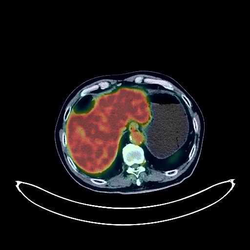
Liver Cancer PET/CT (case 983827-000105 from PETWB-REP)
3 views9 days agoWhole-body 18F-FDG PET/CT scan in a patient with Liver Cancer taken from the PETWB-REP dataset. The following English report (translated from original Chinese) is taken verbatim from the public dataset and has not been modified or otherwise checked for accuracy (see the end for citation). Impression a. Nodular hypermetabolic foci on FDG in the left lateral lobe of the liver, suggestive of a neoplastic lesion; enhanced MRI is recommended for further examination. Multiple liver cysts. b. Reactive hyperplasia of the hilar lymph nodes, hilar space, and retroperitoneal lymph nodes is highly probable; follow-up is recommended to rule out other possibilities. a. Multiple ground-glass nodules in both lungs, FDG metabolism normal, suggestive of chronic inflammatory nodules or atypical adenomatous hyperplasia; annual follow-up with HRCT is recommended. b. Chronic inflammatory nodules (solid) in the right upper lobe and left lower lobe. Pulmonary fibrosis, calcification in the right lower lobe. Emphysema in the right middle lobe. Partial arteriosclerosis (including coronary arteries). Gallstones, chronic cholecystitis. Left renal cyst. Prostatic calcification; follow-up with PSA is recommended. Increased FDG metabolism in parts of the stomach wall and intestines, possibly due to physiological uptake or chronic inflammation; please follow up with endoscopy. Spinal degeneration. L3/4, L4/5, and L5/S1 intervertebral disc bulges. Bilateral hip periarthritis. Bilateral deep lacunar infarcts, senile encephalopathy. This case is from PETWB-REP, a curated dataset of whole-body 18F-FDG PET/CT scans and corresponding radiology reports from 490 patients with a broad spectrum of malignancies. The data were retrospectively collected from patients who underwent clinically indicated whole-body 18F-FDG PET/CT scans at the Shanghai Universal Medical Imaging Diagnostic Center between 2021 and 2024. License: Creative Commons Attribution 4.0 International (CC BY 4.0) Citation: Xue, L., Feng, G., Wenbo, Z., Zhang, Y., Li, L., Wang, S., Peng, L., Peng, S., & Gao, X. (2026). PETWB-REP: A Multi-Cancer Whole-Body FDG PET/CT Dataset with Corresponding Radiology Reports [Data set]. Zenodo. https://doi.org/10.5281/zenodo.18670487
Whole BodyPET/CT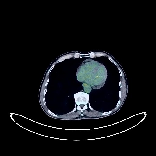
Lung Cancer PET/CT (case 983827-000092 from PETWB-REP)
3 views9 days agoWhole-body 18F-FDG PET/CT scan in a patient with Lung Cancer taken from the PETWB-REP dataset. The following English report (translated from original Chinese) is taken verbatim from the public dataset and has not been modified or otherwise checked for accuracy (see the end for citation). Impression a. Mediastinal soft tissue mass with elevated FDG metabolism, suggestive of malignancy, possible metastasis, mediastinal lung cancer to be ruled out. Right cardiophrenic angle nodule with elevated FDG metabolism, suggestive of metastasis. b. Cerebellar mass, one on the right side with elevated FDG metabolism, suggestive of metastasis, please combine with enhanced MRI for comprehensive analysis. Slightly low-density mass in the tail of the pancreas with elevated FDG metabolism, suggestive of malignancy, pancreatic cancer is highly likely, metastasis to be ruled out, please combine clinical and enhanced MRI for comprehensive analysis. a. Cystic cavity in the posterior segment of the left lower lobe with surrounding mixed ground-glass opacity, low FDG metabolism, suggestive of chronic inflammation, cystic lung cancer to be ruled out, close observation recommended. b. Multiple pure ground-glass nodules in both lungs, low FDG metabolism, suggestive of atypical adenomatous hyperplasia or chronic inflammatory nodules. Multiple solid nodules in both lungs, low FDG metabolism, high probability of chronic inflammatory nodules. c. Bronchiectasis in the right middle lobe with mucus plug formation. Scattered chronic inflammation and sequelae in both lungs. Emphysema in both lungs, bullae in the right upper lobe. Calcification of some arterial walls (including coronary arteries). Pericardial effusion. Gallstones. Accessory spleen. Hydrocele of the left testis. Schistosomiasis intestinal infection. A few ischemic foci in the deep brain. Age-related brain changes. A few chronic inflammations of the right maxillary sinus and left sphenoid sinus. Osteophyte formation in some vertebrae. L4/5 disc bulge. This case is from PETWB-REP, a curated dataset of whole-body 18F-FDG PET/CT scans and corresponding radiology reports from 490 patients with a broad spectrum of malignancies. The data were retrospectively collected from patients who underwent clinically indicated whole-body 18F-FDG PET/CT scans at the Shanghai Universal Medical Imaging Diagnostic Center between 2021 and 2024. License: Creative Commons Attribution 4.0 International (CC BY 4.0) Citation: Xue, L., Feng, G., Wenbo, Z., Zhang, Y., Li, L., Wang, S., Peng, L., Peng, S., & Gao, X. (2026). PETWB-REP: A Multi-Cancer Whole-Body FDG PET/CT Dataset with Corresponding Radiology Reports [Data set]. Zenodo. https://doi.org/10.5281/zenodo.18670487
Whole BodyPET/CT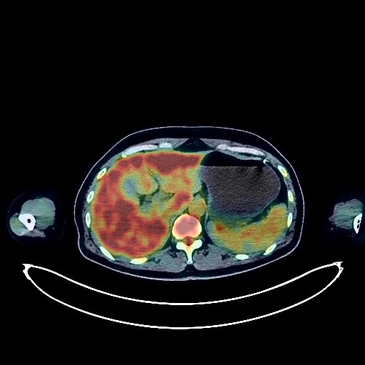
Lymphoma PET/CT (case 983827-000233 from PETWB-REP)
3 views9 days agoWhole-body 18F-FDG PET/CT scan in a patient with Lymphoma taken from the PETWB-REP dataset. The following English report (translated from original Chinese) is taken verbatim from the public dataset and has not been modified or otherwise checked for accuracy (see the end for citation). Impression After lymphoma treatment, no obvious space-occupying lesions were found in the oropharynx and epiglottis, and FDG metabolism was normal; multiple small lymph nodes were observed throughout the body, with some showing mild FDG uptake (see description for details). This suggests that tumor activity was suppressed after treatment, and follow-up is recommended. Deauville score: 1 point. Increased FDG metabolism in the bone marrow cavity throughout the body, suggesting reactive proliferative changes; please correlate with clinical findings. Chronic inflammatory micronodules in the upper lobe of the left lung. A few post-inflammatory lesions in both lungs. Mild anemia, and slight arteriosclerosis in some arteries. Calcification in the right lobe of the liver, and a liver cyst. Chronic inflammatory changes in the antrum of the stomach; please correlate with endoscopic follow-up. Mild osteophyte formation in the spine, with L4/5 and L5/S1 intervertebral disc bulges. No obvious abnormalities were found on cranial scintigraphy. Chronic inflammation of the right maxillary sinus. This case is from PETWB-REP, a curated dataset of whole-body 18F-FDG PET/CT scans and corresponding radiology reports from 490 patients with a broad spectrum of malignancies. The data were retrospectively collected from patients who underwent clinically indicated whole-body 18F-FDG PET/CT scans at the Shanghai Universal Medical Imaging Diagnostic Center between 2021 and 2024. License: Creative Commons Attribution 4.0 International (CC BY 4.0) Citation: Xue, L., Feng, G., Wenbo, Z., Zhang, Y., Li, L., Wang, S., Peng, L., Peng, S., & Gao, X. (2026). PETWB-REP: A Multi-Cancer Whole-Body FDG PET/CT Dataset with Corresponding Radiology Reports [Data set]. Zenodo. https://doi.org/10.5281/zenodo.18670487
Whole BodyPET/CT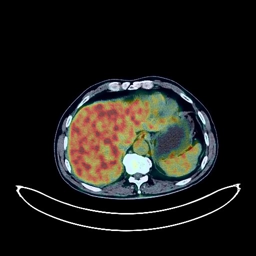
Pancreatic Cancer PET/CT (case 983827-000115 from PETWB-REP)
3 views9 days agoWhole-body 18F-FDG PET/CT scan in a patient with Pancreatic Cancer taken from the PETWB-REP dataset. The following English report (translated from original Chinese) is taken verbatim from the public dataset and has not been modified or otherwise checked for accuracy (see the end for citation). Impression A soft tissue mass around the celiac trunk (L1 level) in the retroperitoneum with increased FDG metabolism; multiple retroperitoneal lymph nodes also showing increased FDG metabolism. This is considered a possible malignant tumor with lymph node metastasis. The boundary with the adjacent pancreas is unclear; please rule out other possibilities based on clinical and pathological findings. Pelvic effusion. Chronic inflammatory nodules in both lungs; CT follow-up is recommended. Calcification in the lower lobe of the left lung. A few post-inflammatory remnants in both lungs. Slight pleural thickening bilaterally. Partial arteriosclerosis. Accessory spleen. Small cyst in the left kidney. Calcification in the prostate. Possible chronic inflammatory changes in the gastric wall; please follow up with endoscopy. Degenerative changes in the spine; L4/5 and L5/S1 intervertebral disc bulges. Age-related brain; deep lacunar infarcts. This case is from PETWB-REP, a curated dataset of whole-body 18F-FDG PET/CT scans and corresponding radiology reports from 490 patients with a broad spectrum of malignancies. The data were retrospectively collected from patients who underwent clinically indicated whole-body 18F-FDG PET/CT scans at the Shanghai Universal Medical Imaging Diagnostic Center between 2021 and 2024. License: Creative Commons Attribution 4.0 International (CC BY 4.0) Citation: Xue, L., Feng, G., Wenbo, Z., Zhang, Y., Li, L., Wang, S., Peng, L., Peng, S., & Gao, X. (2026). PETWB-REP: A Multi-Cancer Whole-Body FDG PET/CT Dataset with Corresponding Radiology Reports [Data set]. Zenodo. https://doi.org/10.5281/zenodo.18670487
Whole BodyPET/CT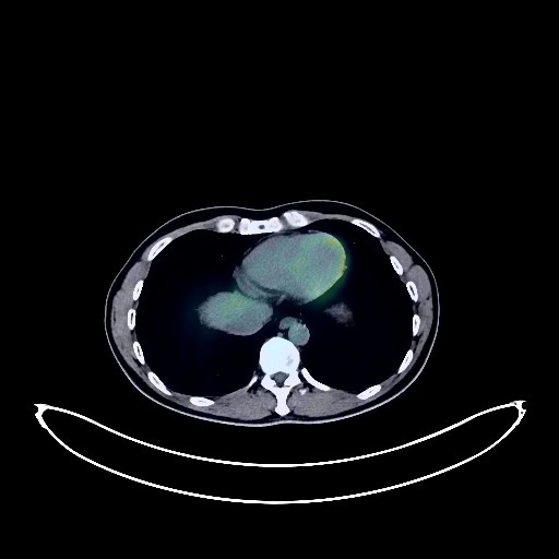
Prostate Cancer PET/CT (case 983827-000242 from PETWB-REP)
3 views9 days agoWhole-body 18F-FDG PET/CT scan in a patient with Prostate Cancer taken from the PETWB-REP dataset. The following English report (translated from original Chinese) is taken verbatim from the public dataset and has not been modified or otherwise checked for accuracy (see the end for citation). Impression Uneven prostate density with punctate calcifications; FDG uptake normal; no clear space-occupying lesion. Please analyze in conjunction with contrast-enhanced MRI images from another hospital; biopsy may be necessary. Postoperative rectal cancer surgery; no obvious signs of tumor recurrence. Scattered chronic inflammatory micronodules (solid) in both lungs. Tracheal diverticulum. Calcification in the right lobe of the liver. Calcification in the tunica vaginalis of the right testis. Spinal degenerative changes. L3/4, L4/5, L5/S1 intervertebral disc bulge. Nodular goiter; ultrasound follow-up recommended. Chronic inflammation of bilateral ethmoid and maxillary sinuses. Cranial FDG imaging normal. This case is from PETWB-REP, a curated dataset of whole-body 18F-FDG PET/CT scans and corresponding radiology reports from 490 patients with a broad spectrum of malignancies. The data were retrospectively collected from patients who underwent clinically indicated whole-body 18F-FDG PET/CT scans at the Shanghai Universal Medical Imaging Diagnostic Center between 2021 and 2024. License: Creative Commons Attribution 4.0 International (CC BY 4.0) Citation: Xue, L., Feng, G., Wenbo, Z., Zhang, Y., Li, L., Wang, S., Peng, L., Peng, S., & Gao, X. (2026). PETWB-REP: A Multi-Cancer Whole-Body FDG PET/CT Dataset with Corresponding Radiology Reports [Data set]. Zenodo. https://doi.org/10.5281/zenodo.18670487
Whole BodyPET/CT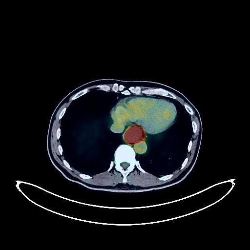
Esophageal Cancer PET/CT (case 983827-000264 from PETWB-REP)
2 views9 days agoWhole-body 18F-FDG PET/CT scan in a patient with Esophageal Cancer taken from the PETWB-REP dataset. The following English report (translated from original Chinese) is taken verbatim from the public dataset and has not been modified or otherwise checked for accuracy (see the end for citation). Impression Space-occupying lesions in the mid-thoracic and lower-thoracic segments of the esophagus with increased FDG metabolism, consistent with esophageal cancer based on pathology; multiple lymph node metastases in the paraesophageal, perigastric, hepatogastric, and right supraclavicular fossa. Chronic inflammatory micronodules in both lungs; CT follow-up is recommended. Emphysema with bullae formation in both lungs. A few post-inflammatory lesions in both lungs. Slight pleural thickening bilaterally. Calcification of some arterial walls (including coronary arteries). Gallbladder sludge-like stones or contrast agent residue. Left adrenal hyperplasia. Contrast agent residue in the urinary tract. Benign prostatic hyperplasia with calcification. Small amount of hydrocele in both testes. Chronic inflammatory changes or physiological changes in some gastric and duodenal walls; endoscopic follow-up is recommended. Slight lumbar scoliosis, degenerative changes in the spine, mild anterior slippage of the L4 vertebral body. L4/5 intervertebral disc bulge. Age-related brain changes. Chronic inflammation of the left maxillary sinus. This case is from PETWB-REP, a curated dataset of whole-body 18F-FDG PET/CT scans and corresponding radiology reports from 490 patients with a broad spectrum of malignancies. The data were retrospectively collected from patients who underwent clinically indicated whole-body 18F-FDG PET/CT scans at the Shanghai Universal Medical Imaging Diagnostic Center between 2021 and 2024. License: Creative Commons Attribution 4.0 International (CC BY 4.0) Citation: Xue, L., Feng, G., Wenbo, Z., Zhang, Y., Li, L., Wang, S., Peng, L., Peng, S., & Gao, X. (2026). PETWB-REP: A Multi-Cancer Whole-Body FDG PET/CT Dataset with Corresponding Radiology Reports [Data set]. Zenodo. https://doi.org/10.5281/zenodo.18670487
Whole BodyPET/CT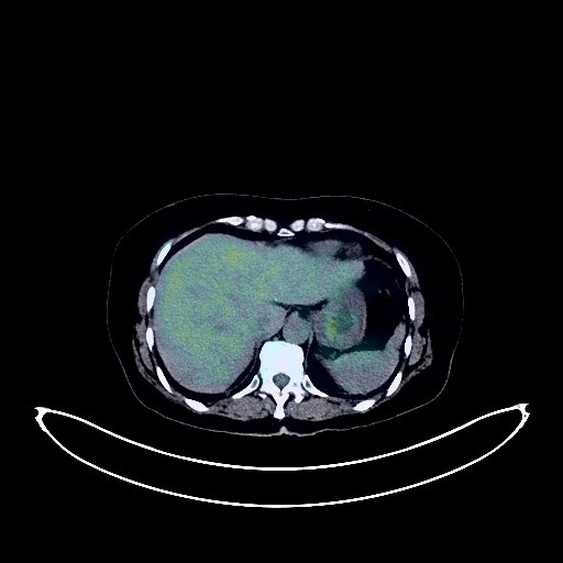
Esophageal Cancer PET/CT (case 983827-000108 from PETWB-REP)
4 views9 days agoWhole-body 18F-FDG PET/CT scan in a patient with Esophageal Cancer taken from the PETWB-REP dataset. The following English report (translated from original Chinese) is taken verbatim from the public dataset and has not been modified or otherwise checked for accuracy (see the end for citation). Impression A mass in the lower thoracic esophagus with elevated FDG metabolism, suggestive of esophageal cancer. Metastasis to the mediastinal paratracheal and left supraclavicular lymph nodes. Left lung infection. Minor chronic inflammation and old lesions in both lungs. Calcification of some arterial walls (including coronary arteries). Post-thyroidectomy changes. Low-density nodule in the left medial lobe of the liver, normal FDG metabolism, suggestive of hemangioma or cyst. A pseudo-lesion near the falciform ligament needs further investigation; enhanced MRI is recommended. Accessory spleen. Right renal cyst. Left adrenal hyperplasia. Degenerative changes in the spine. L4/5 and L5/S1 intervertebral disc bulges. Softening lesions in the right basal ganglia region, suggestive of age-related brain changes. This case is from PETWB-REP, a curated dataset of whole-body 18F-FDG PET/CT scans and corresponding radiology reports from 490 patients with a broad spectrum of malignancies. The data were retrospectively collected from patients who underwent clinically indicated whole-body 18F-FDG PET/CT scans at the Shanghai Universal Medical Imaging Diagnostic Center between 2021 and 2024. License: Creative Commons Attribution 4.0 International (CC BY 4.0) Citation: Xue, L., Feng, G., Wenbo, Z., Zhang, Y., Li, L., Wang, S., Peng, L., Peng, S., & Gao, X. (2026). PETWB-REP: A Multi-Cancer Whole-Body FDG PET/CT Dataset with Corresponding Radiology Reports [Data set]. Zenodo. https://doi.org/10.5281/zenodo.18670487
Whole BodyPET/CT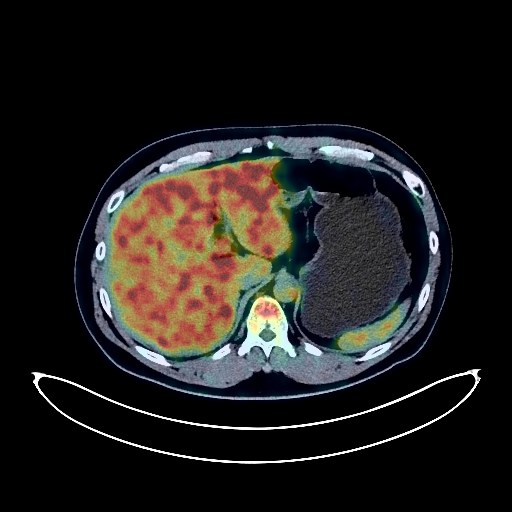
Renal Cancer PET/CT (case 983827-000037 from PETWB-REP)
3 views9 days agoWhole-body 18F-FDG PET/CT scan in a patient with Renal Cancer taken from the PETWB-REP dataset. The following English report (translated from original Chinese) is taken verbatim from the public dataset and has not been modified or otherwise checked for accuracy (see the end for citation). Impression a. Mass at the lower pole of the left kidney, with uneven FDG metabolism, highly suggestive of a neoplastic lesion; please confirm with contrast-enhanced MRI. b. Reactive hyperplasia of the retroperitoneal, mesenteric, and bilateral inguinal lymph nodes is highly likely; please follow up. Chronic inflammatory nodule in the lower lobe of the left lung; please confirm with CT. Fatty liver, no abnormal FDG metabolic foci seen in the liver parenchyma. Prostatic calcification. Increased FDG metabolism in parts of the gastric wall and intestinal tract, possibly due to physiological uptake or chronic inflammation; please confirm with endoscopy. Partial vertebral osteophyte formation. L4/5 disc bulge, L5/S1 disc herniation with posterior margin calcification. Uneven thyroid density; please confirm with ultrasound. No obvious abnormalities seen on cranial scintigraphy. Chronic inflammation of the nasopharynx, bilateral palatine tonsils, and base of the tongue. Reactive hyperplasia of the bilateral deep cervical spaces and submandibular lymph nodes. This case is from PETWB-REP, a curated dataset of whole-body 18F-FDG PET/CT scans and corresponding radiology reports from 490 patients with a broad spectrum of malignancies. The data were retrospectively collected from patients who underwent clinically indicated whole-body 18F-FDG PET/CT scans at the Shanghai Universal Medical Imaging Diagnostic Center between 2021 and 2024. License: Creative Commons Attribution 4.0 International (CC BY 4.0) Citation: Xue, L., Feng, G., Wenbo, Z., Zhang, Y., Li, L., Wang, S., Peng, L., Peng, S., & Gao, X. (2026). PETWB-REP: A Multi-Cancer Whole-Body FDG PET/CT Dataset with Corresponding Radiology Reports [Data set]. Zenodo. https://doi.org/10.5281/zenodo.18670487
Whole BodyPET/CT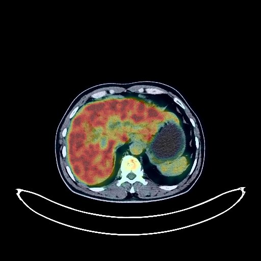
Cholangiocarcinoma PET/CT (case 983827-000199 from PETWB-REP)
3 views9 days agoWhole-body 18F-FDG PET/CT scan in a patient with Cholangiocarcinoma taken from the PETWB-REP dataset. The following English report (translated from original Chinese) is taken verbatim from the public dataset and has not been modified or otherwise checked for accuracy (see the end for citation). Impression a. Mass in the hepatic hilum, increased FDG metabolism, intrahepatic bile duct dilation, suggestive of hilar cholangiocarcinoma. b. Reactive hyperplasia of the lymph nodes around the pancreatic head is highly probable, metastasis to be ruled out. No signs of distant metastasis were observed systemically. Several small, solid, chronic inflammatory nodules in both lungs. A few chronic inflammations and old lesions in both lungs. Emphysema and bullae in the upper lobes of both lungs. Calcification of some arterial walls (including coronary arteries). Chronic cholecystitis. Gallstones. Right renal cyst. Benign prostatic hyperplasia with calcification. Continuous increased FDG metabolism in parts of the colon and rectum, likely due to inflammatory uptake; colonoscopy follow-up is recommended. Degenerative changes in the spine. L4/5 and L5/S1 intervertebral disc bulges. Age-related brain changes. This case is from PETWB-REP, a curated dataset of whole-body 18F-FDG PET/CT scans and corresponding radiology reports from 490 patients with a broad spectrum of malignancies. The data were retrospectively collected from patients who underwent clinically indicated whole-body 18F-FDG PET/CT scans at the Shanghai Universal Medical Imaging Diagnostic Center between 2021 and 2024. License: Creative Commons Attribution 4.0 International (CC BY 4.0) Citation: Xue, L., Feng, G., Wenbo, Z., Zhang, Y., Li, L., Wang, S., Peng, L., Peng, S., & Gao, X. (2026). PETWB-REP: A Multi-Cancer Whole-Body FDG PET/CT Dataset with Corresponding Radiology Reports [Data set]. Zenodo. https://doi.org/10.5281/zenodo.18670487
Whole BodyPET/CT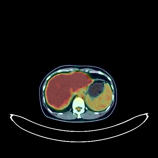
Colon Cancer PET/CT (case 983827-000033 from PETWB-REP)
2 views9 days agoWhole-body 18F-FDG PET/CT scan in a patient with Colon Cancer taken from the PETWB-REP dataset. The following English report (translated from original Chinese) is taken verbatim from the public dataset and has not been modified or otherwise checked for accuracy (see the end for citation). Impression Post-colon cancer surgery: a. No obvious signs of tumor recurrence at the anastomosis site. Pelvic effusion. b. Cystic-solid mass in the right adnexal region, with increased FDG metabolism in the solid component, suggesting a high probability of ovarian neoplasia (metastasis to be ruled out). The boundary with the post-rectal blind end is unclear; please combine laboratory tests and enhanced MRI for comprehensive judgment. A few old lesions in the right upper lung. Mild osteophyte formation in some vertebral bodies, L4/5 and L5/S1 disc herniation. No obvious abnormalities seen on brain scintigraphy. This case is from PETWB-REP, a curated dataset of whole-body 18F-FDG PET/CT scans and corresponding radiology reports from 490 patients with a broad spectrum of malignancies. The data were retrospectively collected from patients who underwent clinically indicated whole-body 18F-FDG PET/CT scans at the Shanghai Universal Medical Imaging Diagnostic Center between 2021 and 2024. License: Creative Commons Attribution 4.0 International (CC BY 4.0) Citation: Xue, L., Feng, G., Wenbo, Z., Zhang, Y., Li, L., Wang, S., Peng, L., Peng, S., & Gao, X. (2026). PETWB-REP: A Multi-Cancer Whole-Body FDG PET/CT Dataset with Corresponding Radiology Reports [Data set]. Zenodo. https://doi.org/10.5281/zenodo.18670487
Whole BodyPET/CT