Loading...
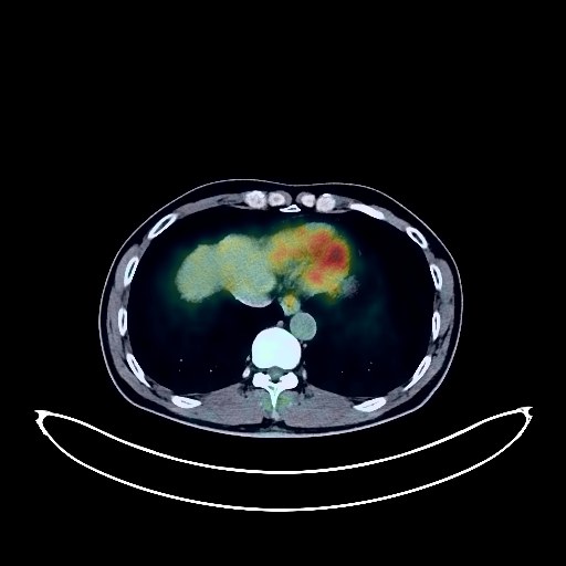
Renal Cancer PET/CT (case 983777-000001 from PETWB-REP)
15 views9 days agoWhole-body 18F-FDG PET/CT scan in a patient with Renal Cancer taken from the PETWB-REP dataset. The following English report (translated from original Chinese) is taken verbatim from the public dataset and has not been modified or otherwise checked for accuracy (see the end for citation). Impression a. Thickening of the rectal wall in the middle and upper segments, with increased FDG metabolism, suggestive of rectal cancer. Multiple lymph node metastases in the perirectal space, bilateral common iliac vessels, and retroperitoneum. b. Continuous increased FDG metabolism in the remaining colon and rectum, suggestive of inflammation or physiological changes; endoscopic follow-up is recommended. Emphysema and bullae in the upper lobes of both lungs, old tuberculous lesions in the right upper lobe, bronchiectasis in the left upper lobe, multiple chronic inflammatory micronodules in both lungs, and scattered post-inflammatory lesions in both lungs. Chronic gastritis; endoscopic follow-up is recommended. Cholestasis in the gallbladder; ultrasound follow-up is recommended. Small amount of hydrocele in both testes. Mild anterior slippage of the L4 vertebral body. Spinal osteophyte formation. L3/4, L4/5, and L5/S1 intervertebral disc bulging. Degenerative changes with inflammatory metabolism in the right L4/5 facet joint are the primary consideration; CT follow-up is recommended to rule out other possibilities. No obvious abnormalities were found on cranial imaging. Right maxillary sinusitis. This case is from PETWB-REP, a curated dataset of whole-body 18F-FDG PET/CT scans and corresponding radiology reports from 490 patients with a broad spectrum of malignancies. The data were retrospectively collected from patients who underwent clinically indicated whole-body 18F-FDG PET/CT scans at the Shanghai Universal Medical Imaging Diagnostic Center between 2021 and 2024. License: Creative Commons Attribution 4.0 International (CC BY 4.0) Citation: Xue, L., Feng, G., Wenbo, Z., Zhang, Y., Li, L., Wang, S., Peng, L., Peng, S., & Gao, X. (2026). PETWB-REP: A Multi-Cancer Whole-Body FDG PET/CT Dataset with Corresponding Radiology Reports [Data set]. Zenodo. https://doi.org/10.5281/zenodo.18670487
Whole BodyPET/CT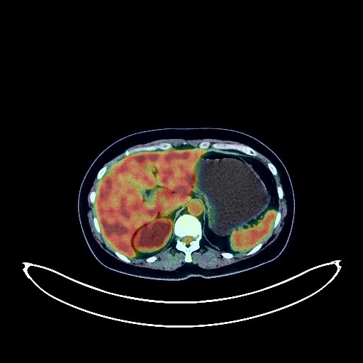
Renal Cancer PET/CT (case 983777-000007 from PETWB-REP)
9 views9 days agoWhole-body 18F-FDG PET/CT scan in a patient with Renal Cancer taken from the PETWB-REP dataset. The following English report (translated from original Chinese) is taken verbatim from the public dataset and has not been modified or otherwise checked for accuracy (see the end for citation). Impression Irregular thickening of the mid-rectal wall with elevated FDG metabolism, suggestive of rectal cancer based on medical history. Reactive hyperplasia of small presacral lymph nodes, close observation recommended to rule out metastasis. Small amount of pelvic effusion. Chronic inflammatory micronodule in the upper lobe of the left lung, follow-up CT recommended. A few post-inflammatory lesions in both lungs. Mild anemia, slight arteriosclerosis in some arteries. Small nodule in the right breast, FDG metabolism normal, suggestive of hyperplastic nodule or fibroadenoma, follow-up ultrasound recommended. Small cyst in the left lateral lobe of the liver, hemangioma in the left medial lobe and right anterior lobe of the liver is the first consideration, please combine with MRI. Uterine fibroid. Chronic inflammatory changes in part of the gastric wall, please combine with endoscopy. Degenerative changes in the spine. L4/5 and L5/S1 intervertebral disc bulge. Low-density nodule in the left lobe of the thyroid, FDG metabolism normal, suggestive of adenomatous nodule, please combine with ultrasound. No obvious abnormalities seen on cranial scintigraphy. Bilateral chronic ethmoid sinusitis. This case is from PETWB-REP, a curated dataset of whole-body 18F-FDG PET/CT scans and corresponding radiology reports from 490 patients with a broad spectrum of malignancies. The data were retrospectively collected from patients who underwent clinically indicated whole-body 18F-FDG PET/CT scans at the Shanghai Universal Medical Imaging Diagnostic Center between 2021 and 2024. License: Creative Commons Attribution 4.0 International (CC BY 4.0) Citation: Xue, L., Feng, G., Wenbo, Z., Zhang, Y., Li, L., Wang, S., Peng, L., Peng, S., & Gao, X. (2026). PETWB-REP: A Multi-Cancer Whole-Body FDG PET/CT Dataset with Corresponding Radiology Reports [Data set]. Zenodo. https://doi.org/10.5281/zenodo.18670487
Whole BodyPET/CT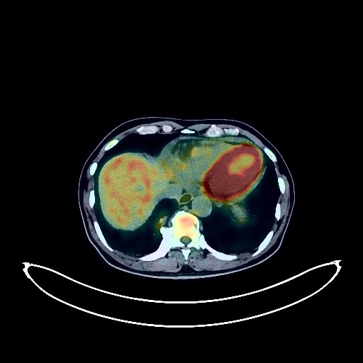
Lung Cancer PET/CT (case 983827-000238 from PETWB-REP)
5 views9 days agoWhole-body 18F-FDG PET/CT scan in a patient with Lung Cancer taken from the PETWB-REP dataset. The following English report (translated from original Chinese) is taken verbatim from the public dataset and has not been modified or otherwise checked for accuracy (see the end for citation). Impression Post-treatment of right lung cancer: a. Soft tissue mass near the hilum of the right upper lobe with increased FDG metabolism, suggesting residual tumor activity, accompanied by minor distal obstructive changes. b. Metastasis to the right hilar and part of the mediastinal lymph nodes. c. Left adrenal metastasis is the primary consideration; follow-up is recommended to rule out other possibilities. Sellar region mass, metastatic tumor to be ruled out; enhanced pituitary MRI is recommended for further examination. Lacunar infarcts in both lobes, senile encephalopathy. Chronic inflammatory micronodules in both lungs, chronic inflammation and sequelae in both lungs, emphysema. Slight pleural thickening bilaterally. Pericardial effusion. Right kidney absent post-surgery; no signs of tumor recurrence were observed in the surgical area. Increased FDG metabolism in parts of the stomach wall and intestines, suggesting physiological uptake or chronic inflammation. Suspicious nodular protrusions in the mid-abdomen small intestine wall; please follow up with endoscopy. Reactive hyperplasia of bilateral inguinal lymph nodes. Spinal degenerative changes. Slight bulging of L2/3 and L3/4 intervertebral discs; calcification at the posterior margin of L4/5 intervertebral disc. Slightly low-density nodule on the medial side of the left scapula, likely benign; enhanced MRI is recommended. Physiological or inflammatory changes on the left side of the oropharyngeal wall; ENT examination is recommended to rule out other possibilities. Chronic inflammation of bilateral ethmoid sinuses and the left maxillary sinus. Slightly low-density nodule in the right lobe of the thyroid gland; FDG metabolism is normal, suggesting nodular goiter; ultrasound follow-up is recommended. Reactive hyperplasia of bilateral cervical lymph nodes. This case is from PETWB-REP, a curated dataset of whole-body 18F-FDG PET/CT scans and corresponding radiology reports from 490 patients with a broad spectrum of malignancies. The data were retrospectively collected from patients who underwent clinically indicated whole-body 18F-FDG PET/CT scans at the Shanghai Universal Medical Imaging Diagnostic Center between 2021 and 2024. License: Creative Commons Attribution 4.0 International (CC BY 4.0) Citation: Xue, L., Feng, G., Wenbo, Z., Zhang, Y., Li, L., Wang, S., Peng, L., Peng, S., & Gao, X. (2026). PETWB-REP: A Multi-Cancer Whole-Body FDG PET/CT Dataset with Corresponding Radiology Reports [Data set]. Zenodo. https://doi.org/10.5281/zenodo.18670487
Whole BodyPET/CT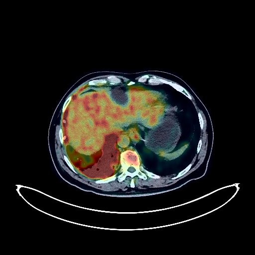
Lung Cancer PET/CT (case 983827-000236 from PETWB-REP)
2 views9 days agoWhole-body 18F-FDG PET/CT scan in a patient with Lung Cancer taken from the PETWB-REP dataset. The following English report (translated from original Chinese) is taken verbatim from the public dataset and has not been modified or otherwise checked for accuracy (see the end for citation). Impression a. Scattered solid nodules and soft tissue masses in the right lung, with increased FDG metabolism, suggestive of malignancy. Right lung cancer with intrapulmonary metastasis is the primary consideration, with right lung cancer lymphangitis being highly probable. Please confirm the diagnosis with pathological examination. b. Metastasis to the right hilar, right internal mammary chain, and right anterior diaphragmatic lymph nodes is the primary consideration. c. Right pleural metastasis is the primary consideration, with right pleural effusion. Increased FDG metabolism at the right chest wall drainage tube puncture site suggests inflammatory changes; tumor infiltration needs to be ruled out. Please follow up. d. Chronic inflammatory nodules and plaque-like foci in the left lung, scattered chronic inflammation in both lungs. Benign prostatic hyperplasia, with unevenly increased FDG metabolism in the parenchyma. Please rule out space-occupying lesions with PSA and enhanced MRI. Reactive hyperplasia of bilateral pelvic wall and inguinal lymph nodes. Please follow up to rule out other possibilities. Hemangioma in the upper right posterior lobe of the liver is the primary consideration; enhanced MRI analysis is recommended. Liver cysts. Bilateral renal cysts. Increased FDG metabolism in part of the gastric wall, suggestive of physiological uptake or chronic inflammation; endoscopic follow-up is recommended. Spinal degenerative changes. L3/4 disc bulge, L4/5 disc herniation. Left sacral islet. Small amount of subdural effusion in the left parietal region, bilateral deep lacunar infarcts, age-related brain changes. Chronic inflammation of the bilateral ethmoid sinuses and right maxillary sinus. Reactive hyperplasia of bilateral cervical lymph nodes. This case is from PETWB-REP, a curated dataset of whole-body 18F-FDG PET/CT scans and corresponding radiology reports from 490 patients with a broad spectrum of malignancies. The data were retrospectively collected from patients who underwent clinically indicated whole-body 18F-FDG PET/CT scans at the Shanghai Universal Medical Imaging Diagnostic Center between 2021 and 2024. License: Creative Commons Attribution 4.0 International (CC BY 4.0) Citation: Xue, L., Feng, G., Wenbo, Z., Zhang, Y., Li, L., Wang, S., Peng, L., Peng, S., & Gao, X. (2026). PETWB-REP: A Multi-Cancer Whole-Body FDG PET/CT Dataset with Corresponding Radiology Reports [Data set]. Zenodo. https://doi.org/10.5281/zenodo.18670487
Whole BodyPET/CT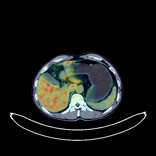
Lung Cancer PET/CT (case 983827-000050 from PETWB-REP)
2 views9 days agoWhole-body 18F-FDG PET/CT scan in a patient with Lung Cancer taken from the PETWB-REP dataset. The following English report (translated from original Chinese) is taken verbatim from the public dataset and has not been modified or otherwise checked for accuracy (see the end for citation). Impression A mass in the posterior segment of the right lower lobe, with elevated FDG metabolism, consistent with lung cancer with obstructive inflammation, involving the right upper lobe; multiple lymph node metastases in the right hilum, mediastinum, and right supraclavicular fossa. Several small, solid, chronic inflammatory nodules in both lungs. A few chronic inflammatory lesions and old lesions in both lungs. Calcification in the left kidney. Accessory spleen. Mild osteophyte formation in the cervical, thoracic, and lumbar spine. A few ischemic lesions in the deep bilateral brain regions. Chronic inflammation of both palatine tonsils. This case is from PETWB-REP, a curated dataset of whole-body 18F-FDG PET/CT scans and corresponding radiology reports from 490 patients with a broad spectrum of malignancies. The data were retrospectively collected from patients who underwent clinically indicated whole-body 18F-FDG PET/CT scans at the Shanghai Universal Medical Imaging Diagnostic Center between 2021 and 2024. License: Creative Commons Attribution 4.0 International (CC BY 4.0) Citation: Xue, L., Feng, G., Wenbo, Z., Zhang, Y., Li, L., Wang, S., Peng, L., Peng, S., & Gao, X. (2026). PETWB-REP: A Multi-Cancer Whole-Body FDG PET/CT Dataset with Corresponding Radiology Reports [Data set]. Zenodo. https://doi.org/10.5281/zenodo.18670487
Whole BodyPET/CT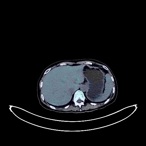
Cervical Cancer PET/CT (case 983827-000161 from PETWB-REP)
2 views9 days agoWhole-body 18F-FDG PET/CT scan in a patient with Cervical Cancer taken from the PETWB-REP dataset. The following English report (translated from original Chinese) is taken verbatim from the public dataset and has not been modified or otherwise checked for accuracy (see the end for citation). Impression a. Cervical mass, involving the uterine body and vagina, with unclear boundaries from the adjacent bladder and rectum, elevated FDG metabolism, consistent with cervical cancer, invading the lower end of the left ureter, causing proximal ureteropelvic dilatation and hydronephrosis. b. Metastasis to the right common iliac, external iliac vessels, and retroperitoneal lymph nodes. Reactive hyperplasia of the left external iliac vessels lymph nodes. Pneumothorax in the uterine cavity. Low-density nodule with multiple calcifications in the right lobe of the thyroid gland, normal FDG metabolism, highly suggestive of nodular goiter, ultrasound follow-up recommended. Ground-glass nodule in the apical segment of the right upper lobe of the lung, normal FDG metabolism, suggestive of inflammatory nodule or atypical adenomatous hyperplasia, annual HRCT follow-up recommended. A few chronic inflammations and old lesions (including calcifications) in both lungs. Bilateral breast proliferative changes. Liver calcifications. Gallbladder cholestasis. Right renal cyst. Spinal degenerative changes. L4/5 and L5/S1 intervertebral disc bulges. No abnormalities found on cranial scintigraphy. This case is from PETWB-REP, a curated dataset of whole-body 18F-FDG PET/CT scans and corresponding radiology reports from 490 patients with a broad spectrum of malignancies. The data were retrospectively collected from patients who underwent clinically indicated whole-body 18F-FDG PET/CT scans at the Shanghai Universal Medical Imaging Diagnostic Center between 2021 and 2024. License: Creative Commons Attribution 4.0 International (CC BY 4.0) Citation: Xue, L., Feng, G., Wenbo, Z., Zhang, Y., Li, L., Wang, S., Peng, L., Peng, S., & Gao, X. (2026). PETWB-REP: A Multi-Cancer Whole-Body FDG PET/CT Dataset with Corresponding Radiology Reports [Data set]. Zenodo. https://doi.org/10.5281/zenodo.18670487
Whole BodyPET/CT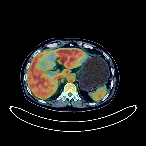
Lung Cancer PET/CT (case 983827-000123 from PETWB-REP)
3 views9 days agoWhole-body 18F-FDG PET/CT scan in a patient with Lung Cancer taken from the PETWB-REP dataset. The following English report (translated from original Chinese) is taken verbatim from the public dataset and has not been modified or otherwise checked for accuracy (see the end for citation). Impression a. A large mass in the posterior segment of the left lower lobe, with increased FDG metabolism, suggestive of lung cancer with surrounding inflammation. Multiple lymph node metastases in the left hilum and mediastinum. T7 vertebral metastasis is highly probable. b. Several small chronic inflammatory nodules (solid) in both lungs are highly probable; CT scan for comparison is recommended. A few chronic inflammations and old lesions in both lungs. Some arterial wall calcification. A few ischemic lesions in the deep bilateral brain; age-related brain, MRI is recommended. Small liver cysts. Fatty infiltration of the pancreas. Increased FDG metabolism in some intestinal segments, suggestive of inflammatory or physiological uptake. Degenerative changes in the spine. L4/5 and L5/S1 intervertebral disc bulges. Reactive hyperplasia of bilateral cervical lymph nodes. This case is from PETWB-REP, a curated dataset of whole-body 18F-FDG PET/CT scans and corresponding radiology reports from 490 patients with a broad spectrum of malignancies. The data were retrospectively collected from patients who underwent clinically indicated whole-body 18F-FDG PET/CT scans at the Shanghai Universal Medical Imaging Diagnostic Center between 2021 and 2024. License: Creative Commons Attribution 4.0 International (CC BY 4.0) Citation: Xue, L., Feng, G., Wenbo, Z., Zhang, Y., Li, L., Wang, S., Peng, L., Peng, S., & Gao, X. (2026). PETWB-REP: A Multi-Cancer Whole-Body FDG PET/CT Dataset with Corresponding Radiology Reports [Data set]. Zenodo. https://doi.org/10.5281/zenodo.18670487
Whole BodyPET/CT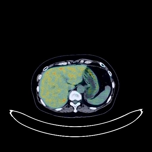
Cervical Cancer PET/CT (case 983827-000005 from PETWB-REP)
2 views9 days agoWhole-body 18F-FDG PET/CT scan in a patient with Cervical Cancer taken from the PETWB-REP dataset. The following English report (translated from original Chinese) is taken verbatim from the public dataset and has not been modified or otherwise checked for accuracy (see the end for citation). Impression a. Cervical mass with elevated FDG metabolism, highly suggestive of cervical cancer; please correlate with clinicopathology. b. Multiple uterine fibroids; please correlate with specialist examination. Chronic inflammatory nodules in both lungs; CT follow-up recommended. A few post-inflammatory lesions in both lungs. Calcification of some arterial walls (including coronary arteries). Possible small cyst in the left kidney. Chronic inflammatory changes or physiological uptake in part of the gastric wall and ascending colon; please correlate with endoscopy. Osteoporosis, mild vertebral osteophyte formation, L4/5 and L5/S1 intervertebral disc bulge. No obvious abnormalities seen on cranial scintigraphy. Right upper alveolar alveolitis. Bilateral palatine tonsillitis. This case is from PETWB-REP, a curated dataset of whole-body 18F-FDG PET/CT scans and corresponding radiology reports from 490 patients with a broad spectrum of malignancies. The data were retrospectively collected from patients who underwent clinically indicated whole-body 18F-FDG PET/CT scans at the Shanghai Universal Medical Imaging Diagnostic Center between 2021 and 2024. License: Creative Commons Attribution 4.0 International (CC BY 4.0) Citation: Xue, L., Feng, G., Wenbo, Z., Zhang, Y., Li, L., Wang, S., Peng, L., Peng, S., & Gao, X. (2026). PETWB-REP: A Multi-Cancer Whole-Body FDG PET/CT Dataset with Corresponding Radiology Reports [Data set]. Zenodo. https://doi.org/10.5281/zenodo.18670487
Whole BodyPET/CT
Lung Cancer PET/CT (case 983827-000130 from PETWB-REP)
2 views9 days agoWhole-body 18F-FDG PET/CT scan in a patient with Lung Cancer taken from the PETWB-REP dataset. The following English report (translated from original Chinese) is taken verbatim from the public dataset and has not been modified or otherwise checked for accuracy (see the end for citation). Impression a. A mass in the right hilum with increased FDG metabolism, highly suggestive of malignancy, but inflammatory granulomatous lesions need to be ruled out. Please correlate with clinicopathology. b. A solid nodule in the apical segment of the right upper lobe, with normal FDG uptake, suggestive of a chronic inflammatory nodule, metastasis needs to be ruled out. Please monitor with CT. Reactive hyperplasia of mediastinal lymph nodes is highly probable. c. A small chronic inflammatory nodule in the anterior basal segment of the right lower lobe. Emphysema in both upper lobes of the lungs. Right frontal sinusitis, bilateral ethmoid sinusitis, and bilateral maxillary sinusitis. No obvious abnormalities were found on cranial scintigraphy. Continuous increased FDG metabolism in the descending colon and sigmoid colon, highly suggestive of inflammatory uptake or polypoid lesions. Colonoscopy is recommended for clarification. Gallstones. Right kidney stone, small kidney stone in the left kidney. Osteophytes in some vertebrae. This case is from PETWB-REP, a curated dataset of whole-body 18F-FDG PET/CT scans and corresponding radiology reports from 490 patients with a broad spectrum of malignancies. The data were retrospectively collected from patients who underwent clinically indicated whole-body 18F-FDG PET/CT scans at the Shanghai Universal Medical Imaging Diagnostic Center between 2021 and 2024. License: Creative Commons Attribution 4.0 International (CC BY 4.0) Citation: Xue, L., Feng, G., Wenbo, Z., Zhang, Y., Li, L., Wang, S., Peng, L., Peng, S., & Gao, X. (2026). PETWB-REP: A Multi-Cancer Whole-Body FDG PET/CT Dataset with Corresponding Radiology Reports [Data set]. Zenodo. https://doi.org/10.5281/zenodo.18670487
Whole BodyPET/CT
Lung Cancer PET/CT (case 983827-000229 from PETWB-REP)
2 views9 days agoWhole-body 18F-FDG PET/CT scan in a patient with Lung Cancer taken from the PETWB-REP dataset. The following English report (translated from original Chinese) is taken verbatim from the public dataset and has not been modified or otherwise checked for accuracy (see the end for citation). Impression a. A soft tissue mass near the hilum of the right upper lobe with increased FDG metabolism, suggestive of malignancy, most likely lung cancer with obstructive changes. Please confirm the diagnosis with pathological examination. Multiple metastatic tumors in the right pleura, right pleural effusion. b. Multiple lymph node metastases in the right hilum and mediastinum. c. Multiple bone metastases throughout the body (see description for details). d. Bilateral adrenal hyperplasia is highly probable; please follow up to rule out metastasis. A few post-inflammatory lesions in both lungs. Partial pulmonary edema in the right lung. Calcification of some arterial walls (including coronary arteries). Calcification in the right lobe of the liver. Small gallstones. Nodular FDG hypermetabolic lesions in the upper abdomen along the course of the small intestine, possibly physiological or inflammatory changes, space-occupying lesion to be ruled out, enhanced CT follow-up recommended. Chronic gastritis changes, gastroscopy follow-up recommended. Spinal degenerative changes. L3/4 intervertebral disc bulge. Bilateral deep cerebral ischemic lesions, mild age-related brain changes, MRI follow-up recommended to rule out micrometastases. Uneven density between the left and right lobes of the thyroid gland; slightly low-density small nodules in the right lobe, FDG metabolism normal, possibly nodular goiter, please follow up with ultrasound. This case is from PETWB-REP, a curated dataset of whole-body 18F-FDG PET/CT scans and corresponding radiology reports from 490 patients with a broad spectrum of malignancies. The data were retrospectively collected from patients who underwent clinically indicated whole-body 18F-FDG PET/CT scans at the Shanghai Universal Medical Imaging Diagnostic Center between 2021 and 2024. License: Creative Commons Attribution 4.0 International (CC BY 4.0) Citation: Xue, L., Feng, G., Wenbo, Z., Zhang, Y., Li, L., Wang, S., Peng, L., Peng, S., & Gao, X. (2026). PETWB-REP: A Multi-Cancer Whole-Body FDG PET/CT Dataset with Corresponding Radiology Reports [Data set]. Zenodo. https://doi.org/10.5281/zenodo.18670487
Whole BodyPET/CT