Loading...
Liver Cancer PET/CT (case 984005-000007 from PETWB-REP)
2 views9 days agoWhole-body 18F-FDG PET/CT scan in a patient with Liver Cancer taken from the PETWB-REP dataset. The following English report (translated from original Chinese) is taken verbatim from the public dataset and has not been modified or otherwise checked for accuracy (see the end for citation). Impression a. A large, slightly low-density mass in the liver with increased FDG metabolism, suggestive of hepatocellular carcinoma with invasion of the left and right branches and main trunk of the portal vein; multiple lymph node metastases in the hepatogastric space and retroperitoneum. b. Increased peritoneal density in the abdominopelvic region with mild FDG metabolism, metastasis to be ruled out; abdominopelvic effusion. c. Liver cirrhosis with splenomegaly. Multiple chronic inflammatory micronodules in both lungs. Slight pleural thickening bilaterally. Mild anemia. Chronic gastritis, endoscopic follow-up recommended. Gallstones and chronic cholecystitis. Accessory spleen. Slight scoliosis with osteophyte formation. No obvious abnormalities seen on cranial imaging. This case is from PETWB-REP, a curated dataset of whole-body 18F-FDG PET/CT scans and corresponding radiology reports from 490 patients with a broad spectrum of malignancies. The data were retrospectively collected from patients who underwent clinically indicated whole-body 18F-FDG PET/CT scans at the Shanghai Universal Medical Imaging Diagnostic Center between 2021 and 2024. License: Creative Commons Attribution 4.0 International (CC BY 4.0) Citation: Xue, L., Feng, G., Wenbo, Z., Zhang, Y., Li, L., Wang, S., Peng, L., Peng, S., & Gao, X. (2026). PETWB-REP: A Multi-Cancer Whole-Body FDG PET/CT Dataset with Corresponding Radiology Reports [Data set]. Zenodo. https://doi.org/10.5281/zenodo.18670487
Whole BodyPET/CT
Liver Cancer PET/CT (case 984005-000006 from PETWB-REP)
2 views9 days agoWhole-body 18F-FDG PET/CT scan in a patient with Liver Cancer taken from the PETWB-REP dataset. The following English report (translated from original Chinese) is taken verbatim from the public dataset and has not been modified or otherwise checked for accuracy (see the end for citation). Impression a. A large, irregular, slightly low-density mass in the right lobe of the liver, with increased FDG metabolism, suggestive of hepatocellular carcinoma with intrahepatic metastases. Small amount of perihepatic and pelvic effusion. b. Metastasis to lymph nodes in the porta hepatis, anterior and posterior to the inferior vena cava, in the right anterior renal space, and around the peritoneal major vessels is the primary consideration. Metastasis to the right 12th rib. c. Calcification in the left medial lobe of the liver. Mild fatty liver. Scattered chronic inflammatory nodules in both lungs; please follow up with CT scans. A few post-inflammatory lesions in both lungs. Gallbladder fundus polyps. Right renal cyst, right renal calculus; ultrasound follow-up recommended. Mild prostatic hyperplasia. Bilateral scrotal calcifications. Chronic gastritis; hemorrhoidal manifestations. Please follow up with endoscopy for the above. Slight reversal of cervical lordosis. L4/5 disc bulge, L5/S1 disc herniation. Left femoral head and right ischial island. Cranial scintigraphy showed no obvious abnormalities. There was slight inflammation of the right sphenoid sinus. Reactive hyperplasia was observed in the bilateral deep cervical spaces, submandibular, and submental lymph nodes. The right lens showed decreased density, but FDG metabolism was not significantly abnormal. Considering the medical history, postoperative changes are suspected; specialist follow-up is recommended. This case is from PETWB-REP, a curated dataset of whole-body 18F-FDG PET/CT scans and corresponding radiology reports from 490 patients with a broad spectrum of malignancies. The data were retrospectively collected from patients who underwent clinically indicated whole-body 18F-FDG PET/CT scans at the Shanghai Universal Medical Imaging Diagnostic Center between 2021 and 2024. License: Creative Commons Attribution 4.0 International (CC BY 4.0) Citation: Xue, L., Feng, G., Wenbo, Z., Zhang, Y., Li, L., Wang, S., Peng, L., Peng, S., & Gao, X. (2026). PETWB-REP: A Multi-Cancer Whole-Body FDG PET/CT Dataset with Corresponding Radiology Reports [Data set]. Zenodo. https://doi.org/10.5281/zenodo.18670487
Whole BodyPET/CT
Liver Cancer PET/CT (case 984005-000005 from PETWB-REP)
2 views9 days agoWhole-body 18F-FDG PET/CT scan in a patient with Liver Cancer taken from the PETWB-REP dataset. The following English report (translated from original Chinese) is taken verbatim from the public dataset and has not been modified or otherwise checked for accuracy (see the end for citation). Impression a. Hepatic parenchyma density is uneven, with a slightly low-density nodule at S4. Increased FDG metabolism, combined with contrast-enhanced MRI from another hospital, suggests hepatocellular carcinoma as the primary consideration. Please correlate with clinical and pathological findings. Reactive hyperplasia of the hepatogastric space, hepatic hilum, and retroperitoneal lymph nodes is highly probable; please follow up. b. Cirrhosis, multiple hepatic cysts. Splenectomy absent, multiple calcifications in the main portal vein and splenic vein walls. Slightly blurred peritoneal spaces in the abdominopelvic cavity, with a small amount of pelvic effusion. Chronic inflammatory micronodules in both lungs, pulmonary fibrosis; please follow up with CT scans. Increased FDG metabolism in parts of the gastric wall and intestines, considered to be physiological uptake or chronic inflammation; please follow up with endoscopy. Spinal degeneration. T12 vertebral wedge deformity. L5/S1 intervertebral disc bulge. Bilateral deep lacunar infarcts, mild age-related brain changes. Right maxillary sinus submucosal cyst. This case is from PETWB-REP, a curated dataset of whole-body 18F-FDG PET/CT scans and corresponding radiology reports from 490 patients with a broad spectrum of malignancies. The data were retrospectively collected from patients who underwent clinically indicated whole-body 18F-FDG PET/CT scans at the Shanghai Universal Medical Imaging Diagnostic Center between 2021 and 2024. License: Creative Commons Attribution 4.0 International (CC BY 4.0) Citation: Xue, L., Feng, G., Wenbo, Z., Zhang, Y., Li, L., Wang, S., Peng, L., Peng, S., & Gao, X. (2026). PETWB-REP: A Multi-Cancer Whole-Body FDG PET/CT Dataset with Corresponding Radiology Reports [Data set]. Zenodo. https://doi.org/10.5281/zenodo.18670487
Whole BodyPET/CT
Liver Cancer PET/CT (case 984005-000004 from PETWB-REP)
2 views9 days agoWhole-body 18F-FDG PET/CT scan in a patient with Liver Cancer taken from the PETWB-REP dataset. The following English report (translated from original Chinese) is taken verbatim from the public dataset and has not been modified or otherwise checked for accuracy (see the end for citation). Impression a. Irregular mixed low-density lesion near the diaphragm in the right lobe of the liver, with increased FDG metabolism, hepatocellular carcinoma is the primary consideration; please correlate with pathology. b. Cirrhosis, splenomegaly. Reactive hyperplasia of the hilar lymph nodes; please follow up to rule out other possibilities. Microhepatic effusion. Chronic inflammatory micronodules in both lungs, chronic inflammation and sequelae (including calcifications) in both lungs; please follow up with CT scans. Partial arteriosclerosis (including coronary arteries). Chronic cholecystitis. Left renal atrophy. Prostatic calcifications. Increased FDG metabolism in parts of the stomach wall and intestines, considered physiological uptake or chronic inflammation, hemorrhoidal changes; please follow up with endoscopy. Scoliosis with degenerative changes. L2/3, L3/4 intervertebral disc bulge, L4/5 and L5/S1 intervertebral disc herniation. Post-fracture changes of the right 11th rib, L1 and L2 transverse processes. No obvious abnormalities were found on cranial scintigraphy. Inflammation of the alveolar bone in the left mandible. This case is from PETWB-REP, a curated dataset of whole-body 18F-FDG PET/CT scans and corresponding radiology reports from 490 patients with a broad spectrum of malignancies. The data were retrospectively collected from patients who underwent clinically indicated whole-body 18F-FDG PET/CT scans at the Shanghai Universal Medical Imaging Diagnostic Center between 2021 and 2024. License: Creative Commons Attribution 4.0 International (CC BY 4.0) Citation: Xue, L., Feng, G., Wenbo, Z., Zhang, Y., Li, L., Wang, S., Peng, L., Peng, S., & Gao, X. (2026). PETWB-REP: A Multi-Cancer Whole-Body FDG PET/CT Dataset with Corresponding Radiology Reports [Data set]. Zenodo. https://doi.org/10.5281/zenodo.18670487
Whole BodyPET/CT
Liver Cancer PET/CT (case 984005-000003 from PETWB-REP)
2 views9 days agoWhole-body 18F-FDG PET/CT scan in a patient with Liver Cancer taken from the PETWB-REP dataset. The following English report (translated from original Chinese) is taken verbatim from the public dataset and has not been modified or otherwise checked for accuracy (see the end for citation). Impression a. A slightly low-density mass in the upper posterior segment of the right lobe of the liver, with increased FDG metabolism. Combined with our center's MRI, hepatocellular carcinoma is suspected. b. Liver cirrhosis, multiple intrahepatic cysts. c. Discontinuous right 7th rib with increased FDG metabolism, suggesting possible fracture; follow-up is recommended to rule out metastasis. Multiple chronic inflammatory nodules and calcifications in both lungs, with a few post-inflammatory remnants in both lungs. Slight thickening of the pleura bilaterally. Calcification of some arterial walls (including coronary arteries). Spinal osteophyte formation. L3/4 and L4/5 intervertebral disc bulge. Possible left parietal meningioma; enhanced MRI follow-up is recommended. Inflammation of the left maxillary and frontal sinuses. This case is from PETWB-REP, a curated dataset of whole-body 18F-FDG PET/CT scans and corresponding radiology reports from 490 patients with a broad spectrum of malignancies. The data were retrospectively collected from patients who underwent clinically indicated whole-body 18F-FDG PET/CT scans at the Shanghai Universal Medical Imaging Diagnostic Center between 2021 and 2024. License: Creative Commons Attribution 4.0 International (CC BY 4.0) Citation: Xue, L., Feng, G., Wenbo, Z., Zhang, Y., Li, L., Wang, S., Peng, L., Peng, S., & Gao, X. (2026). PETWB-REP: A Multi-Cancer Whole-Body FDG PET/CT Dataset with Corresponding Radiology Reports [Data set]. Zenodo. https://doi.org/10.5281/zenodo.18670487
Whole BodyPET/CT
Liver Cancer PET/CT (case 984005-000002 from PETWB-REP)
2 views9 days agoWhole-body 18F-FDG PET/CT scan in a patient with Liver Cancer taken from the PETWB-REP dataset. The following English report (translated from original Chinese) is taken verbatim from the public dataset and has not been modified or otherwise checked for accuracy (see the end for citation). Impression a. Low-density mass in the right lobe of the liver with increased FDG metabolism, suggestive of hepatocellular carcinoma with intrahepatic metastasis. b. Multiple metastatic tumors in both lungs. Left adrenal metastasis. Right upper femoral metastasis. Mild emphysema in both lungs, a few fibrotic lesions in both lungs. Tracheal diverticulum. Cardiomegaly. Cirrhosis, liver cysts. Left renal cyst. Mild fatty infiltration of the pancreas. Prostatic calcification. Degenerative changes in the spine. L2/3 intervertebral disc pneumothorax. L3/4 and L4/5 intervertebral disc bulges. A few ischemic lesions deep in the brain, age-related brain changes. A few chronic inflammations of the right maxillary sinus. This case is from PETWB-REP, a curated dataset of whole-body 18F-FDG PET/CT scans and corresponding radiology reports from 490 patients with a broad spectrum of malignancies. The data were retrospectively collected from patients who underwent clinically indicated whole-body 18F-FDG PET/CT scans at the Shanghai Universal Medical Imaging Diagnostic Center between 2021 and 2024. License: Creative Commons Attribution 4.0 International (CC BY 4.0) Citation: Xue, L., Feng, G., Wenbo, Z., Zhang, Y., Li, L., Wang, S., Peng, L., Peng, S., & Gao, X. (2026). PETWB-REP: A Multi-Cancer Whole-Body FDG PET/CT Dataset with Corresponding Radiology Reports [Data set]. Zenodo. https://doi.org/10.5281/zenodo.18670487
Whole BodyPET/CT
Liver Cancer PET/CT (case 984005-000001 from PETWB-REP)
2 views9 days agoWhole-body 18F-FDG PET/CT scan in a patient with Liver Cancer taken from the PETWB-REP dataset. The following English report (translated from original Chinese) is taken verbatim from the public dataset and has not been modified or otherwise checked for accuracy (see the end for citation). Impression a. Slightly low-density lesion in the right anterior lobe of the liver with increased FDG metabolism, suggestive of malignancy, with hepatocellular carcinoma as the primary consideration. Please combine AFP and enhanced MRI for comprehensive analysis. Multiple solid nodules in both lungs, some with mildly increased FDG metabolism, some metastatic tumors are the primary consideration. Short-term follow-up CT is recommended for comparison. b. Mild cirrhosis, liver cysts. Multiple ground-glass nodules in both lungs, no abnormal FDG uptake, suggestive of atypical adenomatous hyperplasia or chronic inflammatory nodules. Please follow up with CT. Emphysema, chronic inflammation and sequelae in both lungs. Tracheal diverticulum. Calcification of some arterial walls, post-coronary artery stenting. Slightly enlarged cardiac silhouette. Bilateral gynecomastia. Chronic cholecystitis, gallstones. Bilateral renal atrophy, multiple renal cysts and stones. Prostatic calcification. Mild anterior slippage of L3 vertebral body. Spinal osteophyte formation. Pneumodegenerative changes in L3/4 and L4/5 intervertebral discs. Right cerebellar softening lesion, multiple ischemic lesions in the brain, age-related brain changes. This case is from PETWB-REP, a curated dataset of whole-body 18F-FDG PET/CT scans and corresponding radiology reports from 490 patients with a broad spectrum of malignancies. The data were retrospectively collected from patients who underwent clinically indicated whole-body 18F-FDG PET/CT scans at the Shanghai Universal Medical Imaging Diagnostic Center between 2021 and 2024. License: Creative Commons Attribution 4.0 International (CC BY 4.0) Citation: Xue, L., Feng, G., Wenbo, Z., Zhang, Y., Li, L., Wang, S., Peng, L., Peng, S., & Gao, X. (2026). PETWB-REP: A Multi-Cancer Whole-Body FDG PET/CT Dataset with Corresponding Radiology Reports [Data set]. Zenodo. https://doi.org/10.5281/zenodo.18670487
Whole BodyPET/CT
Liver Cancer PET/CT (case 984005-000022 from PETWB-REP)
2 views9 days agoWhole-body 18F-FDG PET/CT scan in a patient with Liver Cancer taken from the PETWB-REP dataset. The following English report (translated from original Chinese) is taken verbatim from the public dataset and has not been modified or otherwise checked for accuracy (see the end for citation). Impression a. A large, slightly low-density mass in the right lobe of the liver with increased FDG metabolism; multiple slightly low-density nodules and masses within the liver with increased FDG metabolism, suggestive of hepatocellular carcinoma with multiple intrahepatic lesions. Metastasis to the perihepatic and hilar lymph nodes is possible. b. Metastatic tumor in the right middle lobe of the lung. Multiple solid micronodules in the remaining two lungs, with normal FDG metabolism; chronic inflammatory nodules are the primary consideration, but some metastases cannot be ruled out. Please review with a CT scan for comparison. c. Multiple bone metastases. A few remnants of chronic inflammation in the right lower lobe. A bulla in the left lower lobe. Calcification of some arterial walls (including coronary arteries). Possible left renal cyst; please review with an MRI. Benign prostatic hyperplasia with calcification; mildly increased FDG metabolism in the right peripheral zone; inflammatory lesions are the primary consideration; please review with a PSA test to rule out other possibilities. Spinal degenerative changes. A few ischemic lesions deep in the brain, age-related brain changes. This case is from PETWB-REP, a curated dataset of whole-body 18F-FDG PET/CT scans and corresponding radiology reports from 490 patients with a broad spectrum of malignancies. The data were retrospectively collected from patients who underwent clinically indicated whole-body 18F-FDG PET/CT scans at the Shanghai Universal Medical Imaging Diagnostic Center between 2021 and 2024. License: Creative Commons Attribution 4.0 International (CC BY 4.0) Citation: Xue, L., Feng, G., Wenbo, Z., Zhang, Y., Li, L., Wang, S., Peng, L., Peng, S., & Gao, X. (2026). PETWB-REP: A Multi-Cancer Whole-Body FDG PET/CT Dataset with Corresponding Radiology Reports [Data set]. Zenodo. https://doi.org/10.5281/zenodo.18670487
Whole BodyPET/CT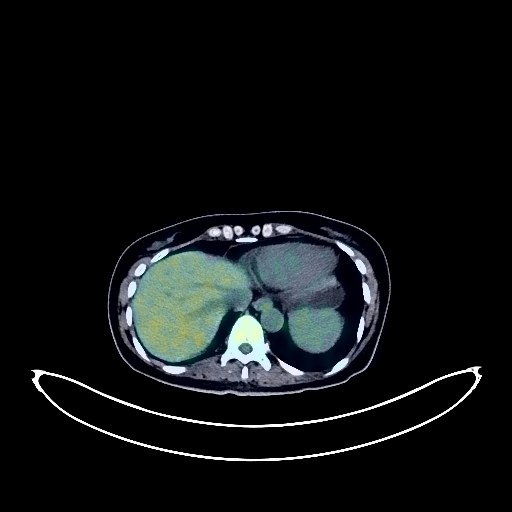
Renal Cancer PET/CT (case 983777-000006 from PETWB-REP)
9 views9 days agoWhole-body 18F-FDG PET/CT scan in a patient with Renal Cancer taken from the PETWB-REP dataset. The following English report (translated from original Chinese) is taken verbatim from the public dataset and has not been modified or otherwise checked for accuracy (see the end for citation). Impression a. Thickening of the lower descending colon near the sigmoid colon with increased FDG metabolism, consistent with colon cancer. Mesenteric lymph node metastasis. Liver metastasis. b. Increased FDG metabolism in the remaining intestinal segment, suggesting physiological changes or inflammatory lesions; chronic gastritis. Please follow up with endoscopy for the above. Multiple ground-glass nodules in both lungs, normal FDG metabolism, atypical adenomatous hyperplasia or early-stage lung cancer (larger ones) are the primary considerations; CT scan follow-up in 3 months is recommended. Chronic inflammatory micronodules (solid) in both lungs. Anemia. Nodular goiter, please follow up with ultrasound. Contrast agent residue or cholestasis in the gallbladder. Contrast agent residue in the intestines. Multiple uterine fibroids, Nabothian cysts in the cervix, please follow up with ultrasound. Small amount of pelvic effusion. Mild osteophyte formation in the spine. No significant abnormalities in FDG metabolism in the brain. This case is from PETWB-REP, a curated dataset of whole-body 18F-FDG PET/CT scans and corresponding radiology reports from 490 patients with a broad spectrum of malignancies. The data were retrospectively collected from patients who underwent clinically indicated whole-body 18F-FDG PET/CT scans at the Shanghai Universal Medical Imaging Diagnostic Center between 2021 and 2024. License: Creative Commons Attribution 4.0 International (CC BY 4.0) Citation: Xue, L., Feng, G., Wenbo, Z., Zhang, Y., Li, L., Wang, S., Peng, L., Peng, S., & Gao, X. (2026). PETWB-REP: A Multi-Cancer Whole-Body FDG PET/CT Dataset with Corresponding Radiology Reports [Data set]. Zenodo. https://doi.org/10.5281/zenodo.18670487
Whole BodyPET/CT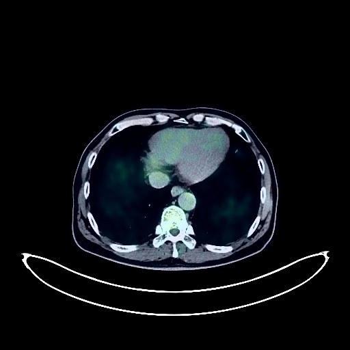
Renal Cancer PET/CT (case 983777-000004 from PETWB-REP)
8 views9 days agoWhole-body 18F-FDG PET/CT scan in a patient with Renal Cancer taken from the PETWB-REP dataset. The following English report (translated from original Chinese) is taken verbatim from the public dataset and has not been modified or otherwise checked for accuracy (see the end for citation). Impression a. Thickening of the rectal wall in the upper and middle segment, involving the adjacent sigmoid colon; increased FDG metabolism, with further increased metabolism after delay, consistent with rectal cancer. b. Metastasis to the surrounding fat spaces and presacral lymph nodes is highly probable; please correlate with clinicopathology. Multiple chronic inflammatory micronodules in both lungs; scattered post-inflammatory remnants in both lungs. Slight thickening of the pleura bilaterally. Lipoma in the subcutaneous intermuscular region of the right shoulder. Chronic gastritis; endoscopic follow-up is recommended. Cholestasis in the gallbladder; ultrasound follow-up is recommended. Left renal cyst. Spinal osteophyte formation. L3-4 vertebral endplate inflammation. L3/4 and L4/5 intervertebral disc bulge. No obvious abnormalities were seen on cranial imaging. Bilateral maxillary sinusitis. This case is from PETWB-REP, a curated dataset of whole-body 18F-FDG PET/CT scans and corresponding radiology reports from 490 patients with a broad spectrum of malignancies. The data were retrospectively collected from patients who underwent clinically indicated whole-body 18F-FDG PET/CT scans at the Shanghai Universal Medical Imaging Diagnostic Center between 2021 and 2024. License: Creative Commons Attribution 4.0 International (CC BY 4.0) Citation: Xue, L., Feng, G., Wenbo, Z., Zhang, Y., Li, L., Wang, S., Peng, L., Peng, S., & Gao, X. (2026). PETWB-REP: A Multi-Cancer Whole-Body FDG PET/CT Dataset with Corresponding Radiology Reports [Data set]. Zenodo. https://doi.org/10.5281/zenodo.18670487
Whole BodyPET/CT