Loading...
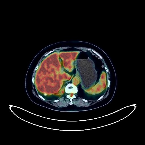
Lung Cancer PET/CT (case 983824-000071 from PETWB-REP)
3 views10 days agoWhole-body 18F-FDG PET/CT scan in a patient with Lung Cancer taken from the PETWB-REP dataset. The following English report (translated from original Chinese) is taken verbatim from the public dataset and has not been modified or otherwise checked for accuracy (see the end for citation). Impression a. Soft tissue mass in the lower lobe of the right lung with increased FDG metabolism, suggestive of lung cancer; bronchoscopy recommended. Obstructive inflammation in the middle lobe of the right lung. b. Multiple solid nodules in both lungs, FDG uptake normal; likely chronic inflammatory nodules; follow-up CT scan recommended to rule out other possible causes. c. Possible right hilar lymph node metastasis. Reactive hyperplasia of mediastinal lymph nodes. Bilateral pleural thickening with multiple calcifications. Calcification of some arterial walls (including coronary arteries). Left renal cyst. Left adrenal hyperplasia. Partial vertebral osteophyte formation. L5/S1 intervertebral disc bulge with pneumothorax and posterior margin calcification. Bilateral palatine tonsillitis. Reactive hyperplasia of bilateral deep cervical lymph nodes. No obvious abnormalities were found on cranial scintigraphy. No obvious space-occupying lesions were found in the sellar region. This case is from PETWB-REP, a curated dataset of whole-body 18F-FDG PET/CT scans and corresponding radiology reports from 490 patients with a broad spectrum of malignancies. The data were retrospectively collected from patients who underwent clinically indicated whole-body 18F-FDG PET/CT scans at the Shanghai Universal Medical Imaging Diagnostic Center between 2021 and 2024. License: Creative Commons Attribution 4.0 International (CC BY 4.0) Citation: Xue, L., Feng, G., Wenbo, Z., Zhang, Y., Li, L., Wang, S., Peng, L., Peng, S., & Gao, X. (2026). PETWB-REP: A Multi-Cancer Whole-Body FDG PET/CT Dataset with Corresponding Radiology Reports [Data set]. Zenodo. https://doi.org/10.5281/zenodo.18670487
Whole BodyPET/CT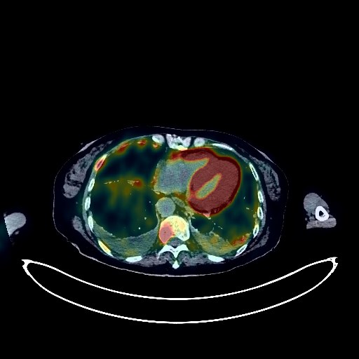
Lung Cancer PET/CT (case 983824-000080 from PETWB-REP)
2 views10 days agoWhole-body 18F-FDG PET/CT scan in a patient with Lung Cancer taken from the PETWB-REP dataset. The following English report (translated from original Chinese) is taken verbatim from the public dataset and has not been modified or otherwise checked for accuracy (see the end for citation). Impression a. A mass in the posterior basal segment of the left lower lobe, with elevated FDG metabolism, suggestive of lung cancer; multiple lymph node metastases in the left hilum, mediastinum, bilateral supraclavicular fossa, and posterior cervical triangle. b. Multiple metastatic tumors in the pleura of both lungs and bilateral interlobar pleura; liver metastases; multiple bone metastases throughout the body. Small amount of pleural effusion bilaterally. Chronic inflammation or fibrosis in both lungs. Chronic cholecystitis. Small renal cysts in both kidneys. Uterine fibroids. Subcutaneous varicose veins on the left anterior pelvic wall. Low-density nodule in the right lobe of the thyroid gland, with normal FDG metabolism, suggestive of nodular goiter or adenoma; ultrasound re-examination recommended. Spinal osteophyte formation, L4/5 and L5/S1 intervertebral disc bulge. Sacral canal cyst. Senile cerebral atrophy. This case is from PETWB-REP, a curated dataset of whole-body 18F-FDG PET/CT scans and corresponding radiology reports from 490 patients with a broad spectrum of malignancies. The data were retrospectively collected from patients who underwent clinically indicated whole-body 18F-FDG PET/CT scans at the Shanghai Universal Medical Imaging Diagnostic Center between 2021 and 2024. License: Creative Commons Attribution 4.0 International (CC BY 4.0) Citation: Xue, L., Feng, G., Wenbo, Z., Zhang, Y., Li, L., Wang, S., Peng, L., Peng, S., & Gao, X. (2026). PETWB-REP: A Multi-Cancer Whole-Body FDG PET/CT Dataset with Corresponding Radiology Reports [Data set]. Zenodo. https://doi.org/10.5281/zenodo.18670487
Whole BodyPET/CT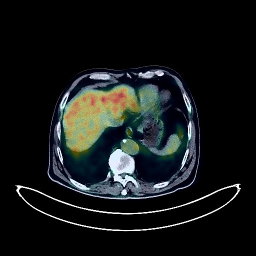
Lung Cancer PET/CT (case 983824-000142 from PETWB-REP)
4 views10 days agoWhole-body 18F-FDG PET/CT scan in a patient with Lung Cancer taken from the PETWB-REP dataset. The following English report (translated from original Chinese) is taken verbatim from the public dataset and has not been modified or otherwise checked for accuracy (see the end for citation). Impression a. Space-occupying lesion in the apical segment of the right upper lobe, with increased FDG metabolism, suggestive of lung cancer; please correlate with clinicopathology. b. Reactive hyperplasia of the right hilar and mediastinal lymph nodes; follow-up is recommended. c. Inflammation in the right upper lobe; ground-glass nodule in the right middle lobe, with normal FDG metabolism, suggestive of inflammatory nodules or atypical adenomatous hyperplasia; annual HRCT follow-up is recommended. d. Calcification of some arterial walls (including coronary arteries). Chronic cholecystitis. Cholecystic stasis. Bilateral renal cysts. Benign prostatic hyperplasia with calcification. Continuous increased FDG metabolism in parts of the colon and rectum, suggestive of inflammatory or physiological uptake; colonoscopy follow-up is recommended. Degenerative changes in the spine. L4/5 and L5/S1 disc bulges. Bilateral iliac bone islands. A few deep cerebral ischemic lesions bilaterally, indicative of age-related cerebral insufficiency. Bilateral chronic maxillary sinusitis. Inflammation around the left maxillary dentition; specialist examination recommended. This case is from PETWB-REP, a curated dataset of whole-body 18F-FDG PET/CT scans and corresponding radiology reports from 490 patients with a broad spectrum of malignancies. The data were retrospectively collected from patients who underwent clinically indicated whole-body 18F-FDG PET/CT scans at the Shanghai Universal Medical Imaging Diagnostic Center between 2021 and 2024. License: Creative Commons Attribution 4.0 International (CC BY 4.0) Citation: Xue, L., Feng, G., Wenbo, Z., Zhang, Y., Li, L., Wang, S., Peng, L., Peng, S., & Gao, X. (2026). PETWB-REP: A Multi-Cancer Whole-Body FDG PET/CT Dataset with Corresponding Radiology Reports [Data set]. Zenodo. https://doi.org/10.5281/zenodo.18670487
Whole BodyPET/CT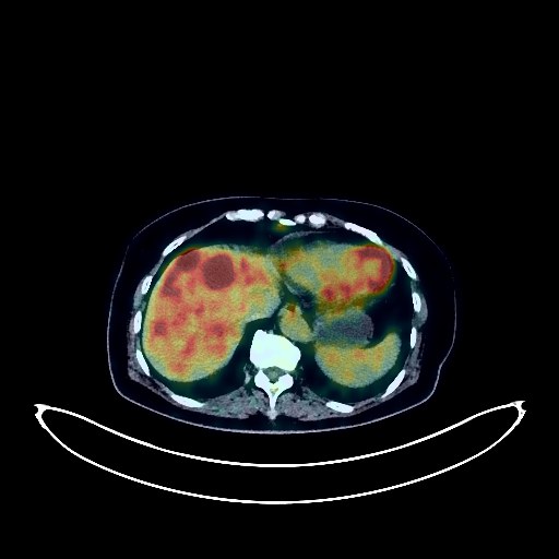
Pancreatic Cancer PET/CT (case 983824-000041 from PETWB-REP)
9 views10 days agoWhole-body 18F-FDG PET/CT scan in a patient with Pancreatic Cancer taken from the PETWB-REP dataset. The following English report (translated from original Chinese) is taken verbatim from the public dataset and has not been modified or otherwise checked for accuracy (see the end for citation). Impression a. Masses in the neck and body of the pancreas, with increased FDG metabolism, suggestive of pancreatic cancer involving surrounding fat spaces and the celiac trunk. Multiple intrahepatic metastases. Right femoral head metastasis. b. Multiple small lymph nodes between the superior mesenteric artery and the hepatogastric ligament, with mild FDG uptake, suggestive of reactive lymph node hyperplasia; follow-up CT scan recommended to rule out metastasis. c. Strip-like thickening of the pelvic floor peritoneum, with mild FDG uptake; metastasis to be ruled out; clinical correlation recommended. a. Multiple nodules in both lungs, with no abnormal FDG uptake, suggestive of inflammatory nodules, with some mixed metastases to be ruled out; close follow-up CT scan recommended. b. Multiple ground-glass nodules in both lungs, with no abnormal FDG metabolism, suggestive of atypical adenomatous hyperplasia or inflammatory nodules. Small cyst in the left lobe of the liver. Right renal cyst. Left adrenal hyperplasia is likely; follow-up examination recommended. Postoperative absence of the uterus. Spinal degenerative changes, L3/4 and L4/5 intervertebral disc bulging, L4/5 and L5/S1 intervertebral disc pneumatosis and degeneration. This case is from PETWB-REP, a curated dataset of whole-body 18F-FDG PET/CT scans and corresponding radiology reports from 490 patients with a broad spectrum of malignancies. The data were retrospectively collected from patients who underwent clinically indicated whole-body 18F-FDG PET/CT scans at the Shanghai Universal Medical Imaging Diagnostic Center between 2021 and 2024. License: Creative Commons Attribution 4.0 International (CC BY 4.0) Citation: Xue, L., Feng, G., Wenbo, Z., Zhang, Y., Li, L., Wang, S., Peng, L., Peng, S., & Gao, X. (2026). PETWB-REP: A Multi-Cancer Whole-Body FDG PET/CT Dataset with Corresponding Radiology Reports [Data set]. Zenodo. https://doi.org/10.5281/zenodo.18670487
Whole BodyPET/CT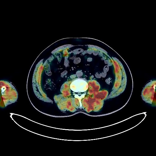
Lymphoma PET/CT (case 983824-000107 from PETWB-REP)
4 views10 days agoWhole-body 18F-FDG PET/CT scan in a patient with Lymphoma taken from the PETWB-REP dataset. The following English report (translated from original Chinese) is taken verbatim from the public dataset and has not been modified or otherwise checked for accuracy (see the end for citation). Impression Bilateral orbital lesions with increased FDG metabolism; multiple enlarged lymph nodes throughout the body with increased FDG metabolism (see description for details). These findings are consistent with lymphoma infiltration; follow-up examination after treatment is recommended. Chronic inflammatory micronodules in both lungs. A few post-inflammatory lesions in both lungs. Calcifications in both pulmonary hilum. Partial arteriosclerosis. Liver cysts. Slightly enlarged spleen, accessory spleen. Left kidney stone, right kidney small cyst. Calcification of the tunica vaginalis in the left testis. Degenerative changes in the spine, multiple lumbar disc herniations. T3 vertebral hemangioma. Bilateral femoral head-neck herniation fossa. Calcification of the falx cerebri; no obvious abnormalities were seen on cranial scintigraphy. This case is from PETWB-REP, a curated dataset of whole-body 18F-FDG PET/CT scans and corresponding radiology reports from 490 patients with a broad spectrum of malignancies. The data were retrospectively collected from patients who underwent clinically indicated whole-body 18F-FDG PET/CT scans at the Shanghai Universal Medical Imaging Diagnostic Center between 2021 and 2024. License: Creative Commons Attribution 4.0 International (CC BY 4.0) Citation: Xue, L., Feng, G., Wenbo, Z., Zhang, Y., Li, L., Wang, S., Peng, L., Peng, S., & Gao, X. (2026). PETWB-REP: A Multi-Cancer Whole-Body FDG PET/CT Dataset with Corresponding Radiology Reports [Data set]. Zenodo. https://doi.org/10.5281/zenodo.18670487
Whole BodyPET/CT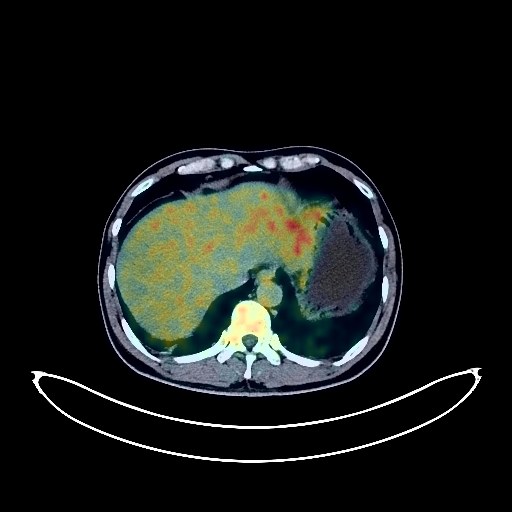
Lung Cancer PET/CT (case 983824-000170 from PETWB-REP)
4 views10 days agoWhole-body 18F-FDG PET/CT scan in a patient with Lung Cancer taken from the PETWB-REP dataset. The following English report (translated from original Chinese) is taken verbatim from the public dataset and has not been modified or otherwise checked for accuracy (see the end for citation). Impression a. Right lower lobe anterior basal segment containing air-filled cavities and soft tissue nodules, with increased FDG metabolism, suggestive of lung cancer. Please confirm the diagnosis with pathological examination. Reactive hyperplasia of hilar and mediastinal lymph nodes bilaterally. b. Patchy and strip-shaped high-density shadows in the upper lobes of both lungs, with increased FDG metabolism, suggestive of infectious lesions. Please compare with previous data and follow up to rule out other possibilities. c. Chronic inflammatory micronodules (solid) in the upper lobes of both lungs and the right lower lobe. Chronic inflammation and sequelae in both lungs, emphysema, bullae. Mild pleural thickening bilaterally. a. Benign prostatic hyperplasia with calcification, with unevenly increased FDG metabolism in the parenchyma; increased FDG metabolism in both seminal vesicles; high FDG metabolism foci in both scrotums. Considering the above, inflammatory changes are possible. Please rule out space-occupying lesions with PSA and enhanced MRI. b. Reactive hyperplasia of bilateral pelvic wall lymph nodes. Fatty liver, no abnormalities in FDG metabolism in liver parenchyma. Left renal cyst. Increased FDG metabolism in part of the gastric wall, considered physiological uptake or chronic inflammation; please follow up with endoscopy. Spinal degenerative changes. Benign bone disease of L4 vertebral body. L4/5 and L5/S1 intervertebral disc bulge. Increased FDG metabolism is observed in multiple medullary cavities, suggesting a high probability of bone marrow proliferative changes. No obvious abnormalities seen on cranial scintigraphy. Increased FDG metabolism on the left lateral nasopharyngeal wall, considered physiological or inflammatory changes; please follow up with specialist examination. Uneven thyroid density, increased FDG metabolism, suggesting possible inflammatory changes; please follow up with thyroid function tests and ultrasound. Reactive hyperplasia of bilateral cervical lymph nodes. This case is from PETWB-REP, a curated dataset of whole-body 18F-FDG PET/CT scans and corresponding radiology reports from 490 patients with a broad spectrum of malignancies. The data were retrospectively collected from patients who underwent clinically indicated whole-body 18F-FDG PET/CT scans at the Shanghai Universal Medical Imaging Diagnostic Center between 2021 and 2024. License: Creative Commons Attribution 4.0 International (CC BY 4.0) Citation: Xue, L., Feng, G., Wenbo, Z., Zhang, Y., Li, L., Wang, S., Peng, L., Peng, S., & Gao, X. (2026). PETWB-REP: A Multi-Cancer Whole-Body FDG PET/CT Dataset with Corresponding Radiology Reports [Data set]. Zenodo. https://doi.org/10.5281/zenodo.18670487
Whole BodyPET/CT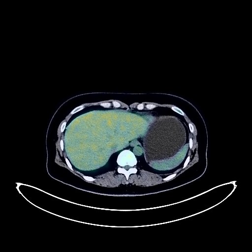
Cervical Cancer PET/CT (case 983824-000036 from PETWB-REP)
9 views10 days agoWhole-body 18F-FDG PET/CT scan in a patient with Cervical Cancer taken from the PETWB-REP dataset. The following English report (translated from original Chinese) is taken verbatim from the public dataset and has not been modified or otherwise checked for accuracy (see the end for citation). Impression a. Cervical mass with increased FDG metabolism, consistent with cervical cancer; reactive hyperplasia of the left iliac lymph nodes is possible, metastasis to be ruled out, follow-up is recommended. b. Nodular FDG uptake foci on the right pelvic wall, possibly due to physiological uptake by the ureter; lymph nodes to be ruled out, MRI is recommended. Physiological uptake in the uterine cavity. Chronic inflammatory micronodules in the right lung. A few post-inflammatory lesions in both lungs. Anemia. Bilateral breast hyperplasia. Small cyst in the right lobe of the liver. Accessory spleen. Possible hemorrhoids. L3/4, L4/5, and L5/S1 intervertebral disc bulges. Bilateral sacroiliac joint condensation osteitis. No obvious abnormalities were found on cranial scintigraphy. This case is from PETWB-REP, a curated dataset of whole-body 18F-FDG PET/CT scans and corresponding radiology reports from 490 patients with a broad spectrum of malignancies. The data were retrospectively collected from patients who underwent clinically indicated whole-body 18F-FDG PET/CT scans at the Shanghai Universal Medical Imaging Diagnostic Center between 2021 and 2024. License: Creative Commons Attribution 4.0 International (CC BY 4.0) Citation: Xue, L., Feng, G., Wenbo, Z., Zhang, Y., Li, L., Wang, S., Peng, L., Peng, S., & Gao, X. (2026). PETWB-REP: A Multi-Cancer Whole-Body FDG PET/CT Dataset with Corresponding Radiology Reports [Data set]. Zenodo. https://doi.org/10.5281/zenodo.18670487
Whole BodyPET/CT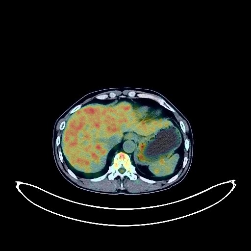
Prostate Cancer PET/CT (case 983824-000154 from PETWB-REP)
4 views10 days agoWhole-body 18F-FDG PET/CT scan in a patient with Prostate Cancer taken from the PETWB-REP dataset. The following English report (translated from original Chinese) is taken verbatim from the public dataset and has not been modified or otherwise checked for accuracy (see the end for citation). Impression Benign prostatic hyperplasia with calcification, prostatic mass with increased FDG metabolism, suggestive of possible prostate cancer invading bilateral seminal vesicles; please correlate with clinicopathology. Bilateral iliac lymph node metastasis. a. Multiple solid nodules in both lungs, with clear borders; some show slightly increased FDG metabolism, some suggest possible metastasis; follow-up CT scan recommended. b. Bilateral emphysema, scattered post-inflammatory lesions in both lungs. Reactive hyperplasia of hilar and mediastinal lymph nodes in both lungs. Calcification of some arterial walls (including coronary arteries). Small liver cysts. Right kidney stone, left kidney calcification. Highly probable chronic antral gastritis; chronic inflammatory changes in the intestines; please correlate with gastroscopy and colonoscopy. Degenerative changes in the spine, multiple bulging lumbar intervertebral discs, and pneumodiscosis of the L5/S1 intervertebral disc. Old fracture of the right clavicle. No obvious abnormalities were found on cranial scintigraphy. This case is from PETWB-REP, a curated dataset of whole-body 18F-FDG PET/CT scans and corresponding radiology reports from 490 patients with a broad spectrum of malignancies. The data were retrospectively collected from patients who underwent clinically indicated whole-body 18F-FDG PET/CT scans at the Shanghai Universal Medical Imaging Diagnostic Center between 2021 and 2024. License: Creative Commons Attribution 4.0 International (CC BY 4.0) Citation: Xue, L., Feng, G., Wenbo, Z., Zhang, Y., Li, L., Wang, S., Peng, L., Peng, S., & Gao, X. (2026). PETWB-REP: A Multi-Cancer Whole-Body FDG PET/CT Dataset with Corresponding Radiology Reports [Data set]. Zenodo. https://doi.org/10.5281/zenodo.18670487
Whole BodyPET/CT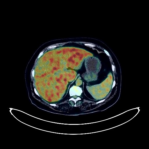
Cervical Cancer PET/CT (case 983824-000055 from PETWB-REP)
6 views10 days agoWhole-body 18F-FDG PET/CT scan in a patient with Cervical Cancer taken from the PETWB-REP dataset. The following English report (translated from original Chinese) is taken verbatim from the public dataset and has not been modified or otherwise checked for accuracy (see the end for citation). Impression a. Cervical mass with increased FDG metabolism, consistent with cervical cancer; reactive hyperplasia of bilateral iliac vessels, retroperitoneum, and bilateral inguinal lymph nodes. b. Nabothian cysts of the cervix, possibly due to physiological uptake within the uterine cavity. a. Multiple solid nodules in both lungs, with clear borders and normal FDG uptake, suggestive of chronic inflammatory nodules, pending exclusion of mixed metastasis; follow-up examination recommended. b. Scattered post-inflammatory lesions in both lungs. Reactive hyperplasia of mediastinal lymph nodes. Minor arteriosclerosis in some arteries. Fatty liver. Left adrenal hyperplasia. Left renal calcification. Chronic inflammatory changes in some gastric wall and intestinal tract; please follow up with endoscopy. Osteoporosis, scoliosis, degenerative changes in the spine, multiple lumbar disc bulges with pneumoconiosis. Subcutaneous calcification in the left buttock. Bilateral frozen shoulder. The thyroid gland shows uneven density and calcifications in both lobes. FDG metabolism is normal, suggesting nodular goiter. Please confirm with ultrasound examination. Cranial scintigraphy showed no obvious abnormalities. Chronic inflammation of the nasopharynx. This case is from PETWB-REP, a curated dataset of whole-body 18F-FDG PET/CT scans and corresponding radiology reports from 490 patients with a broad spectrum of malignancies. The data were retrospectively collected from patients who underwent clinically indicated whole-body 18F-FDG PET/CT scans at the Shanghai Universal Medical Imaging Diagnostic Center between 2021 and 2024. License: Creative Commons Attribution 4.0 International (CC BY 4.0) Citation: Xue, L., Feng, G., Wenbo, Z., Zhang, Y., Li, L., Wang, S., Peng, L., Peng, S., & Gao, X. (2026). PETWB-REP: A Multi-Cancer Whole-Body FDG PET/CT Dataset with Corresponding Radiology Reports [Data set]. Zenodo. https://doi.org/10.5281/zenodo.18670487
Whole BodyPET/CT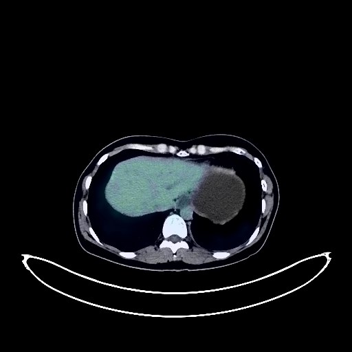
Gastric Cancer PET/CT (case 983824-000200 from PETWB-REP)
4 views10 days agoWhole-body 18F-FDG PET/CT scan in a patient with Gastric Cancer taken from the PETWB-REP dataset. The following English report (translated from original Chinese) is taken verbatim from the public dataset and has not been modified or otherwise checked for accuracy (see the end for citation). Impression a. Irregular thickening of the gastric wall on the lesser curvature of the gastric body with increased FDG metabolism, consistent with gastric cancer. Multiple lymph node metastases in the para-curvature and retroperitoneum. b. Bilateral adnexal lesions with increased FDG metabolism, suggestive of metastatic tumors. Peritoneal seeding metastasis, pelvic effusion. Chronic inflammatory micronodules in both lungs. Incomplete thymic regression. Small liver cysts. Gallbladder cholestasis or gallstones. Left kidney cyst. Physiological uptake in the uterine cavity. Focal increased FDG metabolism in the rectum, likely due to polyps or physiological uptake; endoscopic re-examination is recommended. Low-density thyroid nodules with normal FDG metabolism, suggestive of nodular goiter; ultrasound examination is recommended. Reactive hyperplasia of bilateral deep cervical lymph nodes. No obvious abnormalities were found on cranial scintigraphy. This case is from PETWB-REP, a curated dataset of whole-body 18F-FDG PET/CT scans and corresponding radiology reports from 490 patients with a broad spectrum of malignancies. The data were retrospectively collected from patients who underwent clinically indicated whole-body 18F-FDG PET/CT scans at the Shanghai Universal Medical Imaging Diagnostic Center between 2021 and 2024. License: Creative Commons Attribution 4.0 International (CC BY 4.0) Citation: Xue, L., Feng, G., Wenbo, Z., Zhang, Y., Li, L., Wang, S., Peng, L., Peng, S., & Gao, X. (2026). PETWB-REP: A Multi-Cancer Whole-Body FDG PET/CT Dataset with Corresponding Radiology Reports [Data set]. Zenodo. https://doi.org/10.5281/zenodo.18670487
Whole BodyPET/CT