Loading...
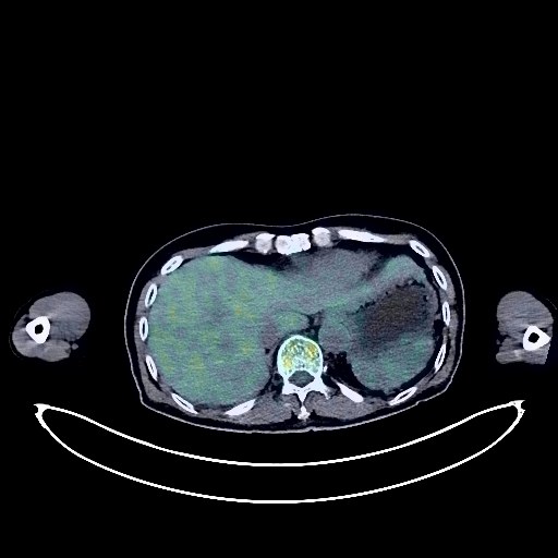
Prostate Cancer PET/CT (case 983824-000163 from PETWB-REP)
2 views10 days agoWhole-body 18F-FDG PET/CT scan in a patient with Prostate Cancer taken from the PETWB-REP dataset. The following English report (translated from original Chinese) is taken verbatim from the public dataset and has not been modified or otherwise checked for accuracy (see the end for citation). Impression a. Prostatic mass with elevated FDG metabolism, suggesting continued activity of prostate cancer after treatment, invading the seminal vesicles. Benign prostatic hyperplasia with calcification. b. Bone metastasis to the S5 vertebral body and left subpubic ramus. Inflammation in the right lower lobe of the lung, chronic inflammatory nodules in both lungs. A few post-inflammatory lesions in both lungs. Slight thickening of the pleura bilaterally. Calcification of some arterial walls (including coronary arteries). Left adrenal hyperplasia. Right renal cyst. Chronic inflammatory changes in part of the gastric wall and intestines; please follow up with endoscopy. Osteoporosis, degenerative changes in the spine, L4/5 intervertebral disc bulge. Bilateral shoulder periarthritis. Age-related brain, deep lacunar infarcts. This case is from PETWB-REP, a curated dataset of whole-body 18F-FDG PET/CT scans and corresponding radiology reports from 490 patients with a broad spectrum of malignancies. The data were retrospectively collected from patients who underwent clinically indicated whole-body 18F-FDG PET/CT scans at the Shanghai Universal Medical Imaging Diagnostic Center between 2021 and 2024. License: Creative Commons Attribution 4.0 International (CC BY 4.0) Citation: Xue, L., Feng, G., Wenbo, Z., Zhang, Y., Li, L., Wang, S., Peng, L., Peng, S., & Gao, X. (2026). PETWB-REP: A Multi-Cancer Whole-Body FDG PET/CT Dataset with Corresponding Radiology Reports [Data set]. Zenodo. https://doi.org/10.5281/zenodo.18670487
Whole BodyPET/CT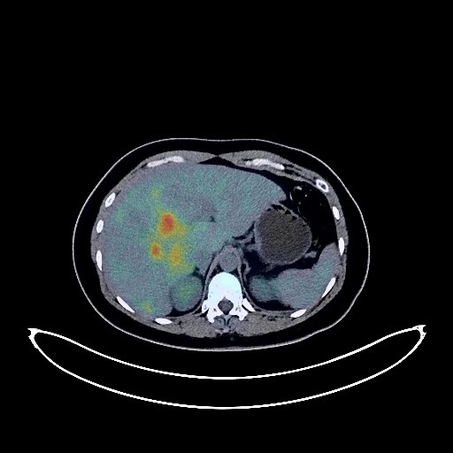
Breast Cancer PET/CT (case 983824-000084 from PETWB-REP)
2 views10 days agoWhole-body 18F-FDG PET/CT scan in a patient with Breast Cancer taken from the PETWB-REP dataset. The following English report (translated from original Chinese) is taken verbatim from the public dataset and has not been modified or otherwise checked for accuracy (see the end for citation). Impression a. Postoperative left breast cancer surgery, no clear signs of tumor recurrence were observed in the surgical area. b. Thickening of soft tissue on the right nasopharyngeal wall, increased FDG metabolism, malignancy to be ruled out, please combine with specialist examination. c. Multiple lesions in the liver, increased FDG metabolism, considered malignant tumor, metastasis is highly likely, enhanced MRI examination is necessary. d. Cystic-solid lesion in the right adnexa with increased FDG metabolism in the solid part, neoplastic lesion is considered possible, physiological changes to be ruled out; cystic lesion in the left adnexa, no increased FDG metabolism; physiological uptake in the uterine cavity is highly likely. Please combine the above with enhanced MRI examination. e. Metastasis to the left retropharyngeal space, bilateral deep cervical spaces, bilateral posterior cervical triangles, bilateral supraclavicular fossa, left internal mammary chain, hilar space and retroperitoneal lymph nodes, and possible metastasis to the right hilar and mediastinal lymph nodes. ? f. Multiple lung metastases. Multiple bone metastases throughout the body. A few fibrotic lesions in the right middle lobe of the lung. Dense glandular tissue in the right breast, reactive hyperplasia of the right axillary lymph nodes. Small amount of pericardial effusion. Signs of anemia. Inflammatory or physiological uptake in some intestinal segments; endoscopic re-examination is necessary if required. Brain parenchyma is not fully developed; no obvious abnormalities were seen on cranial FDG imaging. Physiological uptake is likely in both palatine tonsils. Uneven thyroid density with increased FDG metabolism, suggesting possible nodular goiter or adenoma; please combine with ultrasound and thyroid function tests. This case is from PETWB-REP, a curated dataset of whole-body 18F-FDG PET/CT scans and corresponding radiology reports from 490 patients with a broad spectrum of malignancies. The data were retrospectively collected from patients who underwent clinically indicated whole-body 18F-FDG PET/CT scans at the Shanghai Universal Medical Imaging Diagnostic Center between 2021 and 2024. License: Creative Commons Attribution 4.0 International (CC BY 4.0) Citation: Xue, L., Feng, G., Wenbo, Z., Zhang, Y., Li, L., Wang, S., Peng, L., Peng, S., & Gao, X. (2026). PETWB-REP: A Multi-Cancer Whole-Body FDG PET/CT Dataset with Corresponding Radiology Reports [Data set]. Zenodo. https://doi.org/10.5281/zenodo.18670487
Whole BodyPET/CT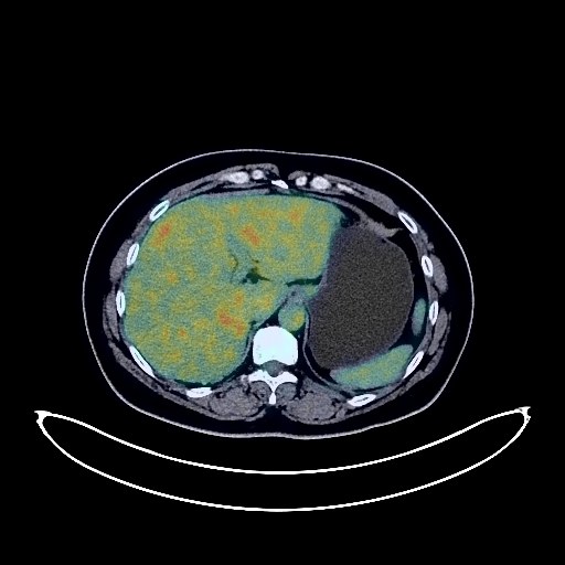
Renal Cancer PET/CT (case 983824-000168 from PETWB-REP)
2 views10 days agoWhole-body 18F-FDG PET/CT scan in a patient with Renal Cancer taken from the PETWB-REP dataset. The following English report (translated from original Chinese) is taken verbatim from the public dataset and has not been modified or otherwise checked for accuracy (see the end for citation). Impression a. Left renal mass with increased FDG metabolism, suggestive of renal cell carcinoma; please correlate with clinicopathology. Reactive hyperplasia of small retroperitoneal lymph nodes. b. Metastatic tumors in the left 8th anterior rib and left vertebral arch of T9. Metastatic tumor in the right parietal bone, to be ruled out. Possible chronic inflammatory nodules in both lungs; CT follow-up is recommended to rule out mixed metastases. Chronic inflammation and post-inflammatory remnants in both lungs. Reactive hyperplasia of hilar and mediastinal lymph nodes in both lungs. Calcification of some arterial walls (including coronary arteries). Cystic lesions in the uterus, possibly cystic degeneration of a fibroid; physiological uptake by the left ovary. Ultrasound follow-up is recommended for all of the above. Fatty liver. Chronic inflammatory changes in the antrum of the stomach; please correlate with endoscopic follow-up. Degenerative changes in the spine, L5/S1 intervertebral disc bulge with pneumoconiosis. Low-density nodule in the right lobe of the thyroid gland; FDG metabolism normal; suggestive of an adenoma-like nodule; please confirm with ultrasound examination. Cranial scintigraphy showed no obvious abnormalities. Left maxillary sinus submucosal cyst. This case is from PETWB-REP, a curated dataset of whole-body 18F-FDG PET/CT scans and corresponding radiology reports from 490 patients with a broad spectrum of malignancies. The data were retrospectively collected from patients who underwent clinically indicated whole-body 18F-FDG PET/CT scans at the Shanghai Universal Medical Imaging Diagnostic Center between 2021 and 2024. License: Creative Commons Attribution 4.0 International (CC BY 4.0) Citation: Xue, L., Feng, G., Wenbo, Z., Zhang, Y., Li, L., Wang, S., Peng, L., Peng, S., & Gao, X. (2026). PETWB-REP: A Multi-Cancer Whole-Body FDG PET/CT Dataset with Corresponding Radiology Reports [Data set]. Zenodo. https://doi.org/10.5281/zenodo.18670487
Whole BodyPET/CT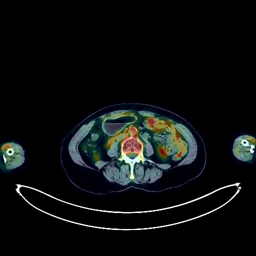
Lymphoma PET/CT (case 983824-000001 from PETWB-REP)
9 views10 days agoWhole-body 18F-FDG PET/CT scan in a patient with Lymphoma taken from the PETWB-REP dataset. The following English report (translated from original Chinese) is taken verbatim from the public dataset and has not been modified or otherwise checked for accuracy (see the end for citation). Impression Following comprehensive treatment for lymphoma, comparing PET/CT images from our center on December 25, 2021: a. Postoperative gastric lesser curvature lymphoma surgery showed slight thickening of the gastric wall in the surgical area with mild FDG uptake, suggesting postoperative changes; the soft tissue nodules in the left posteroinferior mediastinum (medial to the descending aorta at T8 level) decreased in size, with reduced FDG metabolism; the right inguinal lymph nodes also decreased in size, with reduced FDG metabolism. These findings suggest effective lymphoma treatment. b. Multiple small lymph nodes throughout the body (see description for details) showed no significant increase in FDG metabolism, remaining similar to previous findings, suggesting suppressed lesion activity or possibly reactive proliferative lymph nodes after treatment. c. Patchy and strip-like lesions in both lungs with increased FDG metabolism, some adjacent pleural thickening and adhesions, and some newly added lesions suggest a high probability of inflammatory lesions. Please follow up with CT scans to rule out other possibilities. Multiple thin-walled cystic lesions in both lungs, scattered post-inflammatory remnants (including calcifications) in both lungs, roughly similar to previous findings. A small amount of effusion in the pericardial recesses and at their bases. Partial arteriosclerosis. Bilateral breast hyperplasia. Multiple solid lesions in the liver, roughly similar to previous findings, suggestive of hemangioma; enhanced MRI follow-up is recommended. A small nodule in the left adrenal region, roughly similar to previous findings, highly suggestive of adenoma. Chronic inflammatory changes or physiological uptake in some intestinal segments; endoscopic follow-up is recommended. Duodenal diverticulum. Hemorrhoidal changes. Scoliosis with degenerative changes. Post-radiotherapy changes in some thoracic spine segments. Mild L2/3 intervertebral disc bulge. Lipoma and calcifications in the quadrigeminal cistern. A small amount of inflammation in the right maxillary sinus. Thyroid gland density is uneven; FDG metabolism is normal; ultrasound follow-up is recommended. This case is from PETWB-REP, a curated dataset of whole-body 18F-FDG PET/CT scans and corresponding radiology reports from 490 patients with a broad spectrum of malignancies. The data were retrospectively collected from patients who underwent clinically indicated whole-body 18F-FDG PET/CT scans at the Shanghai Universal Medical Imaging Diagnostic Center between 2021 and 2024. License: Creative Commons Attribution 4.0 International (CC BY 4.0) Citation: Xue, L., Feng, G., Wenbo, Z., Zhang, Y., Li, L., Wang, S., Peng, L., Peng, S., & Gao, X. (2026). PETWB-REP: A Multi-Cancer Whole-Body FDG PET/CT Dataset with Corresponding Radiology Reports [Data set]. Zenodo. https://doi.org/10.5281/zenodo.18670487
Whole BodyPET/CT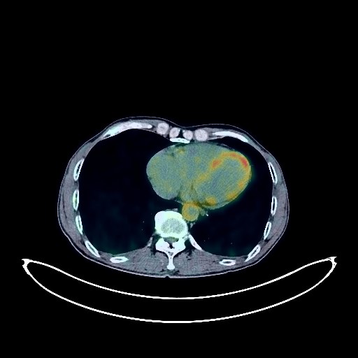
Lung Cancer PET/CT (case 983824-000098 from PETWB-REP)
2 views10 days agoWhole-body 18F-FDG PET/CT scan in a patient with Lung Cancer taken from the PETWB-REP dataset. The following English report (translated from original Chinese) is taken verbatim from the public dataset and has not been modified or otherwise checked for accuracy (see the end for citation). Impression a. A mass in the anterior segment of the left upper lobe, with increased FDG metabolism, suggestive of lung cancer, accompanied by surrounding obstructive changes. Please confirm with pathology. b. Multiple lymph node metastases in the left hilum and mediastinum; right hilar lymph node metastasis to be ruled out. c. Solid nodules and plaque-like lesions in both lungs, with normal FDG metabolism, highly suggestive of chronic inflammatory lesions. Annual CT scan recommended. Sequelae of pneumonia in both lungs. Calcification of some arterial walls (including coronary arteries). Slight thickening of the walls in parts of the gastric body and antrum, with mildly increased FDG uptake, suggestive of chronic gastritis; continuous increased FDG metabolism in parts of the colon and rectum, suggestive of inflammatory or physiological uptake. Recommended follow-up gastroscopy and colonoscopy. Hemorrhoidal changes. Benign prostatic hyperplasia with calcification. Degenerative changes in the spine. L4/5 and L5/S1 intervertebral disc bulges. A few ischemic lesions in the deep bilateral cerebral regions; age-related brain abnormalities. This case is from PETWB-REP, a curated dataset of whole-body 18F-FDG PET/CT scans and corresponding radiology reports from 490 patients with a broad spectrum of malignancies. The data were retrospectively collected from patients who underwent clinically indicated whole-body 18F-FDG PET/CT scans at the Shanghai Universal Medical Imaging Diagnostic Center between 2021 and 2024. License: Creative Commons Attribution 4.0 International (CC BY 4.0) Citation: Xue, L., Feng, G., Wenbo, Z., Zhang, Y., Li, L., Wang, S., Peng, L., Peng, S., & Gao, X. (2026). PETWB-REP: A Multi-Cancer Whole-Body FDG PET/CT Dataset with Corresponding Radiology Reports [Data set]. Zenodo. https://doi.org/10.5281/zenodo.18670487
Whole BodyPET/CT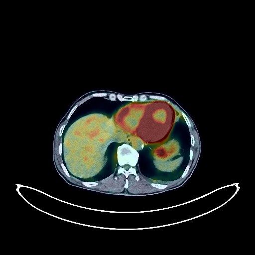
Lung Cancer PET/CT (case 983824-000108 from PETWB-REP)
2 views10 days agoWhole-body 18F-FDG PET/CT scan in a patient with Lung Cancer taken from the PETWB-REP dataset. The following English report (translated from original Chinese) is taken verbatim from the public dataset and has not been modified or otherwise checked for accuracy (see the end for citation). Impression a. Mass in the posterior segment of the left upper lobe, with increased FDG metabolism, suggestive of lung cancer with obstructive changes; carcinomatous lymphangitis of the left upper lobe. b. Multiple lymph node metastases in the left hilum, mediastinum, and left supraclavicular fossa. Chronic inflammatory lymph nodes in the right hilum. c. Several small (solid) chronic inflammatory nodules in both lungs. A small amount of chronic inflammation and old lesions in both lungs. d. Small amount of pleural effusion on the left side. Calcification of some arterial walls (including coronary arteries). Slight thickening of the gastric fundus, part of the gastric body, and antrum walls, with increased FDG uptake, suggestive of chronic gastritis; follow-up with gastroscopy is recommended. Mild dilatation of the pancreatic duct. Benign prostatic hyperplasia, with increased FDG metabolism in the peripheral zone, suggestive of inflammatory or physiological uptake; follow-up with PSA and ultrasound is recommended. Degenerative changes in the spine. L4/5 and L5/S1 intervertebral disc bulges. A few ischemic foci in the deep bilateral cerebral regions, indicative of age-related brain changes. Chronic inflammation of the bilateral maxillary and ethmoid sinuses. Focal FDG hypermetabolism in the genioglossus muscle, suggestive of stress-induced uptake. This case is from PETWB-REP, a curated dataset of whole-body 18F-FDG PET/CT scans and corresponding radiology reports from 490 patients with a broad spectrum of malignancies. The data were retrospectively collected from patients who underwent clinically indicated whole-body 18F-FDG PET/CT scans at the Shanghai Universal Medical Imaging Diagnostic Center between 2021 and 2024. License: Creative Commons Attribution 4.0 International (CC BY 4.0) Citation: Xue, L., Feng, G., Wenbo, Z., Zhang, Y., Li, L., Wang, S., Peng, L., Peng, S., & Gao, X. (2026). PETWB-REP: A Multi-Cancer Whole-Body FDG PET/CT Dataset with Corresponding Radiology Reports [Data set]. Zenodo. https://doi.org/10.5281/zenodo.18670487
Whole BodyPET/CT
Bladder Cancer PET/CT (case 983824-000208 from PETWB-REP)
2 views10 days agoWhole-body 18F-FDG PET/CT scan in a patient with Bladder Cancer taken from the PETWB-REP dataset. The following English report (translated from original Chinese) is taken verbatim from the public dataset and has not been modified or otherwise checked for accuracy (see the end for citation). Impression Post-bladder cancer surgery, no signs of tumor recurrence observed; specialist follow-up recommended. Chronic inflammation and sequelae in the right lower lobe of the lung. Slight localized pleural thickening bilaterally. Calcification of some arterial walls. Physiological or inflammatory uptake of the colorectal tissue; endoscopic follow-up recommended. (The right hydronephrosis and right renal cystic lesion shown on ultrasound from another hospital were not clearly visualized.) Partial vertebral osteophyte formation. L4/5 and L5/S1 intervertebral disc bulge with posterior margin calcification. No obvious abnormalities seen on cranial FDG scintigraphy. This case is from PETWB-REP, a curated dataset of whole-body 18F-FDG PET/CT scans and corresponding radiology reports from 490 patients with a broad spectrum of malignancies. The data were retrospectively collected from patients who underwent clinically indicated whole-body 18F-FDG PET/CT scans at the Shanghai Universal Medical Imaging Diagnostic Center between 2021 and 2024. License: Creative Commons Attribution 4.0 International (CC BY 4.0) Citation: Xue, L., Feng, G., Wenbo, Z., Zhang, Y., Li, L., Wang, S., Peng, L., Peng, S., & Gao, X. (2026). PETWB-REP: A Multi-Cancer Whole-Body FDG PET/CT Dataset with Corresponding Radiology Reports [Data set]. Zenodo. https://doi.org/10.5281/zenodo.18670487
Whole BodyPET/CT
Prostate Cancer PET/CT (case 983824-000013 from PETWB-REP)
7 views10 days agoWhole-body 18F-FDG PET/CT scan in a patient with Prostate Cancer taken from the PETWB-REP dataset. The following English report (translated from original Chinese) is taken verbatim from the public dataset and has not been modified or otherwise checked for accuracy (see the end for citation). Impression a. Prostatic mass with elevated FDG metabolism, consistent with prostate cancer; multiple lymph node metastases bilaterally to the iliac vessels, retroperitoneum, and bilateral posterior diaphragmatic crura. b. Bone metastases to the right scapula, left transverse process of T11, and right side of L2 vertebral body. Thickening and roughening of the right bladder wall, with normal FDG metabolism, suggestive of chronic inflammation; further specialist examination is required. Chronic inflammatory nodules in both lungs. Paraseptal emphysema in the right upper lobe, with a few post-inflammatory lesions in both lungs. Pleural thickening bilaterally. Reactive hyperplasia of mediastinal lymph nodes. Mild anemia, with partial calcification of arterial walls (including coronary arteries). Bilateral renal cysts, with angiomyolipoma of the right upper pole of the kidney to be ruled out. Bilateral inguinal hernias are possible. Small amount of hydrocele in both testes. Chronic gastritis. Degenerative changes in the spine, with L4/5 and L5/S1 intervertebral disc bulges. Left hip periarthritis. Age-related brain abnormalities, deep lacunar infarcts; MRI follow-up recommended. Chronic inflammation of the left maxillary sinus. Right vocal cord laxity. This case is from PETWB-REP, a curated dataset of whole-body 18F-FDG PET/CT scans and corresponding radiology reports from 490 patients with a broad spectrum of malignancies. The data were retrospectively collected from patients who underwent clinically indicated whole-body 18F-FDG PET/CT scans at the Shanghai Universal Medical Imaging Diagnostic Center between 2021 and 2024. License: Creative Commons Attribution 4.0 International (CC BY 4.0) Citation: Xue, L., Feng, G., Wenbo, Z., Zhang, Y., Li, L., Wang, S., Peng, L., Peng, S., & Gao, X. (2026). PETWB-REP: A Multi-Cancer Whole-Body FDG PET/CT Dataset with Corresponding Radiology Reports [Data set]. Zenodo. https://doi.org/10.5281/zenodo.18670487
Whole BodyPET/CT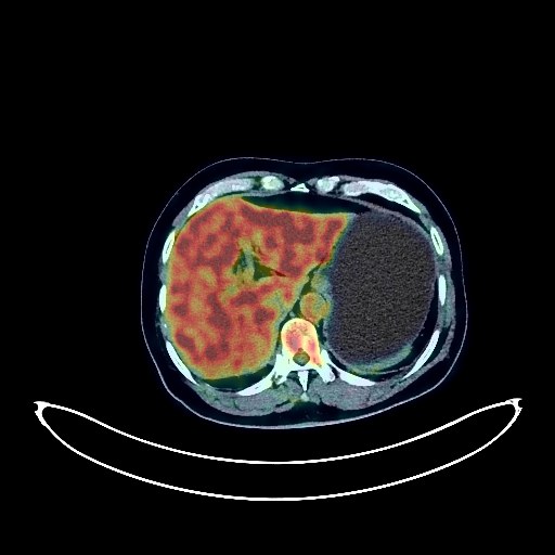
Lung Cancer PET/CT (case 983824-000179 from PETWB-REP)
1 views10 days agoWhole-body 18F-FDG PET/CT scan in a patient with Lung Cancer taken from the PETWB-REP dataset. The following English report (translated from original Chinese) is taken verbatim from the public dataset and has not been modified or otherwise checked for accuracy (see the end for citation). Impression a. Postoperative right lung cancer surgery, no obvious signs of tumor recurrence in the surgical area. b. Right frontal lobe mass, FDG uptake deficiency, suggestive of neoplastic lesion, metastasis is the primary consideration, glioma to be ruled out, further examination with contrast-enhanced MRI recommended. Multiple chronic inflammatory micronodules in both lungs, a few post-inflammatory lesions in both lungs. Slight thickening of the pleura bilaterally. Full cardiac silhouette. Partial calcification of the aorta and coronary artery walls. Small cyst in the right posterior lobe of the liver. Left kidney stone, thinning of the left renal parenchyma, mild dilation of the left ureter. Postoperative changes after right inguinal hernia repair. Small amount of hydrocele in the right testis. Spinal degenerative changes. Mild anterior slippage of the L4 vertebral body. L3/4 and L4/5 intervertebral disc bulge. Right frozen shoulder. Inflammation of the right ethmoid sinus, bilateral maxillary sinuses, and bilateral sphenoid sinuses. This case is from PETWB-REP, a curated dataset of whole-body 18F-FDG PET/CT scans and corresponding radiology reports from 490 patients with a broad spectrum of malignancies. The data were retrospectively collected from patients who underwent clinically indicated whole-body 18F-FDG PET/CT scans at the Shanghai Universal Medical Imaging Diagnostic Center between 2021 and 2024. License: Creative Commons Attribution 4.0 International (CC BY 4.0) Citation: Xue, L., Feng, G., Wenbo, Z., Zhang, Y., Li, L., Wang, S., Peng, L., Peng, S., & Gao, X. (2026). PETWB-REP: A Multi-Cancer Whole-Body FDG PET/CT Dataset with Corresponding Radiology Reports [Data set]. Zenodo. https://doi.org/10.5281/zenodo.18670487
Whole BodyPET/CT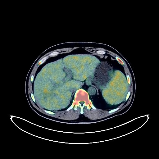
Rectal Cancer PET/CT (case 983824-000066 from PETWB-REP)
2 views10 days agoWhole-body 18F-FDG PET/CT scan in a patient with Rectal Cancer taken from the PETWB-REP dataset. The following English report (translated from original Chinese) is taken verbatim from the public dataset and has not been modified or otherwise checked for accuracy (see the end for citation). Impression a. Post-rectal cancer treatment: Slight thickening of the rectal wall, no increase in FDG metabolism, suggesting suppressed tumor activity after treatment. Please follow up with colonoscopy. b. Right lobe of liver capsule shrinkage with a slightly low-density mass, partially reduced FDG metabolism, suggesting suppressed tumor activity after treatment of liver metastases based on medical history; left lateral lobe of liver nodule with increased FDG metabolism, suggesting an active metastatic lesion; localized increased FDG metabolism in the caudate lobe of the liver, decreasing to background levels after delayed scanning, suggesting physiological uptake. Follow-up with contrast-enhanced MRI is recommended for all of the above. c. Bone destruction of the right 4th rib with a soft tissue mass, lack of FDG metabolism in the central area, and slightly increased FDG metabolism in the peripheral area, suggesting post-treatment changes in bone metastases, with tumor activity largely suppressed. Splenomegaly with slightly increased FDG metabolism, showing widespread increased FDG metabolism throughout the bone marrow cavity, suggesting changes after chemotherapy. Degenerative changes in the spine, multiple bony islands (as described above). Chronic inflammatory miliary foci and small nodular lesions in both lungs. Scattered chronic inflammation and remnants in both lungs. Manifestations of chronic gastritis. Right renal cyst. Reactive hyperplasia of retroperitoneal and bilateral inguinal lymph nodes. No obvious abnormalities on cranial scintigraphy. Dense shadow in the left parapharyngeal space with chronic inflammatory changes on the adjacent left oropharyngeal wall; please correlate with clinical findings. This case is from PETWB-REP, a curated dataset of whole-body 18F-FDG PET/CT scans and corresponding radiology reports from 490 patients with a broad spectrum of malignancies. The data were retrospectively collected from patients who underwent clinically indicated whole-body 18F-FDG PET/CT scans at the Shanghai Universal Medical Imaging Diagnostic Center between 2021 and 2024. License: Creative Commons Attribution 4.0 International (CC BY 4.0) Citation: Xue, L., Feng, G., Wenbo, Z., Zhang, Y., Li, L., Wang, S., Peng, L., Peng, S., & Gao, X. (2026). PETWB-REP: A Multi-Cancer Whole-Body FDG PET/CT Dataset with Corresponding Radiology Reports [Data set]. Zenodo. https://doi.org/10.5281/zenodo.18670487
Whole BodyPET/CT