Loading...
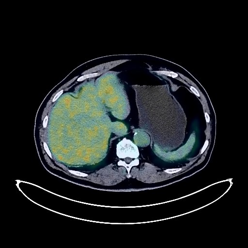
Lung Cancer PET/CT (case 983824-000172 from PETWB-REP)
4 views10 days agoWhole-body 18F-FDG PET/CT scan in a patient with Lung Cancer taken from the PETWB-REP dataset. The following English report (translated from original Chinese) is taken verbatim from the public dataset and has not been modified or otherwise checked for accuracy (see the end for citation). Impression a. A mass at the bronchial opening in the anterior segment of the left upper lobe of the lung, fused with the left hilar lymph nodes, with increased FDG metabolism, consistent with central lung cancer. Reactive hyperplasia of the right hilar, mediastinal, and left axillary lymph nodes is highly probable; follow-up is recommended. b. Several ground-glass opacities in the apical-posterior segment of the left upper lobe, suggestive of chronic inflammatory nodules or atypical adenomatous hyperplasia; annual HRCT follow-up is recommended. c. Chronic inflammatory nodules (solid) in the apical-posterior segment of the left upper lobe and the lateral basal segment of the right lower lobe. Scattered chronic inflammation and remnants in both lungs. Partial arteriosclerosis (including coronary arteries). Manifestations of liver cirrhosis. Subcapsular calcification in the right posterior lobe of the liver. Mild prostatic hyperplasia. Bilateral hydrocele. Postoperative changes following left inguinal hernia repair. Chronic antral gastritis. Degenerative changes in the spine. L1 vertebral body compression with changes following bone cement injection. L4/5 and L5/S1 intervertebral disc bulging. Age-related brain changes, deep lacunar infarcts in the brain. Minor inflammation of bilateral ethmoid and maxillary sinuses. Bilateral mastoid hypoplasia. This case is from PETWB-REP, a curated dataset of whole-body 18F-FDG PET/CT scans and corresponding radiology reports from 490 patients with a broad spectrum of malignancies. The data were retrospectively collected from patients who underwent clinically indicated whole-body 18F-FDG PET/CT scans at the Shanghai Universal Medical Imaging Diagnostic Center between 2021 and 2024. License: Creative Commons Attribution 4.0 International (CC BY 4.0) Citation: Xue, L., Feng, G., Wenbo, Z., Zhang, Y., Li, L., Wang, S., Peng, L., Peng, S., & Gao, X. (2026). PETWB-REP: A Multi-Cancer Whole-Body FDG PET/CT Dataset with Corresponding Radiology Reports [Data set]. Zenodo. https://doi.org/10.5281/zenodo.18670487
Whole BodyPET/CT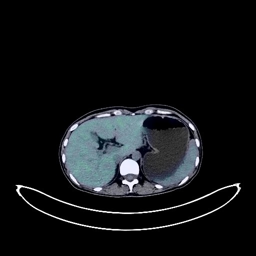
Cervical Cancer PET/CT (case 983824-000174 from PETWB-REP)
2 views10 days agoWhole-body 18F-FDG PET/CT scan in a patient with Cervical Cancer taken from the PETWB-REP dataset. The following English report (translated from original Chinese) is taken verbatim from the public dataset and has not been modified or otherwise checked for accuracy (see the end for citation). Impression a. A cystic-solid mixed-density mass is seen at the cervical-body junction, with unevenly increased FDG metabolism in the solid portion, suggestive of malignancy, accompanied by pelvic lymph node metastasis. Please confirm the diagnosis with pathological examination. b. Strip-like areas of increased FDG metabolism within the uterine cavity, suggestive of physiological changes. Further enhanced MRI is recommended to rule out endometrial lesions. Irregular mixed ground-glass opacities in the lateral/posterior basal segment of the right lower lobe, suggestive of lung cancer. A few chronic inflammations and remnants in both lungs. a. Several slightly high-density nodules and mass-like shadows in the transverse colon lumen, with increased FDG metabolism, suggestive of intestinal contents. Follow-up colonoscopy is recommended to rule out other possibilities. b. Increased FDG metabolism in the remaining colon and rectum, suggestive of physiological uptake or chronic inflammatory changes. L5/S1 intervertebral disc bulge. No obvious abnormalities were found on cranial scintigraphy. Bilateral inferior turbinate hypertrophy was observed. This case is from PETWB-REP, a curated dataset of whole-body 18F-FDG PET/CT scans and corresponding radiology reports from 490 patients with a broad spectrum of malignancies. The data were retrospectively collected from patients who underwent clinically indicated whole-body 18F-FDG PET/CT scans at the Shanghai Universal Medical Imaging Diagnostic Center between 2021 and 2024. License: Creative Commons Attribution 4.0 International (CC BY 4.0) Citation: Xue, L., Feng, G., Wenbo, Z., Zhang, Y., Li, L., Wang, S., Peng, L., Peng, S., & Gao, X. (2026). PETWB-REP: A Multi-Cancer Whole-Body FDG PET/CT Dataset with Corresponding Radiology Reports [Data set]. Zenodo. https://doi.org/10.5281/zenodo.18670487
Whole BodyPET/CT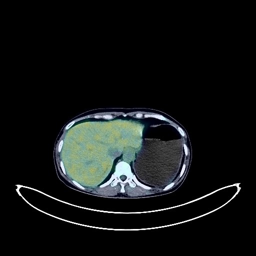
Nasopharyngeal Cancer PET/CT (case 983824-000129 from PETWB-REP)
3 views10 days agoWhole-body 18F-FDG PET/CT scan in a patient with Nasopharyngeal Cancer taken from the PETWB-REP dataset. The following English report (translated from original Chinese) is taken verbatim from the public dataset and has not been modified or otherwise checked for accuracy (see the end for citation). Impression A mass on the right lateral wall of the nasopharynx, with elevated FDG metabolism, consistent with nasopharyngeal carcinoma, possibly involving the right tonsil; multiple lymph node metastases in the right retropharyngeal space, right deep cervical space, and right supraclavicular fossa. Several small, solid, chronic inflammatory nodules in both lungs; follow-up CT scan recommended to rule out other involvement. A small amount of chronic inflammation and old lesions in both lungs. Calcifications in the liver. Small amount of pelvic effusion. Physiological uptake in the uterine cavity. A left adnexal ovarian cyst is highly probable; follow-up ultrasound is recommended. No abnormalities were found on cranial scintigraphy. This case is from PETWB-REP, a curated dataset of whole-body 18F-FDG PET/CT scans and corresponding radiology reports from 490 patients with a broad spectrum of malignancies. The data were retrospectively collected from patients who underwent clinically indicated whole-body 18F-FDG PET/CT scans at the Shanghai Universal Medical Imaging Diagnostic Center between 2021 and 2024. License: Creative Commons Attribution 4.0 International (CC BY 4.0) Citation: Xue, L., Feng, G., Wenbo, Z., Zhang, Y., Li, L., Wang, S., Peng, L., Peng, S., & Gao, X. (2026). PETWB-REP: A Multi-Cancer Whole-Body FDG PET/CT Dataset with Corresponding Radiology Reports [Data set]. Zenodo. https://doi.org/10.5281/zenodo.18670487
Whole BodyPET/CT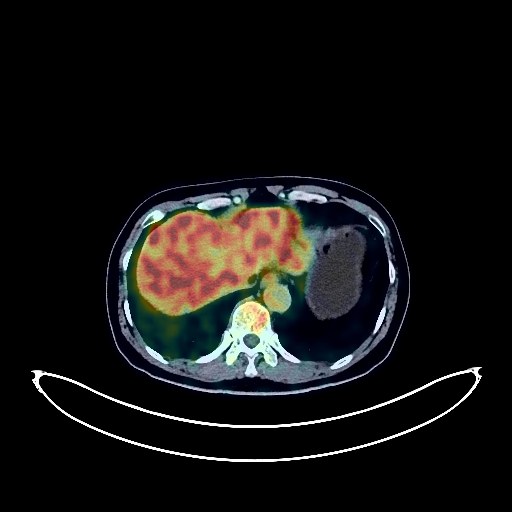
Renal Cancer PET/CT (case 983824-000201 from PETWB-REP)
2 views10 days agoWhole-body 18F-FDG PET/CT scan in a patient with Renal Cancer taken from the PETWB-REP dataset. The following English report (translated from original Chinese) is taken verbatim from the public dataset and has not been modified or otherwise checked for accuracy (see the end for citation). Impression Right renal mass with increased FDG metabolism, renal cell carcinoma is suspected; enhanced MRI is recommended to rule out benign tumors. Reactive hyperplasia of retroperitoneal lymph nodes. Chronic inflammatory micronodules in the right lung; follow-up CT is recommended. Bilateral emphysema, scattered post-inflammatory lesions in both lungs, small calcification in the lower lobe of the left lung. Partial pleural thickening bilaterally. Reactive hyperplasia of hilar and mediastinal lymph nodes bilaterally. Partial arteriosclerosis. Small liver cyst. Left renal cyst. Residual contrast agent in the urinary tract. Mild vertebral osteophyte formation. L4/5 and L5/S1 intervertebral disc bulge. No obvious abnormalities on cranial scintigraphy. Physiological uptake in the glottic region. This case is from PETWB-REP, a curated dataset of whole-body 18F-FDG PET/CT scans and corresponding radiology reports from 490 patients with a broad spectrum of malignancies. The data were retrospectively collected from patients who underwent clinically indicated whole-body 18F-FDG PET/CT scans at the Shanghai Universal Medical Imaging Diagnostic Center between 2021 and 2024. License: Creative Commons Attribution 4.0 International (CC BY 4.0) Citation: Xue, L., Feng, G., Wenbo, Z., Zhang, Y., Li, L., Wang, S., Peng, L., Peng, S., & Gao, X. (2026). PETWB-REP: A Multi-Cancer Whole-Body FDG PET/CT Dataset with Corresponding Radiology Reports [Data set]. Zenodo. https://doi.org/10.5281/zenodo.18670487
Whole BodyPET/CT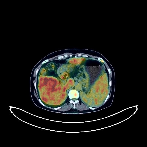
Cervical Cancer PET/CT (case 983824-000078 from PETWB-REP)
44 views10 days agoWhole-body 18F-FDG PET/CT scan in a patient with Cervical Cancer taken from the PETWB-REP dataset. The following English report (translated from original Chinese) is taken verbatim from the public dataset and has not been modified or otherwise checked for accuracy (see the end for citation). Impression Cervical mass with elevated FDG metabolism, consistent with cervical cancer; lymph node metastasis bilaterally to the iliac vessels, bilaterally to the supraclavicular fossa, and at the mediastinal-thoracic inlet. Chronic inflammatory micronodules in the right lung. A few post-inflammatory lesions in both lungs. Anemia. Bilateral breast hyperplasia, calcification in the left breast. Liver cirrhosis, uneven FDG metabolism in the liver; please combine with MRI to rule out occult lesions. Portal hypertension with collateral circulation formation, splenomegaly. Reactive hyperplasia of the hilar lymph nodes. Pelvic effusion, mesenteric turbidity. Chronic inflammatory or physiological changes in part of the gastric wall and intestines. Mild vertebral osteophyte formation, L4/5 and L5/S1 intervertebral disc bulge. Post-fracture changes in the left iliac bone; please combine with clinical history. No obvious abnormalities were found on cranial scintigraphy. This case is from PETWB-REP, a curated dataset of whole-body 18F-FDG PET/CT scans and corresponding radiology reports from 490 patients with a broad spectrum of malignancies. The data were retrospectively collected from patients who underwent clinically indicated whole-body 18F-FDG PET/CT scans at the Shanghai Universal Medical Imaging Diagnostic Center between 2021 and 2024. License: Creative Commons Attribution 4.0 International (CC BY 4.0) Citation: Xue, L., Feng, G., Wenbo, Z., Zhang, Y., Li, L., Wang, S., Peng, L., Peng, S., & Gao, X. (2026). PETWB-REP: A Multi-Cancer Whole-Body FDG PET/CT Dataset with Corresponding Radiology Reports [Data set]. Zenodo. https://doi.org/10.5281/zenodo.18670487
Whole BodyPET/CT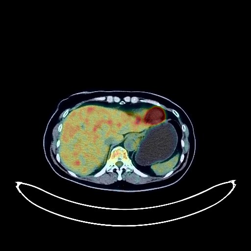
Cervical Cancer PET/CT (case 983824-000094 from PETWB-REP)
1 views10 days agoWhole-body 18F-FDG PET/CT scan in a patient with Cervical Cancer taken from the PETWB-REP dataset. The following English report (translated from original Chinese) is taken verbatim from the public dataset and has not been modified or otherwise checked for accuracy (see the end for citation). Impression a. Cervical mass with increased FDG metabolism, consistent with cervical cancer. b. Reactive hyperplasia of small lymph nodes in the retroperitoneum, bilateral iliac vessels, and groin. Uterine fibroids with calcification. a. Irregular mixed ground-glass opacity in the posterior segment of the left lower lobe with increased FDG metabolism, highly suggestive of lung cancer; please confirm with pathology. b. Chronic inflammatory micronodules in the remaining lungs. A few post-inflammatory remnants in both lungs. Partial thickening of the left pleura. Anemia changes, partial arteriosclerosis. Chronic inflammatory changes in the antrum of the stomach and part of the intestine; please confirm with endoscopic follow-up. Degenerative changes in the spine, L4/5 and L5/S1 intervertebral disc bulges. Subcutaneous calcification in the left buttock. Low-density nodule in the right lobe of the thyroid gland; FDG metabolism normal; suggestive of nodular goiter with cystic degeneration; follow-up ultrasound recommended. Cranial scintigraphy showed no obvious abnormalities. Inflammation of the left lateral nasopharyngeal wall. Bilateral chronic inflammatory lymph nodes in the neck. This case is from PETWB-REP, a curated dataset of whole-body 18F-FDG PET/CT scans and corresponding radiology reports from 490 patients with a broad spectrum of malignancies. The data were retrospectively collected from patients who underwent clinically indicated whole-body 18F-FDG PET/CT scans at the Shanghai Universal Medical Imaging Diagnostic Center between 2021 and 2024. License: Creative Commons Attribution 4.0 International (CC BY 4.0) Citation: Xue, L., Feng, G., Wenbo, Z., Zhang, Y., Li, L., Wang, S., Peng, L., Peng, S., & Gao, X. (2026). PETWB-REP: A Multi-Cancer Whole-Body FDG PET/CT Dataset with Corresponding Radiology Reports [Data set]. Zenodo. https://doi.org/10.5281/zenodo.18670487
Whole BodyPET/CT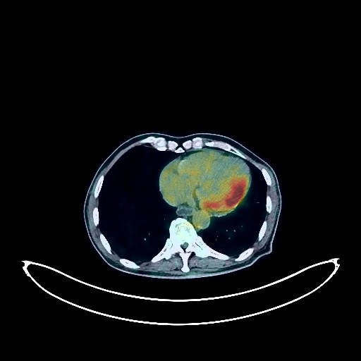
Gallbladder Cancer PET/CT (case 983824-000024 from PETWB-REP)
8 views10 days agoWhole-body 18F-FDG PET/CT scan in a patient with Gallbladder Cancer taken from the PETWB-REP dataset. The following English report (translated from original Chinese) is taken verbatim from the public dataset and has not been modified or otherwise checked for accuracy (see the end for citation). Impression a. Changes after comprehensive treatment for gallbladder cancer; no clear signs of tumor recurrence were seen in the surgical area. Slight local dilation of intrahepatic bile ducts. b. High probability of metastasis to the right quadratus femoris muscle involving the adjacent femur; please confirm with pathology. a. After treatment for brain metastases, no clear space-occupying lesions were seen in the brain; lacunar ischemic lesions in the deep cerebral region and brainstem bilaterally, senile brain changes. b. Liver cysts; no obvious abnormal density shadows were seen in the remaining liver parenchyma; no abnormal increase in FDG metabolism was observed. Please follow up with enhanced MRI for the above. Benign prostatic hyperplasia; unevenly increased FDG metabolism; please analyze comprehensively with PSA and MRI. A few chronic pulmonary lesions and remnants. Reactive hyperplasia of mediastinal lymph nodes. Small amount of pericardial effusion. Calcification of some arterial walls. Mild pancreatic atrophy. Multiple renal cysts. Mildly increased FDG metabolism in parts of the gastric wall; slight thickening of the rectal wall with increased FDG metabolism, suggestive of physiological uptake or chronic inflammatory changes; please follow up with endoscopy. Decreased FDG metabolism in parts of the thoracic and lumbar spine, suggestive of post-radiotherapy changes. Spinal degeneration. L5/S1 vertebral endplate inflammation. Minor inflammation of bilateral ethmoid sinuses. This case is from PETWB-REP, a curated dataset of whole-body 18F-FDG PET/CT scans and corresponding radiology reports from 490 patients with a broad spectrum of malignancies. The data were retrospectively collected from patients who underwent clinically indicated whole-body 18F-FDG PET/CT scans at the Shanghai Universal Medical Imaging Diagnostic Center between 2021 and 2024. License: Creative Commons Attribution 4.0 International (CC BY 4.0) Citation: Xue, L., Feng, G., Wenbo, Z., Zhang, Y., Li, L., Wang, S., Peng, L., Peng, S., & Gao, X. (2026). PETWB-REP: A Multi-Cancer Whole-Body FDG PET/CT Dataset with Corresponding Radiology Reports [Data set]. Zenodo. https://doi.org/10.5281/zenodo.18670487
Whole BodyPET/CT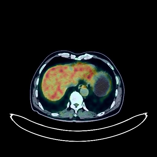
Pancreatic Cancer PET/CT (case 983824-000156 from PETWB-REP)
3 views10 days agoWhole-body 18F-FDG PET/CT scan in a patient with Pancreatic Cancer taken from the PETWB-REP dataset. The following English report (translated from original Chinese) is taken verbatim from the public dataset and has not been modified or otherwise checked for accuracy (see the end for citation). Impression After chemotherapy for pancreatic cancer, irregular soft tissue density shadows were observed in the pancreatic body, with slightly increased FDG metabolism; small lymph nodes around the pancreas and in the retroperitoneum showed no abnormalities in FDG metabolism. This suggests that most tumor activity is suppressed after treatment; comparison with previous imaging data and follow-up are recommended. Chronic inflammatory nodules in both lungs; CT follow-up is recommended. Bilateral emphysema. A few post-inflammatory remnants in both lungs. Reactive hyperplasia of hilar and mediastinal lymph nodes in both lungs. Calcification of some arterial walls (including coronary arteries). A port-a-cath has been placed in the right clavicular region. Cyst in the left lobe of the liver; no other obvious space-occupying lesions were seen in the liver, and FDG metabolism was normal. MRI is recommended. Left adrenal hyperplasia. Chronic inflammatory changes in part of the gastric wall and intestinal tract; endoscopic follow-up is recommended. Osteoporosis, degenerative changes in the spine, L5/S1 disc bulge. Age-related brain abnormalities, deep lacunar infarcts; please include MRI. Bilateral chronic maxillary sinusitis. This case is from PETWB-REP, a curated dataset of whole-body 18F-FDG PET/CT scans and corresponding radiology reports from 490 patients with a broad spectrum of malignancies. The data were retrospectively collected from patients who underwent clinically indicated whole-body 18F-FDG PET/CT scans at the Shanghai Universal Medical Imaging Diagnostic Center between 2021 and 2024. License: Creative Commons Attribution 4.0 International (CC BY 4.0) Citation: Xue, L., Feng, G., Wenbo, Z., Zhang, Y., Li, L., Wang, S., Peng, L., Peng, S., & Gao, X. (2026). PETWB-REP: A Multi-Cancer Whole-Body FDG PET/CT Dataset with Corresponding Radiology Reports [Data set]. Zenodo. https://doi.org/10.5281/zenodo.18670487
Whole BodyPET/CT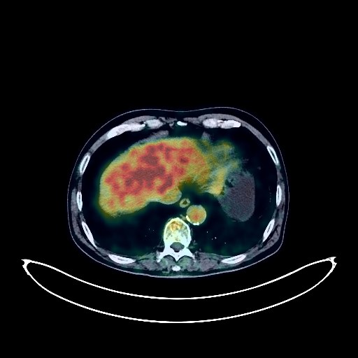
Lung Cancer PET/CT (case 983824-000004 from PETWB-REP)
10 views10 days agoWhole-body 18F-FDG PET/CT scan in a patient with Lung Cancer taken from the PETWB-REP dataset. The following English report (translated from original Chinese) is taken verbatim from the public dataset and has not been modified or otherwise checked for accuracy (see the end for citation). Impression a. Irregular nodule in the apical segment of the right upper lobe, with elevated FDG metabolism, suggestive of peripheral lung cancer; further clinical pathology is recommended. b. Several miliary chronic inflammatory nodules in both lungs. A few old lesions in both lungs. Emphysema in both lungs. Calcification of some arterial walls (including coronary arteries). Cystic mass in the head and neck of the pancreas, cystadenoma to be ruled out; further examination with contrast-enhanced MRI is recommended. Multiple cysts in the liver. Bilateral hydrocele. Degenerative changes in the spine. L4/5 and L5/S1 intervertebral disc bulge. Fat deposition in the right hip bone. Age-related brain changes. This case is from PETWB-REP, a curated dataset of whole-body 18F-FDG PET/CT scans and corresponding radiology reports from 490 patients with a broad spectrum of malignancies. The data were retrospectively collected from patients who underwent clinically indicated whole-body 18F-FDG PET/CT scans at the Shanghai Universal Medical Imaging Diagnostic Center between 2021 and 2024. License: Creative Commons Attribution 4.0 International (CC BY 4.0) Citation: Xue, L., Feng, G., Wenbo, Z., Zhang, Y., Li, L., Wang, S., Peng, L., Peng, S., & Gao, X. (2026). PETWB-REP: A Multi-Cancer Whole-Body FDG PET/CT Dataset with Corresponding Radiology Reports [Data set]. Zenodo. https://doi.org/10.5281/zenodo.18670487
Whole BodyPET/CT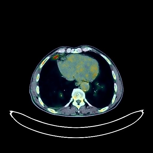
Lung Cancer PET/CT (case 983824-000195 from PETWB-REP)
4 views10 days agoWhole-body 18F-FDG PET/CT scan in a patient with Lung Cancer taken from the PETWB-REP dataset. The following English report (translated from original Chinese) is taken verbatim from the public dataset and has not been modified or otherwise checked for accuracy (see the end for citation). Impression a. Stenosis at the bronchial opening in the right middle lobe with soft tissue density mass formation and increased FDG metabolism, suggestive of central lung cancer in the right middle lobe with obstructive changes in the right upper and middle lobes. Metastasis to the right hilar, mediastinal, and right supraclavicular fossa lymph nodes. Minimal right pleural effusion. b. Scattered chronic inflammatory nodules and calcifications in the remaining lungs; please follow up with CT scans. A few post-inflammatory remnants in both lungs. Partial arteriosclerosis. Multiple cysts in the left lobe of the liver. Cyst in the right kidney. Benign prostatic hyperplasia with calcification. (No definite space-occupying lesion seen in the left upper quadrant.) Chronic gastritis; please follow up with endoscopy. Degenerative changes in the spine. Schmorl's nodes at the lower margin of the T8 vertebral body and the upper margin of the T10 vertebral body. L4/5 and L5/S1 intervertebral disc bulges. Right ischial insula. Age-related brain changes, deep lacunar insufficiency in the brain. Minor inflammation of the bilateral ethmoid and maxillary sinuses. This case is from PETWB-REP, a curated dataset of whole-body 18F-FDG PET/CT scans and corresponding radiology reports from 490 patients with a broad spectrum of malignancies. The data were retrospectively collected from patients who underwent clinically indicated whole-body 18F-FDG PET/CT scans at the Shanghai Universal Medical Imaging Diagnostic Center between 2021 and 2024. License: Creative Commons Attribution 4.0 International (CC BY 4.0) Citation: Xue, L., Feng, G., Wenbo, Z., Zhang, Y., Li, L., Wang, S., Peng, L., Peng, S., & Gao, X. (2026). PETWB-REP: A Multi-Cancer Whole-Body FDG PET/CT Dataset with Corresponding Radiology Reports [Data set]. Zenodo. https://doi.org/10.5281/zenodo.18670487
Whole BodyPET/CT