Loading...
Nasopharyngeal Cancer PET/CT (case 983824-000140 from PETWB-REP)
4 views10 days agoWhole-body 18F-FDG PET/CT scan in a patient with Nasopharyngeal Cancer taken from the PETWB-REP dataset. The following English report (translated from original Chinese) is taken verbatim from the public dataset and has not been modified or otherwise checked for accuracy (see the end for citation). Impression a. Nasopharyngeal mass with elevated FDG metabolism, consistent with nasopharyngeal carcinoma. b. Right retropharyngeal lymph node metastasis. Bilateral deep cervical lymph node reactive hyperplasia is highly probable; follow-up is recommended. Minor chronic inflammation in the left lower lobe of the lung. Incomplete thymic regression. Bilateral breast hyperplasia. Calcification in the right lobe of the liver. Small amount of pelvic effusion. L3/4 intervertebral disc bulge. No obvious abnormalities seen on cranial scintigraphy. Physiological uptake in the glottic region. Chronic inflammation of the right ethmoid sinus and right maxillary sinus, submucosal cyst of the right maxillary sinus. Right middle otitis media and mastoiditis. This case is from PETWB-REP, a curated dataset of whole-body 18F-FDG PET/CT scans and corresponding radiology reports from 490 patients with a broad spectrum of malignancies. The data were retrospectively collected from patients who underwent clinically indicated whole-body 18F-FDG PET/CT scans at the Shanghai Universal Medical Imaging Diagnostic Center between 2021 and 2024. License: Creative Commons Attribution 4.0 International (CC BY 4.0) Citation: Xue, L., Feng, G., Wenbo, Z., Zhang, Y., Li, L., Wang, S., Peng, L., Peng, S., & Gao, X. (2026). PETWB-REP: A Multi-Cancer Whole-Body FDG PET/CT Dataset with Corresponding Radiology Reports [Data set]. Zenodo. https://doi.org/10.5281/zenodo.18670487
Whole BodyPET/CT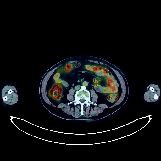
Lymphoma PET/CT (case 983824-000014 from PETWB-REP)
9 views10 days agoWhole-body 18F-FDG PET/CT scan in a patient with Lymphoma taken from the PETWB-REP dataset. The following English report (translated from original Chinese) is taken verbatim from the public dataset and has not been modified or otherwise checked for accuracy (see the end for citation). Impression a. After treatment for splenic lymphoma, no obvious space-occupying lesion was found in the spleen, and FDG metabolism was normal. Tumor activity is considered suppressed. It is recommended to compare previous imaging data and follow up. b. Left pleural thickening and adhesions, slightly increased FDG metabolism, and a small amount of pleural effusion on the left side suggest that tumor activity is likely largely suppressed. It is recommended to compare previous imaging data and, in conjunction with clinical findings, rule out residual tumor activity. a. Irregular patchy lesions in the lower lingular segment of the left upper lobe and the subpleural region of the left lower lobe, accompanied by slightly increased FDG metabolism, suggest possible chronic inflammation or atelectasis. It is recommended to compare previous imaging data and follow up. b. Ground-glass nodules in the apical segment of the right upper lobe, with normal FDG metabolism, suggest inflammation or atypical adenomatous hyperplasia. Annual HRCT follow-up is recommended. c. Chronic inflammatory micronodules in the right lung. Chronic inflammation and post-inflammatory remnants in both lungs. Slight pericardial thickening, mild anemia changes, and partial calcification of arterial walls (including coronary arteries). Bilateral breast hyperplasia and calcifications; ultrasound follow-up is recommended. Mild fatty liver. Accessory spleen. Left adrenal hyperplasia. Possible chronic inflammatory changes in the lower esophagus and part of the intestine; endoscopic follow-up is recommended. Osteoporosis, degenerative changes in the spine, L4/5 and L5/S1 disc bulges. Bilateral frozen shoulder. Uneven thyroid density; normal FDG metabolism; ultrasound follow-up is recommended. Deep lacunar infarcts in the brain; mild age-related encephalopathy. Minor chronic inflammation of both ethmoid sinuses. This case is from PETWB-REP, a curated dataset of whole-body 18F-FDG PET/CT scans and corresponding radiology reports from 490 patients with a broad spectrum of malignancies. The data were retrospectively collected from patients who underwent clinically indicated whole-body 18F-FDG PET/CT scans at the Shanghai Universal Medical Imaging Diagnostic Center between 2021 and 2024. License: Creative Commons Attribution 4.0 International (CC BY 4.0) Citation: Xue, L., Feng, G., Wenbo, Z., Zhang, Y., Li, L., Wang, S., Peng, L., Peng, S., & Gao, X. (2026). PETWB-REP: A Multi-Cancer Whole-Body FDG PET/CT Dataset with Corresponding Radiology Reports [Data set]. Zenodo. https://doi.org/10.5281/zenodo.18670487
Whole BodyPET/CT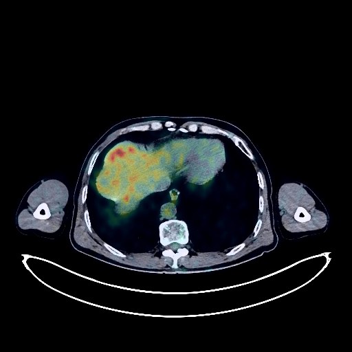
Lung Cancer PET/CT (case 983824-000166 from PETWB-REP)
6 views10 days agoWhole-body 18F-FDG PET/CT scan in a patient with Lung Cancer taken from the PETWB-REP dataset. The following English report (translated from original Chinese) is taken verbatim from the public dataset and has not been modified or otherwise checked for accuracy (see the end for citation). Impression a. Right upper lobe lung mass, elevated FDG metabolism, suggestive of peripheral lung cancer. b. Mediastinal lymph node metastasis. c. Multiple solid nodules in both lungs, normal FDG metabolism, suggestive of chronic inflammatory nodules, some metastasis to be ruled out; regular CT scans for comparison are recommended. d. Minor emphysema and remnants of chronic inflammation in both lungs. Some arterial wall calcification (including coronary arteries). Minor ischemic lesions deep in the brain. Age-related brain changes; MRI is recommended. Chronic inflammation of the right ethmoid and maxillary sinuses. Partial vertebral osteophyte formation. L2-S1 intervertebral disc bulge. Low-density nodule in the left lobe of the thyroid gland, normal FDG metabolism, suggestive of adenoma; ultrasound is recommended. This case is from PETWB-REP, a curated dataset of whole-body 18F-FDG PET/CT scans and corresponding radiology reports from 490 patients with a broad spectrum of malignancies. The data were retrospectively collected from patients who underwent clinically indicated whole-body 18F-FDG PET/CT scans at the Shanghai Universal Medical Imaging Diagnostic Center between 2021 and 2024. License: Creative Commons Attribution 4.0 International (CC BY 4.0) Citation: Xue, L., Feng, G., Wenbo, Z., Zhang, Y., Li, L., Wang, S., Peng, L., Peng, S., & Gao, X. (2026). PETWB-REP: A Multi-Cancer Whole-Body FDG PET/CT Dataset with Corresponding Radiology Reports [Data set]. Zenodo. https://doi.org/10.5281/zenodo.18670487
Whole BodyPET/CT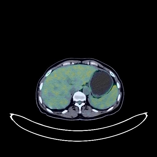
Cervical Cancer PET/CT (case 983824-000194 from PETWB-REP)
4 views10 days agoWhole-body 18F-FDG PET/CT scan in a patient with Cervical Cancer taken from the PETWB-REP dataset. The following English report (translated from original Chinese) is taken verbatim from the public dataset and has not been modified or otherwise checked for accuracy (see the end for citation). Impression a. Cervical mass with elevated FDG metabolism, consistent with cervical cancer, involving the lower part of the uterine body; multiple lymph node metastases in the bilateral pelvic walls, bilateral iliac vessels, and retroperitoneum. b. Uterine cavity effusion, indwelling IUD. Uterine fibroids. Chronic inflammatory micronodules in both lungs; CT follow-up is recommended. Segmental atelectasis in the left upper lobe, and a pneumocystic cavity in the right lower lobe. A few fibrotic lesions in both lungs. Anemic changes, calcification of some arterial walls (including coronary arteries). Small cyst in the right lobe of the liver. Chronic cholecystitis. Calcified nodules in the splenic hilum. Chronic inflammatory changes in the antrum of the stomach; please follow up with endoscopy. Degenerative changes in the spine, multiple lumbar disc herniations. Subcutaneous calcification in the right buttock. Uneven thyroid density with elevated FDG metabolism suggests inflammation; please follow up with ultrasound and thyroid function tests. Cranial scintigraphy showed no obvious abnormalities. Inflammation is present at the base of the tongue and bilateral palatine tonsils. This case is from PETWB-REP, a curated dataset of whole-body 18F-FDG PET/CT scans and corresponding radiology reports from 490 patients with a broad spectrum of malignancies. The data were retrospectively collected from patients who underwent clinically indicated whole-body 18F-FDG PET/CT scans at the Shanghai Universal Medical Imaging Diagnostic Center between 2021 and 2024. License: Creative Commons Attribution 4.0 International (CC BY 4.0) Citation: Xue, L., Feng, G., Wenbo, Z., Zhang, Y., Li, L., Wang, S., Peng, L., Peng, S., & Gao, X. (2026). PETWB-REP: A Multi-Cancer Whole-Body FDG PET/CT Dataset with Corresponding Radiology Reports [Data set]. Zenodo. https://doi.org/10.5281/zenodo.18670487
Whole BodyPET/CT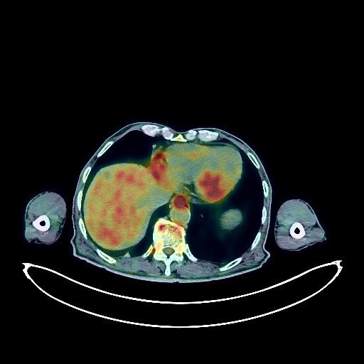
Lung Cancer PET/CT (case 983824-000120 from PETWB-REP)
6 views10 days agoWhole-body 18F-FDG PET/CT scan in a patient with Lung Cancer taken from the PETWB-REP dataset. The following English report (translated from original Chinese) is taken verbatim from the public dataset and has not been modified or otherwise checked for accuracy (see the end for citation). Impression a. A mass in the left upper lobe of the lung with increased FDG metabolism, consistent with lung cancer. Multiple lymph node metastases in the left hilum, mediastinum, and left supraclavicular fossa. b. Bilateral lung metastases. Scattered post-inflammatory lesions in both lungs. c. Anemia, partial arterial wall calcification (including coronary arteries). Liver calcifications, multiple liver cysts. Bilateral kidney stones, left renal cyst. Benign prostatic hyperplasia with calcification. Postoperative changes in the stomach; chronic inflammatory changes in the middle and lower esophagus; possible rectal polyps, malignancy to be ruled out. Follow-up gastroscopy and colonoscopy are recommended. a. Osteoporosis, degenerative changes in the spine, L4/5 and L5/S1 intervertebral disc bulges. b. Thickening of the soft tissue in the chin with increased FDG metabolism suggests possible inflammation or physiological uptake; clinical correlation is recommended. Low-density nodule with calcification in the right lobe of the thyroid gland; FDG metabolism is normal, suggesting possible nodular goiter; malignancy is a possibility. Further ultrasound examination is recommended. Elderly brain with deep lacunar infarcts. Chronic inflammation of both ethmoid sinuses and the left maxillary sinus. Calcification of the right palatine tonsil. This case is from PETWB-REP, a curated dataset of whole-body 18F-FDG PET/CT scans and corresponding radiology reports from 490 patients with a broad spectrum of malignancies. The data were retrospectively collected from patients who underwent clinically indicated whole-body 18F-FDG PET/CT scans at the Shanghai Universal Medical Imaging Diagnostic Center between 2021 and 2024. License: Creative Commons Attribution 4.0 International (CC BY 4.0) Citation: Xue, L., Feng, G., Wenbo, Z., Zhang, Y., Li, L., Wang, S., Peng, L., Peng, S., & Gao, X. (2026). PETWB-REP: A Multi-Cancer Whole-Body FDG PET/CT Dataset with Corresponding Radiology Reports [Data set]. Zenodo. https://doi.org/10.5281/zenodo.18670487
Whole BodyPET/CT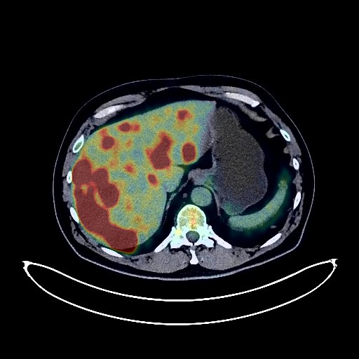
Lung Cancer PET/CT (case 983824-000125 from PETWB-REP)
6 views10 days agoWhole-body 18F-FDG PET/CT scan in a patient with Lung Cancer taken from the PETWB-REP dataset. The following English report (translated from original Chinese) is taken verbatim from the public dataset and has not been modified or otherwise checked for accuracy (see the end for citation). Impression a. Space-occupying lesion in the apical segment of the right upper lobe, with increased FDG metabolism, suggestive of lung cancer. b. Multiple lymph node metastases in the right hilum, mediastinum, and right supraclavicular fossa. Multiple metastatic tumors in both lungs. Diffuse liver metastases. Multiple bone metastases throughout the body. c. Small amount of pericardial effusion. Calcification of some arterial walls (including coronary arteries). d. Chronic inflammation and old lesions in the remaining lungs. Right renal cyst. Degenerative changes in the spine. L4/5 and L5/S1 intervertebral disc bulges. No abnormalities found on cranial scintigraphy. This case is from PETWB-REP, a curated dataset of whole-body 18F-FDG PET/CT scans and corresponding radiology reports from 490 patients with a broad spectrum of malignancies. The data were retrospectively collected from patients who underwent clinically indicated whole-body 18F-FDG PET/CT scans at the Shanghai Universal Medical Imaging Diagnostic Center between 2021 and 2024. License: Creative Commons Attribution 4.0 International (CC BY 4.0) Citation: Xue, L., Feng, G., Wenbo, Z., Zhang, Y., Li, L., Wang, S., Peng, L., Peng, S., & Gao, X. (2026). PETWB-REP: A Multi-Cancer Whole-Body FDG PET/CT Dataset with Corresponding Radiology Reports [Data set]. Zenodo. https://doi.org/10.5281/zenodo.18670487
Whole BodyPET/CT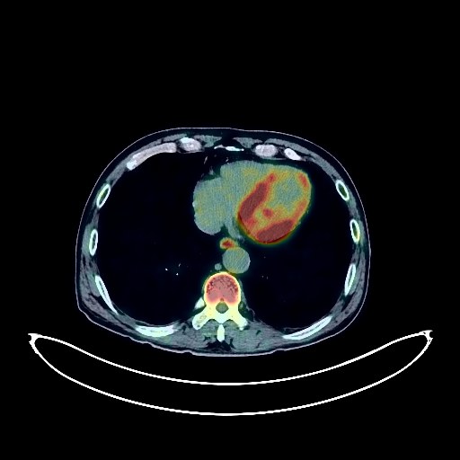
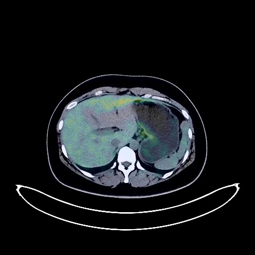
Liver Cancer PET/CT (case 983824-000095 from PETWB-REP)
4 views10 days agoWhole-body 18F-FDG PET/CT scan in a patient with Liver Cancer taken from the PETWB-REP dataset. The following English report (translated from original Chinese) is taken verbatim from the public dataset and has not been modified or otherwise checked for accuracy (see the end for citation). Impression a. An irregular soft tissue mass with increased FDG metabolism in the left inner lobe of the liver; patchy, slightly low-density lesions in the right posterior lobe of the liver, with FDG metabolism slightly higher than the liver background. Combined with contrast-enhanced MRI from another hospital, primary liver cancer is suspected. Please correlate with clinical findings. b. Liver cirrhosis, splenomegaly, pelvic effusion. Reactive hyperplasia of the hilar, hilar space, and retroperitoneal lymph nodes is highly probable. c. A roundish low-density lesion behind the left psoas major muscle (approximately at the L5/S1 intervertebral space), with increased FDG metabolism, is suspected to be a neurogenic tumor. Other possibilities need to be ruled out. Contrast-enhanced MRI is recommended for follow-up. Chronic inflammatory nodules in both lungs. Follow-up CT is recommended. A few post-inflammatory lesions in both lungs. Calcification of some arterial walls (including coronary arteries). Chronic cholecystitis, gallstones. Left renal cyst. Benign prostatic hyperplasia with calcification. The mid-esophageal wall is slightly thickened with increased FDG metabolism, suggesting possible inflammatory changes. A follow-up gastroscopy is recommended to rule out a space-occupying lesion. Chronic inflammatory changes are present in parts of the stomach wall and intestines. Degenerative changes in the spine, with L4/5 and L5/S1 intervertebral disc bulges. Age-related brain abnormalities, with deep lacunar infarcts; MRI is recommended. This case is from PETWB-REP, a curated dataset of whole-body 18F-FDG PET/CT scans and corresponding radiology reports from 490 patients with a broad spectrum of malignancies. The data were retrospectively collected from patients who underwent clinically indicated whole-body 18F-FDG PET/CT scans at the Shanghai Universal Medical Imaging Diagnostic Center between 2021 and 2024. License: Creative Commons Attribution 4.0 International (CC BY 4.0) Citation: Xue, L., Feng, G., Wenbo, Z., Zhang, Y., Li, L., Wang, S., Peng, L., Peng, S., & Gao, X. (2026). PETWB-REP: A Multi-Cancer Whole-Body FDG PET/CT Dataset with Corresponding Radiology Reports [Data set]. Zenodo. https://doi.org/10.5281/zenodo.18670487
Whole BodyPET/CT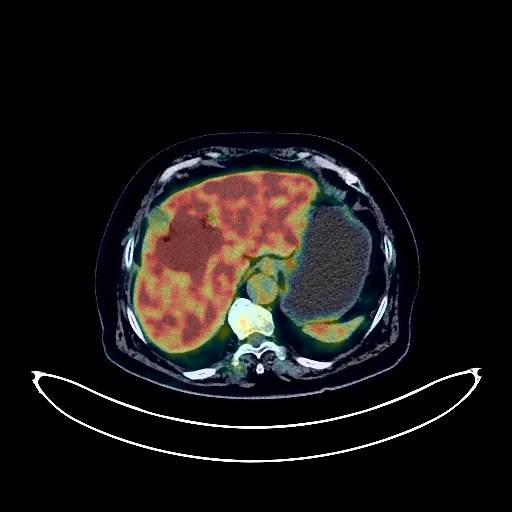
Liver Cancer PET/CT (case 983824-000038 from PETWB-REP)
10 views10 days agoWhole-body 18F-FDG PET/CT scan in a patient with Liver Cancer taken from the PETWB-REP dataset. The following English report (translated from original Chinese) is taken verbatim from the public dataset and has not been modified or otherwise checked for accuracy (see the end for citation). Impression A mass between the right anterior lobe and left medial lobe of the liver, with increased FDG metabolism, suggestive of malignancy, most likely cholangiocarcinoma. Metastasis to the hilar lymph nodes is also highly probable. Schistosomiasis-related liver disease. Chronic cholecystitis. Gallstones. Post-inferior vena cava filter placement changes. Several small, solid, chronic inflammatory nodules in both lungs. A few chronic inflammations and old lesions in both lungs. A full cardiac silhouette with partial calcification of the arterial walls (including the coronary arteries). Continuous increased FDG metabolism in the sigmoid colon and rectum, suggestive of inflammatory or physiological uptake; colonoscopy follow-up is recommended. Degenerative changes in the spine. L3-S1 intervertebral disc bulge and pneumatosis. Inflammatory or physiological uptake in the right premandibular region; clinical correlation is recommended. A few ischemic lesions in the deep bilateral cerebral regions, suggestive of age-related encephalopathy. Reactive hyperplasia of bilateral cervical lymph nodes. This case is from PETWB-REP, a curated dataset of whole-body 18F-FDG PET/CT scans and corresponding radiology reports from 490 patients with a broad spectrum of malignancies. The data were retrospectively collected from patients who underwent clinically indicated whole-body 18F-FDG PET/CT scans at the Shanghai Universal Medical Imaging Diagnostic Center between 2021 and 2024. License: Creative Commons Attribution 4.0 International (CC BY 4.0) Citation: Xue, L., Feng, G., Wenbo, Z., Zhang, Y., Li, L., Wang, S., Peng, L., Peng, S., & Gao, X. (2026). PETWB-REP: A Multi-Cancer Whole-Body FDG PET/CT Dataset with Corresponding Radiology Reports [Data set]. Zenodo. https://doi.org/10.5281/zenodo.18670487
Whole BodyPET/CT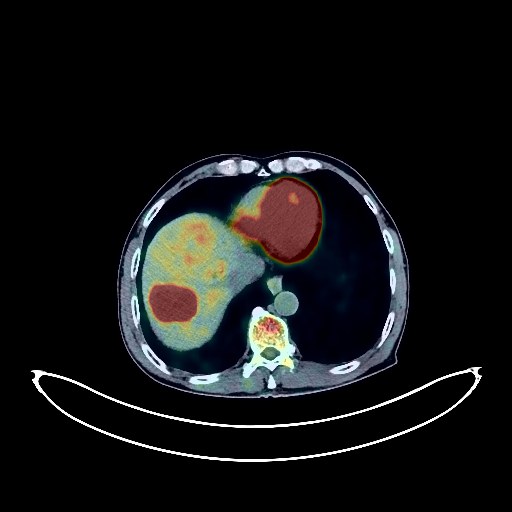
Liver Cancer PET/CT (case 983824-000133 from PETWB-REP)
7 views10 days agoWhole-body 18F-FDG PET/CT scan in a patient with Liver Cancer taken from the PETWB-REP dataset. The following English report (translated from original Chinese) is taken verbatim from the public dataset and has not been modified or otherwise checked for accuracy (see the end for citation). Impression a. Multiple space-occupying lesions in the liver with elevated FDG metabolism, suggestive of malignancy, with hepatocellular carcinoma being a strong possibility. Please combine tumor markers for comprehensive analysis. b. Changes associated with cirrhosis. Reactive hyperplasia of lymph nodes in the hepatogastric space and retroperitoneum; follow-up is recommended. Chronic inflammatory nodules in both lungs. Emphysema in both lungs, scattered post-inflammatory lesions in both lungs. Calcification of some arterial walls (including coronary arteries). Chronic cholecystitis. Benign prostatic hyperplasia with calcification. Chronic inflammatory changes in the lower esophagus and gastric antrum; please follow up with endoscopy. Degenerative changes in the spine, bilateral isthmic fracture of the L5 vertebral body with slight anterior slippage of the vertebral body. L4/5 disc herniation. Age-related brain abnormalities, deep lacunar infarcts; please include MRI. Chronic inflammation of the left nasopharyngeal wall and right ethmoid sinus. This case is from PETWB-REP, a curated dataset of whole-body 18F-FDG PET/CT scans and corresponding radiology reports from 490 patients with a broad spectrum of malignancies. The data were retrospectively collected from patients who underwent clinically indicated whole-body 18F-FDG PET/CT scans at the Shanghai Universal Medical Imaging Diagnostic Center between 2021 and 2024. License: Creative Commons Attribution 4.0 International (CC BY 4.0) Citation: Xue, L., Feng, G., Wenbo, Z., Zhang, Y., Li, L., Wang, S., Peng, L., Peng, S., & Gao, X. (2026). PETWB-REP: A Multi-Cancer Whole-Body FDG PET/CT Dataset with Corresponding Radiology Reports [Data set]. Zenodo. https://doi.org/10.5281/zenodo.18670487
Whole BodyPET/CT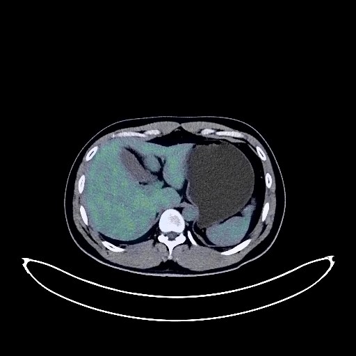
Nasopharyngeal Cancer PET/CT (case 983824-000019 from PETWB-REP)
35 views10 days agoWhole-body 18F-FDG PET/CT scan in a patient with Nasopharyngeal Cancer taken from the PETWB-REP dataset. The following English report (translated from original Chinese) is taken verbatim from the public dataset and has not been modified or otherwise checked for accuracy (see the end for citation). Impression a. Space-occupying lesions on the right lateral and posterior walls of the nasopharynx, with elevated FDG metabolism, consistent with active nasopharyngeal carcinoma, involving the posterior nasal aperture and sphenoid sinus. b. Left deep cervical lymph node metastasis. Reactive hyperplasia of the right deep cervical lymph nodes and bilateral submandibular lymph nodes is possible; partial metastasis needs to be ruled out, follow-up is recommended. c. Paranasal sinusitis. A few fibrotic lesions in both lungs. Mild fatty liver. No abnormalities were found on cranial scintigraphy. This case is from PETWB-REP, a curated dataset of whole-body 18F-FDG PET/CT scans and corresponding radiology reports from 490 patients with a broad spectrum of malignancies. The data were retrospectively collected from patients who underwent clinically indicated whole-body 18F-FDG PET/CT scans at the Shanghai Universal Medical Imaging Diagnostic Center between 2021 and 2024. License: Creative Commons Attribution 4.0 International (CC BY 4.0) Citation: Xue, L., Feng, G., Wenbo, Z., Zhang, Y., Li, L., Wang, S., Peng, L., Peng, S., & Gao, X. (2026). PETWB-REP: A Multi-Cancer Whole-Body FDG PET/CT Dataset with Corresponding Radiology Reports [Data set]. Zenodo. https://doi.org/10.5281/zenodo.18670487
Whole BodyPET/CT