Loading...
Orbital floor and rim fracture (EDNeurorad Case 43)
48 views8 months agoThere is a displaced fracture of the right orbital floor, which involved the infra-orbital canal (@Key Finding 1). The fracture extends anteriorly where there is comminution as it extends to the inferior orbital rim and the adjacent anterior wall of the right maxillary sinus (@Key Finding 2).No herniation of the inferior rectus muscle or evidence of injury to the globe on soft tissue reconstructions.
HeadCT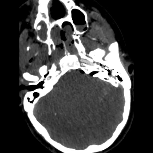
Right carotid traumatic injury (EDNeurorad Case 42)
112 views8 months agoThis is a followup of EDNeuroRad Case 41. See the prior case for discussion of additional extensive traumatic findings.Inferior to the skull base, there is an intimo-medial flap in the right internal carotid artery which extends to the junction of the cervical and petrous segments (@Key Finding 1). There is near occlusion of the vessel prior to skull base entry (@Key Finding 2). Further superiorly, there is an area of what appears to be moderate to severe narrowing of the right ICA in the distal cavernous segment immediately adjacent to the anterior clinoid process, suspicious for an additional site of injury (@Key Finding 3).
NeckCT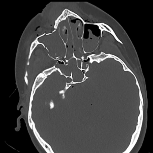
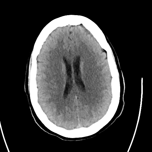
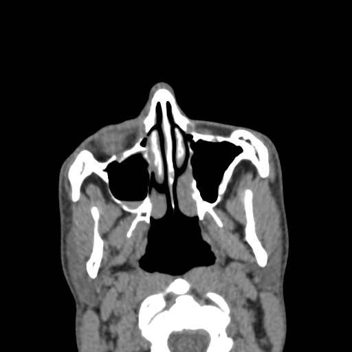
Complex trauma (EDNeurorad Case 41)
138 views8 months agoThis is a busy case with many findings. I always start with the brain and intracranial findings before getting bogged down on all the fractures. There are multifocal areas of parenchymal hemorrhage (@Key Finding 1) with hemorrhages in the left frontal lobe inferiorly annotated on the above panel. Additional multifocal subarachnoid hemorrhages are also present (blue arrows) shown in particular in the medial left middle cranial fossa (@Key Finding 23), the left sylvian fissure @Key Finding 24, the left frontal lobe @Key Finding 25, and the left upper cervical spine @Key Finding 26. Finally, there is subdural blood and gas in the left temporo-occipital junction convexity (green arrow @Key Finding 27).Additional findings are the presence of subarachnoid gas in the basilar cisterns as well as diffuse effacement of the sulci and particularly the basilar cisterns suspicious for cerebral edema. Now on to the fractures!Knowing the basic fracture patterns can be helpful in cases like this, more than anything to know what fractures tend to occur together and to be more concise in our impressions.The components of a naso-orbito-ethmoid (NOE) fracture pattern are present including fracture of the nasal bone (right sided, shown on axial @Key Finding 2), medial orbital walls @Key Finding 4, adjacent ethmoid sinuses (@Key Finding 4 the exact fractures are difficult to see but the substantial opacification implies fractures), and the nasal septum (seen best on the coronal plane, both anteriorly @Key Finding 3 and posteriorly @Key Finding 5).On the left, there are fractures of the pterygoid plates (red arrow on axials @Key Finding 6). Whenever, you see pterygoid plate fractures, look for the other component of LeFort fracture patterns. On the coronal images, a horizontal fracture of the left anterolateral nasal aperture with associated horizontal fracture of the left lateral maxillary sinus wall are present completing a LeFort I pattern (red arrows @Key Finding 7).There is fracture of the anteromedial orbital wall, the inferior orbital rim and floor, which combined with the nasal bone and lateral maxillary sinus wall fractures form the components of a LeFort II pattern @Key Finding 8. However, I do not think that the nasal bone fracture @Key Finding 9 has the characteristic transverse orientation you're supposed to have for a LeFort II and it seems separate and not extending to the medial orbital wall fracture. Therefore, I would just describe the components here and not specifically mention that there is a LeFort II fracture (this study was however read as having a LeFort II fracture so not everyone always agrees).There is no lateral orbital rim or zygomatic arch fractures on the left so no LeFort III. No pterygoid plate fracture on the right means no LeFort fracture on that side.The components of a ZMC fracture are present on the right: fracture of the zygomatic arch and mild diasthasis at the zygomatico-temporal suture (@Key Finding 10 and @Key Finding 11), comminuted fracture of the antero-medial maxillary sinus walls near the zygomatico-maxillary suture (@Key Finding 12), fracture of the zygomatico-sphenoid suture (@Key Finding 13), and fracture at the level of the zygomatico-frontal suture (@Key Finding 14).Once I've done a pattern based search for the fractures, I then go through the paranasal sinuses, nasal cavity, orbits, and skull base and see what's fractured.In the paranasal sinuses, there is fracture of the right frontal sinus inner wall with intracranial extension (@Key Finding 15). All the walls of the sphenoid sinuses and the sphenoid septum have been fractured (@Key Finding 16). A fracture fragment protrudes to the right superior orbital fissure (rightmost blue arrow in @Key Finding 16).When looking at the sphenoid and ethmoid sinuses, it's important to look at the roofs, best seen on coronal and sagittal planes. Here, the ethmoid roof and the planum sphenoidale are fractured, predisposing the patient to CSF leakage (@Key Finding 17).All the walls of the maxillary sinuses are fractured (green arrows @Key Finding 18), some of which we have already discussed as part of the fracture patterns.In the orbits, on the right, all the walls are fractured (@Key Finding 19), including the roof with intracranial extension and pneumocephalus. The orbital floor fracture is comminuted and highly depressed. On soft tissue recons, there is herniation of orbital fat and inferior rectus muscle through the fracture defect (@Key Finding 20). The inferior rectus muscle is thickened compared to the contralateral side consistent with intramuscular hematoma. Clinical assessment for the presence of entrapment is likely prudent. Also mention how much orbital hemorrhage there is, here mainly extra-conal and in the supero-lateral orbit (green arrow @Key Finding 20).On the left, there is also a fracture of the roof. The medial and floor fractures we have already described. There is not much displacement of the fractures and not as much intra-orbital hemorrhage.The globes should be assessed and here there is no evidence of injury. There is of course peri-orbital soft tissue swelling and gas.Finally, looking at the remaining skull base/calvarium, one sees that the right sphenoid sinus fractures have a component extending along the greater wing of the right sphenoid (red arrow @Key Finding 21).In the nasal cavity, there is a fracture of the right hard palate (blue arrow @Key Finding 22). This and palate fractures in general are also very well seen on coronal reformations.Interrogating the mastoids and middle ears, there is most likely a left mastoid fracture given the large effusion in the mastoids and middle ear (green arrow @Key Finding 22). However, the fracture line is not evident on these images.
HeadCT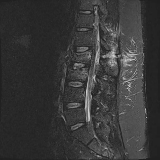
Lumbar fractures with posterior ligamentous complex injury (EDNeurorad Case 40)
131 views8 months agoSagittal STIR images show the marrow edema associated with the fractures with saw on CT. There is clearly edema within the interspinous space at L1-L2 (@Key Finding 1) consistent with injury to the interspinous ligament. On the left, there is focal disruption of the ligamentum flavum at the L1-L2 level consistent with traumatic injury/rupture (@Key Finding 2). On the right, the curvilinear STIR hyperintensity is likely within the bone and corresponds to the fracture line we saw on the prior CT (@Key Finding 3).One final finding in this case is the prominence of the posterior epidural fat as seen on sagittal T1 images (red arrows @Key Finding 4) which when correlated with the STIR image, demonstrates superimposed edema. The result is severe crowding in the thecal sack as can be seen on the axial images (orange arrow @Key Finding 5)
Lumbar spineMRI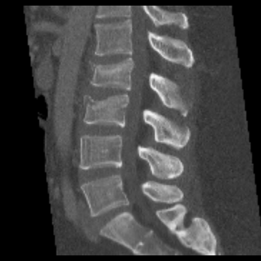
Complex lumbar spine fractures, suspected posterior ligamentous complex injury (EDNeurorad Case 39)
145 views8 months agoClearly there are compression fractures of the L2 and L3 superior endplates (@Key Finding 1). The sclerotic lines in the upper portion of the vertebral bodies are due to compressed bone. The upper part of the posterior cortices is also buckled to the spinal canal. If you want to use the Thoraco-Lumbar Injury Classification and Severity (TLICS) terminology, you may call the morphology of this fracture as "burst" but there are more findings to consider. In particular, the interspinous distance at L1-L2 appears increased compared to the remaining interspinous distances, a finding that should raise suspicious (but is not definitive for) injury to the posterior ligamentous complex.@Key Finding 2 (Sagittal image through patient's right) shows a horizontally oriented fracture through the posterior elements at L2 on the right side. @Key Finding 3 on the other hand shows fractures of the left superior articular process of L2 with mild widening of the left L1-L2 facet joint compared to the right. Based on these additional fracture, one can make the case that the fracture is best described as a distraction pattern or "Chance" fracture. Regardless, these additional fractures make an injury to the posterior ligamentous complex much more likely and so an MRI is warranted to assess the ligaments.The L2 left transverse process is also fractured but it's not of much concern!
Lumbar spineCT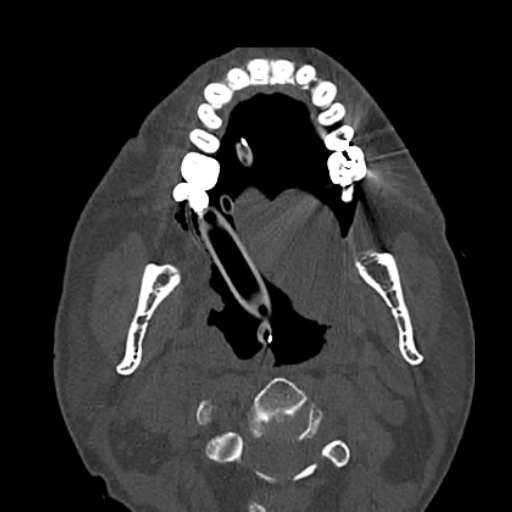
Maxillary alveolus fracture (EDNeurorad Case 38)
110 views8 months agoThere is a minimally displaced fracture of the left maxillary alveolus involving the socket to the left medial incisor tooth (@Key Finding 1 and @Key Finding 2). These fractures are easily missed on imaging. Clinically they are more apparent because the involved tooth is often loose and may be bleeding.
HeadCT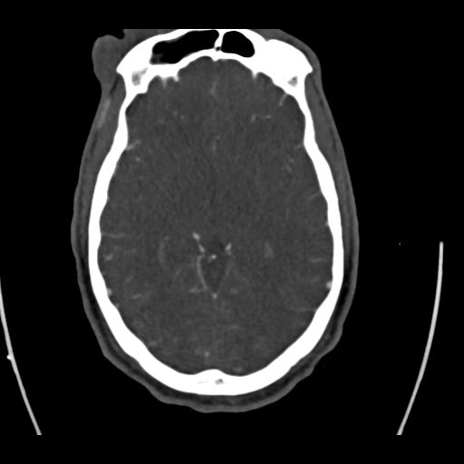
Traumatic superior sagittal sinus thrombosis (EDNeurorad Case 37)
125 views8 months agoCTV images show that the posterior half of the superior sagittal sinus is occluded consistent with traumatic thrombosis. @Key Finding 1 shows that the occlusion extends further inferior to the epicenter of the gunshot fracture. @Key Finding 2 shows the approximate upper and lower margins of the thrombosis (red arrows)
HeadCTA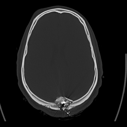
Communited gunshot fracture, subdural hematomas, concern for superior sagittal sinus injury (EDNeurorad Case 36)
100 views8 months agoThere is a comminuted and depressed gunshot fracture centered in the inferior part of the sagittal suture with fracture lines in both parietal bones as well as in the upper part of the occipital bone. The fracture lines also involve the upper part of the sigmoid sutures (red arrows in @Key Finding 1).The immediate vicinity of the gunshot fracture is suboptimally assessed due to the streak artifact. But scrolling above the gunshot fragments, and using subdural windows, there are bilateral extra-axial hemorrhages (red arrows in @Key Finding 2 and @Key Finding 3) likely subdural in location.One important point is that the location of the gunshot injury and depressed fracture fragments at the sagittal suture should raise concern for injury to the superior sagittal sinus. Therefore, a CTV is likely prudent as a next imaging step.
HeadCT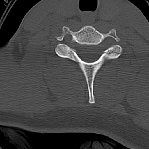
Bilateral facet fractures (EDNeurorad Case 35)
107 views8 months agoThere is a comminuted fracture of the left C7 superior articular process with intra-articular extension to the C6-C7 facet joint (@Key Finding 1 and @Key Finding 2).An additional minimally displaced fracture is also present on the right involving the tip of the superior articular process as best shown on sagittal images (@Key Finding 3 and @Key Finding 4).Finally, a very subtle non-displaced fracture of the C6 posterior spinous process (@Key Finding 5 and @Key Finding 6)
Cervical spineCT