Loading...
Lung Cancer PET/CT (case 983824-000043 from PETWB-REP)
8 views10 days agoWhole-body 18F-FDG PET/CT scan in a patient with Lung Cancer taken from the PETWB-REP dataset. The following English report (translated from original Chinese) is taken verbatim from the public dataset and has not been modified or otherwise checked for accuracy (see the end for citation). Impression Postoperative changes following a left brain lesion; a slightly high-density nodule in the left temporoparietal-occipital lobe with increased FDG metabolism, suggestive of a metastatic tumor. a. Lung cancer in the apical-posterior segment of the left upper lobe; lung cancer or metastatic lymph nodes in the left hilar region. b. Chronic inflammation and sequelae in both lungs. Paraseptal emphysema in both lungs. Mild osteophyte formation in some vertebral bodies. L4/5 intervertebral disc bulge. This case is from PETWB-REP, a curated dataset of whole-body 18F-FDG PET/CT scans and corresponding radiology reports from 490 patients with a broad spectrum of malignancies. The data were retrospectively collected from patients who underwent clinically indicated whole-body 18F-FDG PET/CT scans at the Shanghai Universal Medical Imaging Diagnostic Center between 2021 and 2024. License: Creative Commons Attribution 4.0 International (CC BY 4.0) Citation: Xue, L., Feng, G., Wenbo, Z., Zhang, Y., Li, L., Wang, S., Peng, L., Peng, S., & Gao, X. (2026). PETWB-REP: A Multi-Cancer Whole-Body FDG PET/CT Dataset with Corresponding Radiology Reports [Data set]. Zenodo. https://doi.org/10.5281/zenodo.18670487
Whole BodyPET/CT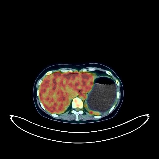
Colon Cancer PET/CT (case 983824-000010 from PETWB-REP)
7 views10 days agoWhole-body 18F-FDG PET/CT scan in a patient with Colon Cancer taken from the PETWB-REP dataset. The following English report (translated from original Chinese) is taken verbatim from the public dataset and has not been modified or otherwise checked for accuracy (see the end for citation). Impression a. A cystic-solid mass in the pelvic cavity with increased FDG metabolism at the periphery, suggesting a high probability of neoplastic lesions (metastasis is more likely than primary malignant ovarian tumors). Inflammatory lesions (tuberculosis) need to be ruled out. Please combine laboratory tests and clinicopathological examination. b. Pelvic effusion, extensive thickening and blurring of the perihepatic and splenic capsule and peritoneum in the abdominopelvic cavity, with multiple small lymph nodes visible in the mesentery, and increased FDG metabolism, suggesting extensive involvement of the pelvic and abdominal cavities. c. No obvious abnormal FDG metabolism was observed in the pancreas and biliary system (further CA19-9 and enhanced MRI follow-up are recommended). Scattered ground-glass nodules in the lateral basal segment of the left lower lobe and the apical segment of the right upper lobe, with no increased FDG metabolism, suggestive of atypical adenomatous hyperplasia or chronic inflammatory nodules. A small chronic inflammatory nodule in the posterior segment of the right upper lobe, please follow up with CT. Small amount of pleural effusion in the left side. Bilateral solid breast nodules, with no increased FDG metabolism, suggestive of fibroadenomas, please combine with ultrasound follow-up. Small hepatic cysts. Accessory spleen. Increased FDG metabolism in the rectum and anal canal, likely due to inflammatory uptake; please confirm clinical findings and repeat colonoscopy to rule out the possibility of a low-lying tumor. Partial vertebral osteophyte formation. Nuchal ligament calcification. No obvious abnormalities found on cranial scintigraphy. This case is from PETWB-REP, a curated dataset of whole-body 18F-FDG PET/CT scans and corresponding radiology reports from 490 patients with a broad spectrum of malignancies. The data were retrospectively collected from patients who underwent clinically indicated whole-body 18F-FDG PET/CT scans at the Shanghai Universal Medical Imaging Diagnostic Center between 2021 and 2024. License: Creative Commons Attribution 4.0 International (CC BY 4.0) Citation: Xue, L., Feng, G., Wenbo, Z., Zhang, Y., Li, L., Wang, S., Peng, L., Peng, S., & Gao, X. (2026). PETWB-REP: A Multi-Cancer Whole-Body FDG PET/CT Dataset with Corresponding Radiology Reports [Data set]. Zenodo. https://doi.org/10.5281/zenodo.18670487
Whole BodyPET/CT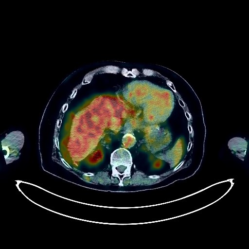
Lung Cancer PET/CT (case 983824-000093 from PETWB-REP)
3 views10 days agoWhole-body 18F-FDG PET/CT scan in a patient with Lung Cancer taken from the PETWB-REP dataset. The following English report (translated from original Chinese) is taken verbatim from the public dataset and has not been modified or otherwise checked for accuracy (see the end for citation). Impression a. Mass in the posterior segment of the left lower lobe, with increased FDG metabolism, suggestive of lung cancer. b. Mixed ground-glass nodules in the anterior segment of the right upper lobe, with mild FDG uptake, highly suggestive of lung cancer; inflammation to be ruled out. Short-term HRCT follow-up is recommended for comparison. c. Several small, solid, chronic inflammatory nodules in both lungs. Scattered chronic inflammation and old lesions in both lungs. Emphysema in both lungs. d. Calcification of some arterial walls (including coronary arteries). Degenerative changes in the spine. L4/5 and L5/S1 intervertebral disc bulges. Bilateral deep cerebral ischemia, softening lesions in the right basal ganglia, age-related brain. Chronic inflammation of the right maxillary sinus. This case is from PETWB-REP, a curated dataset of whole-body 18F-FDG PET/CT scans and corresponding radiology reports from 490 patients with a broad spectrum of malignancies. The data were retrospectively collected from patients who underwent clinically indicated whole-body 18F-FDG PET/CT scans at the Shanghai Universal Medical Imaging Diagnostic Center between 2021 and 2024. License: Creative Commons Attribution 4.0 International (CC BY 4.0) Citation: Xue, L., Feng, G., Wenbo, Z., Zhang, Y., Li, L., Wang, S., Peng, L., Peng, S., & Gao, X. (2026). PETWB-REP: A Multi-Cancer Whole-Body FDG PET/CT Dataset with Corresponding Radiology Reports [Data set]. Zenodo. https://doi.org/10.5281/zenodo.18670487
Whole BodyPET/CT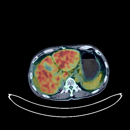
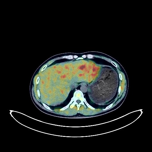
Liver Cancer PET/CT (case 983824-000096 from PETWB-REP)
4 views10 days agoWhole-body 18F-FDG PET/CT scan in a patient with Liver Cancer taken from the PETWB-REP dataset. The following English report (translated from original Chinese) is taken verbatim from the public dataset and has not been modified or otherwise checked for accuracy (see the end for citation). Impression a. Multiple lesions in the right lobe and left inner lobe of the liver, some fused, with FDG background metabolism, combined with the enhanced MRI report from another hospital, strongly suggest primary liver cancer. b. Multiple lymph nodes in the hepatogastric space, porta hepatis, and retroperitoneum, with no increased FDG metabolism, suggest possible reactive lymph node hyperplasia, with partial metastasis not ruled out. Peripheral thickening around the liver, spleen, and bilateral paracolic gutter, with slightly increased FDG metabolism, suggests possible peritoneal seeding metastasis. Please follow up clinically. c. Liver cirrhosis, portal hypertension with collateral circulation, and abdominopelvic effusion. Chronic inflammatory changes in the lower esophagus, stomach, and part of the intestine; endoscopic re-examination is necessary if needed. Chronic inflammatory micronodules in the apical segment of the right upper lobe of the lung. Partial arteriosclerosis. Degenerative changes in the spine, slight posterior slippage of the L4 vertebral body, and L4/5 disc herniation. No obvious abnormalities were found on cranial scintigraphy. Postoperative changes in the left nasal cavity, and minor chronic inflammation in the right ethmoid and maxillary sinuses. This case is from PETWB-REP, a curated dataset of whole-body 18F-FDG PET/CT scans and corresponding radiology reports from 490 patients with a broad spectrum of malignancies. The data were retrospectively collected from patients who underwent clinically indicated whole-body 18F-FDG PET/CT scans at the Shanghai Universal Medical Imaging Diagnostic Center between 2021 and 2024. License: Creative Commons Attribution 4.0 International (CC BY 4.0) Citation: Xue, L., Feng, G., Wenbo, Z., Zhang, Y., Li, L., Wang, S., Peng, L., Peng, S., & Gao, X. (2026). PETWB-REP: A Multi-Cancer Whole-Body FDG PET/CT Dataset with Corresponding Radiology Reports [Data set]. Zenodo. https://doi.org/10.5281/zenodo.18670487
Whole BodyPET/CT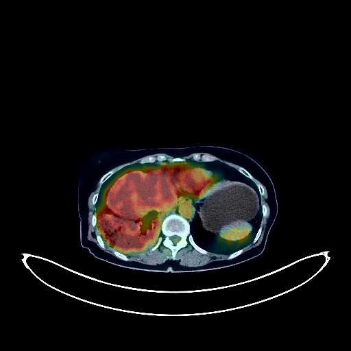
Lung Cancer PET/CT (case 983824-000030 from PETWB-REP)
7 views10 days agoWhole-body 18F-FDG PET/CT scan in a patient with Lung Cancer taken from the PETWB-REP dataset. The following English report (translated from original Chinese) is taken verbatim from the public dataset and has not been modified or otherwise checked for accuracy (see the end for citation). Impression a. Space-occupying lesion in the lower lobe of the right lung, with increased FDG metabolism, suggestive of lung cancer. b. Multiple lymph node metastases in the right hilum and mediastinum. Small amount of pleural effusion on the right side. c. Multiple intracranial metastases. Multiple bone metastases throughout the body. d. Multiple metastases in both lungs. A few fibrotic lesions in the left lung. Some arterial wall calcification. Nodular goiter in the left lobe of the thyroid gland is highly probable; ultrasound re-examination is recommended to rule out malignancy. Inflammation of the left ethmoid sinus. Spinal osteophyte formation, L4/5 intervertebral disc bulge. This case is from PETWB-REP, a curated dataset of whole-body 18F-FDG PET/CT scans and corresponding radiology reports from 490 patients with a broad spectrum of malignancies. The data were retrospectively collected from patients who underwent clinically indicated whole-body 18F-FDG PET/CT scans at the Shanghai Universal Medical Imaging Diagnostic Center between 2021 and 2024. License: Creative Commons Attribution 4.0 International (CC BY 4.0) Citation: Xue, L., Feng, G., Wenbo, Z., Zhang, Y., Li, L., Wang, S., Peng, L., Peng, S., & Gao, X. (2026). PETWB-REP: A Multi-Cancer Whole-Body FDG PET/CT Dataset with Corresponding Radiology Reports [Data set]. Zenodo. https://doi.org/10.5281/zenodo.18670487
Whole BodyPET/CT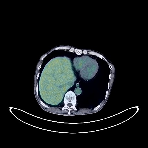
Lung Cancer PET/CT (case 983824-000034 from PETWB-REP)
8 views10 days agoWhole-body 18F-FDG PET/CT scan in a patient with Lung Cancer taken from the PETWB-REP dataset. The following English report (translated from original Chinese) is taken verbatim from the public dataset and has not been modified or otherwise checked for accuracy (see the end for citation). Impression Right lung cancer after chemotherapy and brain metastasis after radiotherapy: a. Right lower lobe posterior segment lesion with increased FDG metabolism in the solid portion, suggesting tumor activity; multiple lymph node metastases in the right hilum, mediastinum, and right supraclavicular fossa; high probability of right pleural metastasis. b. After brain metastasis radiotherapy, patchy low-density shadow in the right cerebellum with lost FDG uptake; patchy slightly low-density lesions in the right frontal lobe, with relatively uniform density in the temporal lobe, and no abnormalities in FDG metabolism. Considering the changes after treatment, the tumor activity is likely to be basically suppressed; enhanced MRI follow-up is recommended. c. Several small chronic inflammatory nodules (solid) in both lungs. Scattered chronic inflammation and old lesions in both lungs. Emphysema with small bullae in both lungs. Calcification of some arterial walls (including coronary arteries). Localized thickening of the left pleura. Diverticulum in the gastric fundus. Inflammatory or physiological uptake in some intestinal segments. Hemorrhoids. Calcification in the right lobe of the liver. Benign prostatic hyperplasia, with uneven FDG metabolism in the gland; PSA and ultrasound follow-up are recommended. Left scrotal calcification. Degenerative changes in the spine. Multiple Schmorl's nodes in the thoracic and lumbar vertebrae. L4/5 and L5/S1 intervertebral disc bulges. Benign osteopathy of the left iliac bone. This case is from PETWB-REP, a curated dataset of whole-body 18F-FDG PET/CT scans and corresponding radiology reports from 490 patients with a broad spectrum of malignancies. The data were retrospectively collected from patients who underwent clinically indicated whole-body 18F-FDG PET/CT scans at the Shanghai Universal Medical Imaging Diagnostic Center between 2021 and 2024. License: Creative Commons Attribution 4.0 International (CC BY 4.0) Citation: Xue, L., Feng, G., Wenbo, Z., Zhang, Y., Li, L., Wang, S., Peng, L., Peng, S., & Gao, X. (2026). PETWB-REP: A Multi-Cancer Whole-Body FDG PET/CT Dataset with Corresponding Radiology Reports [Data set]. Zenodo. https://doi.org/10.5281/zenodo.18670487
Whole BodyPET/CT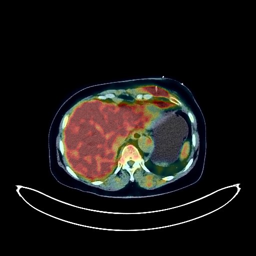
Lung Cancer PET/CT (case 983824-000063 from PETWB-REP)
3 views10 days agoWhole-body 18F-FDG PET/CT scan in a patient with Lung Cancer taken from the PETWB-REP dataset. The following English report (translated from original Chinese) is taken verbatim from the public dataset and has not been modified or otherwise checked for accuracy (see the end for citation). Impression a. A mass in the right upper lobe of the lung with increased FDG uptake, suggestive of right upper lobe lung cancer with obstructive changes; multiple lymph node metastases in the right hilum, mediastinum, and left supraclavicular fossa; multiple bone metastases; pericardial metastases with pericardial effusion; subcutaneous metastases in the left shoulder and back. b. Changes after pericardiocentesis. Inflammation of the left chest wall soft tissue is highly probable. c. A few fibrotic lesions in both lungs. Right pleural effusion, slight thickening of the left pleura. Senile cerebral atrophy. Inflammation of the left maxillary sinus, likely due to fungal infection. Chronic cholecystitis, gallstones. Left adrenal hyperplasia. Spinal degeneration, L3/4, L4/5, L5/S1 intervertebral disc bulge, L5/S1 intervertebral disc pneumodegenerative changes. This case is from PETWB-REP, a curated dataset of whole-body 18F-FDG PET/CT scans and corresponding radiology reports from 490 patients with a broad spectrum of malignancies. The data were retrospectively collected from patients who underwent clinically indicated whole-body 18F-FDG PET/CT scans at the Shanghai Universal Medical Imaging Diagnostic Center between 2021 and 2024. License: Creative Commons Attribution 4.0 International (CC BY 4.0) Citation: Xue, L., Feng, G., Wenbo, Z., Zhang, Y., Li, L., Wang, S., Peng, L., Peng, S., & Gao, X. (2026). PETWB-REP: A Multi-Cancer Whole-Body FDG PET/CT Dataset with Corresponding Radiology Reports [Data set]. Zenodo. https://doi.org/10.5281/zenodo.18670487
Whole BodyPET/CT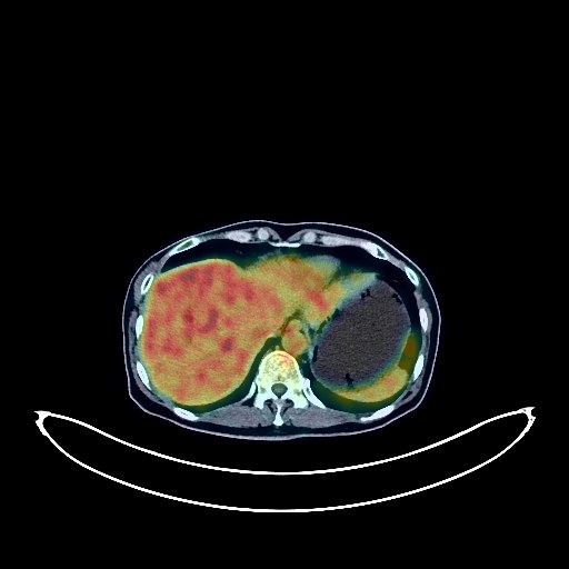
Breast Cancer PET/CT (case 983824-000153 from PETWB-REP)
2 views10 days agoWhole-body 18F-FDG PET/CT scan in a patient with Breast Cancer taken from the PETWB-REP dataset. The following English report (translated from original Chinese) is taken verbatim from the public dataset and has not been modified or otherwise checked for accuracy (see the end for citation). Impression a. Left breast mass with increased FDG metabolism, consistent with breast cancer. b. Multiple lymph node metastases in the left axilla. Possible multiple lymph node metastases in the right supraclavicular and pretracheal spaces. c. Post-operative changes following right breast cancer resection. a. Pancreatic duct dilation, uneven density in the head, neck, and body of the pancreas, with increased FDG metabolism; cystic shadow in the pancreatic neck, with absent FDG metabolism; IPMN is highly likely, but malignancy cannot be ruled out. Please combine clinical findings with contrast-enhanced MRI. Right iliac lymph node metastasis is highly probable. b. Intrahepatic and extrahepatic bile duct dilation, gallbladder enlargement. Multiple chronic inflammatory nodules in both lungs. Scattered chronic inflammation and remnants (including plaque nodules and calcifications) in both lungs, more prominent in the apical segment of the right upper lobe. Interstitial changes in the lower lobes of both lungs. Slight thickening of the pleura in some areas. Reactive hyperplasia of small mediastinal lymph nodes. Chronic inflammatory changes in the gastric antrum; endoscopic re-examination is necessary if required. Degenerative changes in the spine. L5 and S1 endplate inflammation. L4/5 and L5/S1 intervertebral disc herniation. No obvious abnormalities were found on cranial scintigraphy. This case is from PETWB-REP, a curated dataset of whole-body 18F-FDG PET/CT scans and corresponding radiology reports from 490 patients with a broad spectrum of malignancies. The data were retrospectively collected from patients who underwent clinically indicated whole-body 18F-FDG PET/CT scans at the Shanghai Universal Medical Imaging Diagnostic Center between 2021 and 2024. License: Creative Commons Attribution 4.0 International (CC BY 4.0) Citation: Xue, L., Feng, G., Wenbo, Z., Zhang, Y., Li, L., Wang, S., Peng, L., Peng, S., & Gao, X. (2026). PETWB-REP: A Multi-Cancer Whole-Body FDG PET/CT Dataset with Corresponding Radiology Reports [Data set]. Zenodo. https://doi.org/10.5281/zenodo.18670487
Whole BodyPET/CT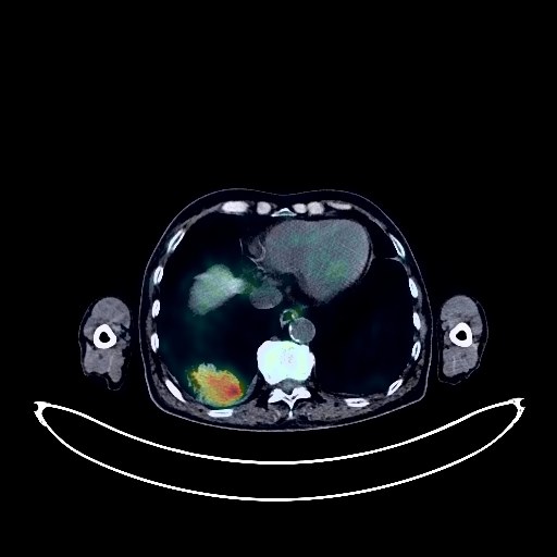
Lung Cancer PET/CT (case 983824-000152 from PETWB-REP)
3 views10 days agoWhole-body 18F-FDG PET/CT scan in a patient with Lung Cancer taken from the PETWB-REP dataset. The following English report (translated from original Chinese) is taken verbatim from the public dataset and has not been modified or otherwise checked for accuracy (see the end for citation). Impression a. Space-occupying lesion in the posterior basal segment of the right lower lobe, with increased FDG metabolism, consistent with lung cancer. b. Right hilar lymph node metastasis. Reactive hyperplasia of mediastinal lymph nodes. Small amount of pleural effusion bilaterally. c. Scattered chronic inflammation and old lesions in both lungs. Emphysema with bullae in both lungs. Calcification of some arterial walls (including coronary arteries). Softening lesions in the right cerebellum and bilateral basal ganglia, age-related brain changes. Chronic inflammation of bilateral maxillary sinuses. Left renal cyst. Right renal calculus. Small amount of hydrocephalus bilaterally. Degenerative changes in the spine. L4/5, L5/S1 intervertebral disc bulge. Multiple old compression fractures of the thoracic and lumbar vertebrae, most notably T7 and L5. Osteoporosis. Soft tissue nodule in the left parotid gland with increased FDG metabolism, suggestive of adenolymphoma, MRI follow-up recommended. This case is from PETWB-REP, a curated dataset of whole-body 18F-FDG PET/CT scans and corresponding radiology reports from 490 patients with a broad spectrum of malignancies. The data were retrospectively collected from patients who underwent clinically indicated whole-body 18F-FDG PET/CT scans at the Shanghai Universal Medical Imaging Diagnostic Center between 2021 and 2024. License: Creative Commons Attribution 4.0 International (CC BY 4.0) Citation: Xue, L., Feng, G., Wenbo, Z., Zhang, Y., Li, L., Wang, S., Peng, L., Peng, S., & Gao, X. (2026). PETWB-REP: A Multi-Cancer Whole-Body FDG PET/CT Dataset with Corresponding Radiology Reports [Data set]. Zenodo. https://doi.org/10.5281/zenodo.18670487
Whole BodyPET/CT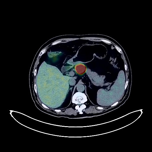
Liver Cancer PET/CT (case 983824-000151 from PETWB-REP)
2 views10 days agoWhole-body 18F-FDG PET/CT scan in a patient with Liver Cancer taken from the PETWB-REP dataset. The following English report (translated from original Chinese) is taken verbatim from the public dataset and has not been modified or otherwise checked for accuracy (see the end for citation). Impression a. Multiple intrahepatic lesions with elevated FDG metabolism, suggestive of malignancy, most likely hepatocellular carcinoma with intrahepatic metastasis; tumor thrombus formation near the right atrium in the left portal vein, hepatic vein, and inferior vena cava; peripancreatic lymph node metastasis; small amount of pelvic effusion. b. Cirrhosis, slightly enlarged spleen. Reactive hyperplasia of small retroperitoneal lymph nodes. A few post-inflammatory lesions in both lungs. Bilateral pleural thickening. Partial calcification of the aorta and coronary artery walls. Absence after cholecystectomy. Small cyst in the left kidney. Calcification lesion in the prostate. Small amount of hydrocele in both testes. Low-density nodules in both lobes of the thyroid gland, with enlargement of the left lobe and local FDG metabolism loss in the left lobe, suggestive of nodular goiter; ultrasound and thyroid function tests are recommended. Spinal degenerative changes. T12-L1 vertebral body wedge deformity with localized kyphosis, L2 vertebral body instability. T10/11 and T11/12 intervertebral disc pneumoconiosis. Right basal ganglia softening lesion, age-related brain changes. Bilateral maxillary and ethmoid sinusitis. This case is from PETWB-REP, a curated dataset of whole-body 18F-FDG PET/CT scans and corresponding radiology reports from 490 patients with a broad spectrum of malignancies. The data were retrospectively collected from patients who underwent clinically indicated whole-body 18F-FDG PET/CT scans at the Shanghai Universal Medical Imaging Diagnostic Center between 2021 and 2024. License: Creative Commons Attribution 4.0 International (CC BY 4.0) Citation: Xue, L., Feng, G., Wenbo, Z., Zhang, Y., Li, L., Wang, S., Peng, L., Peng, S., & Gao, X. (2026). PETWB-REP: A Multi-Cancer Whole-Body FDG PET/CT Dataset with Corresponding Radiology Reports [Data set]. Zenodo. https://doi.org/10.5281/zenodo.18670487
Whole BodyPET/CT