Loading...
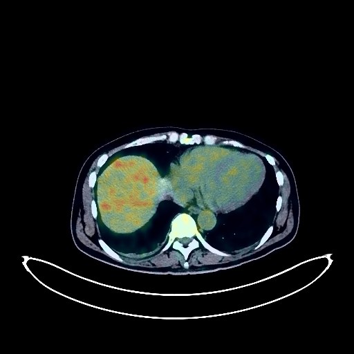
Liver Cancer PET/CT (case 983824-000058 from PETWB-REP)
2 views10 days agoWhole-body 18F-FDG PET/CT scan in a patient with Liver Cancer taken from the PETWB-REP dataset. The following English report (translated from original Chinese) is taken verbatim from the public dataset and has not been modified or otherwise checked for accuracy (see the end for citation). Impression a. Multiple masses and patchy shadows in the liver, with increased FDG metabolism, suggestive of malignancy, possibly hepatocellular carcinoma with intrahepatic metastasis, but metastatic tumors cannot be ruled out. Please combine tumor markers for comprehensive analysis. Lymph node metastasis in the pancreatic head region. Small amount of pelvic hemorrhage. b. Localized decreased density in the pancreatic head region, focal FDG uptake, MRI is recommended to rule out neoplastic lesions. Reactive hyperplasia of multiple retroperitoneal lymph nodes. c. Liver cirrhosis, small cyst in the left lobe of the liver. Absence of the right kidney, compensatory enlargement of the left kidney, suspicious space-occupying lesion, enhanced MRI is recommended; bilateral renal pelvis on the left, postoperative left hydronephrosis, slight thickening of the left perirenal fascia, please correlate with clinical findings. Diverticulum in the horizontal part of the duodenum. a. Two small ground-glass nodules in the upper lobe of the right lung, FDG metabolism normal, suggestive of atypical adenomatous hyperplasia or inflammatory nodules, annual HRCT is recommended. b. Linear lesions in both lungs. Slight thickening of the pleura on both sides, small amount of pleural effusion on the right side. Slightly decreased density in the right lobe of the thyroid gland, with increased FDG metabolism, suggesting possible thyroiditis; ultrasound and thyroid function tests are recommended. Spinal degenerative changes, with L4/5 and L5/S1 intervertebral disc bulges. No obvious abnormalities were found on cranial imaging. This case is from PETWB-REP, a curated dataset of whole-body 18F-FDG PET/CT scans and corresponding radiology reports from 490 patients with a broad spectrum of malignancies. The data were retrospectively collected from patients who underwent clinically indicated whole-body 18F-FDG PET/CT scans at the Shanghai Universal Medical Imaging Diagnostic Center between 2021 and 2024. License: Creative Commons Attribution 4.0 International (CC BY 4.0) Citation: Xue, L., Feng, G., Wenbo, Z., Zhang, Y., Li, L., Wang, S., Peng, L., Peng, S., & Gao, X. (2026). PETWB-REP: A Multi-Cancer Whole-Body FDG PET/CT Dataset with Corresponding Radiology Reports [Data set]. Zenodo. https://doi.org/10.5281/zenodo.18670487
Whole BodyPET/CT
Esophageal Cancer PET/CT (case 983824-000189 from PETWB-REP)
2 views10 days agoWhole-body 18F-FDG PET/CT scan in a patient with Esophageal Cancer taken from the PETWB-REP dataset. The following English report (translated from original Chinese) is taken verbatim from the public dataset and has not been modified or otherwise checked for accuracy (see the end for citation). Impression Post-esophageal tumor surgery, no obvious space-occupying lesions were found in the esophageal wall, and FDG metabolism was normal. A follow-up gastroscopy is recommended. Chronic inflammatory micronodules in both lungs. Emphysema in both lungs, a few post-inflammatory remnants in both lungs. Reactive hyperplasia of hilar and mediastinal lymph nodes in both lungs. Pleural thickening bilaterally. Calcification of some arterial walls (including coronary arteries). Chronic cholecystitis. Benign prostatic hyperplasia with calcification. Widening of the bilateral inguinal canals, inguinal hernia to be ruled out; please correlate with clinical findings. Degenerative changes in the spine, L4/5 and L5/S1 intervertebral disc bulge. Post-cervical spine surgery changes. Multiple old rib fractures bilaterally. Atrophy of the left gluteal and thigh muscles. Senile brain, deep lacunar infarcts. Chronic inflammation of the bilateral maxillary sinuses with submucosal cysts. The left lens is located in the posterior part of the eyeball; a specialist examination is recommended. This case is from PETWB-REP, a curated dataset of whole-body 18F-FDG PET/CT scans and corresponding radiology reports from 490 patients with a broad spectrum of malignancies. The data were retrospectively collected from patients who underwent clinically indicated whole-body 18F-FDG PET/CT scans at the Shanghai Universal Medical Imaging Diagnostic Center between 2021 and 2024. License: Creative Commons Attribution 4.0 International (CC BY 4.0) Citation: Xue, L., Feng, G., Wenbo, Z., Zhang, Y., Li, L., Wang, S., Peng, L., Peng, S., & Gao, X. (2026). PETWB-REP: A Multi-Cancer Whole-Body FDG PET/CT Dataset with Corresponding Radiology Reports [Data set]. Zenodo. https://doi.org/10.5281/zenodo.18670487
Whole BodyPET/CT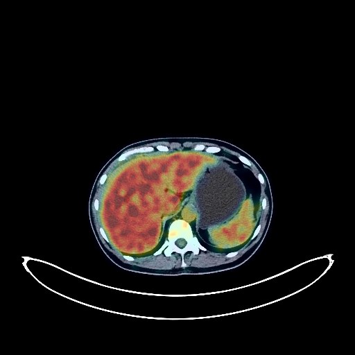
Breast Cancer PET/CT (case 983824-000033 from PETWB-REP)
6 views10 days agoWhole-body 18F-FDG PET/CT scan in a patient with Breast Cancer taken from the PETWB-REP dataset. The following English report (translated from original Chinese) is taken verbatim from the public dataset and has not been modified or otherwise checked for accuracy (see the end for citation). Impression Right breast mass with increased FDG metabolism, consistent with breast cancer based on pathology. Reactive hyperplasia of small lymph nodes in both axillae. a. Bilateral adnexal masses with increased FDG metabolism, suggestive of malignancy, possible metastasis, ovarian cancer to be ruled out; comprehensive analysis with contrast-enhanced MRI is recommended. b. Peritoneal metastasis in the rectouterine pouch. Uterine fibroids. Abdominal and pelvic effusion. c. Bilateral paracolic gutter opacities, no significant abnormalities in FDG uptake; follow-up recommended. d. Heterogeneous bone density in the spine and pelvis, no significant abnormalities in FDG metabolism; follow-up recommended. Ground-glass nodule in the anterior segment of the right upper lobe, no abnormalities in FDG metabolism; suggestive of inflammatory nodule or atypical adenomatous hyperplasia; annual HRCT follow-up recommended. Scattered chronic inflammation in both lower lobes of the lungs. Chronic inflammatory nodule (solid) in the right middle lobe. Anemia. Cervical, thoracic, and lumbar vertebrae osteophyte formation. L4/5 and L5/S1 intervertebral disc bulges. No abnormalities were found on cranial imaging. This case is from PETWB-REP, a curated dataset of whole-body 18F-FDG PET/CT scans and corresponding radiology reports from 490 patients with a broad spectrum of malignancies. The data were retrospectively collected from patients who underwent clinically indicated whole-body 18F-FDG PET/CT scans at the Shanghai Universal Medical Imaging Diagnostic Center between 2021 and 2024. License: Creative Commons Attribution 4.0 International (CC BY 4.0) Citation: Xue, L., Feng, G., Wenbo, Z., Zhang, Y., Li, L., Wang, S., Peng, L., Peng, S., & Gao, X. (2026). PETWB-REP: A Multi-Cancer Whole-Body FDG PET/CT Dataset with Corresponding Radiology Reports [Data set]. Zenodo. https://doi.org/10.5281/zenodo.18670487
Whole BodyPET/CT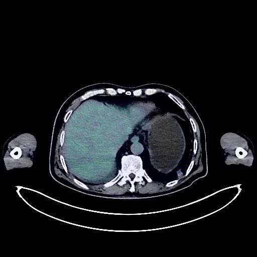
Lung Cancer PET/CT (case 983824-000117 from PETWB-REP)
2 views10 days agoWhole-body 18F-FDG PET/CT scan in a patient with Lung Cancer taken from the PETWB-REP dataset. The following English report (translated from original Chinese) is taken verbatim from the public dataset and has not been modified or otherwise checked for accuracy (see the end for citation). Impression a. Space-occupying lesion in the right upper lobe of the lung, with increased FDG metabolism, suggestive of lung cancer with obstructive changes. b. Metastasis to the right hilar, mediastinal, and right supraclavicular fossa lymph nodes. Multiple small solid nodules in both lungs, suggesting possible metastasis; follow-up examination for comparison is recommended. c. Scattered inflammation in the remaining lungs. Small amount of pleural effusion in the right side. Calcification of some arterial walls (including coronary arteries). Multiple intracranial nodules with increased FDG metabolism, highly suggestive of metastatic tumors. Gallstones. Mild bilateral adrenal hyperplasia. Spinal degenerative changes. L4/5 and L5/S1 intervertebral disc bulges. Bilateral sacroiliac joint degeneration. Subcutaneous calcifications in both buttocks. This case is from PETWB-REP, a curated dataset of whole-body 18F-FDG PET/CT scans and corresponding radiology reports from 490 patients with a broad spectrum of malignancies. The data were retrospectively collected from patients who underwent clinically indicated whole-body 18F-FDG PET/CT scans at the Shanghai Universal Medical Imaging Diagnostic Center between 2021 and 2024. License: Creative Commons Attribution 4.0 International (CC BY 4.0) Citation: Xue, L., Feng, G., Wenbo, Z., Zhang, Y., Li, L., Wang, S., Peng, L., Peng, S., & Gao, X. (2026). PETWB-REP: A Multi-Cancer Whole-Body FDG PET/CT Dataset with Corresponding Radiology Reports [Data set]. Zenodo. https://doi.org/10.5281/zenodo.18670487
Whole BodyPET/CT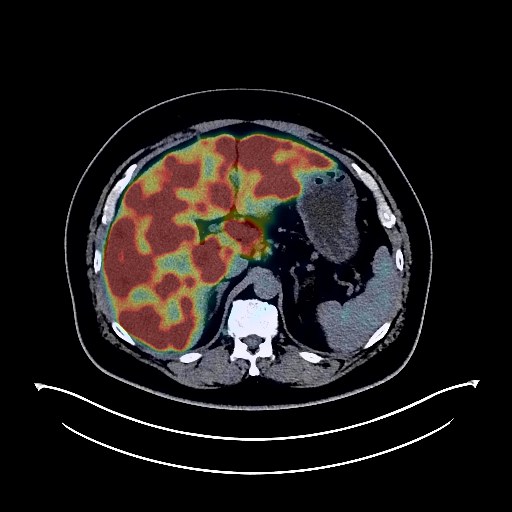
Lung Cancer PET/CT (case 983824-000122 from PETWB-REP)
2 views10 days agoWhole-body 18F-FDG PET/CT scan in a patient with Lung Cancer taken from the PETWB-REP dataset. The following English report (translated from original Chinese) is taken verbatim from the public dataset and has not been modified or otherwise checked for accuracy (see the end for citation). Impression a. Space-occupying lesion in the right upper lobe with increased FDG metabolism, suggestive of lung cancer with obstructive inflammation. b. Multiple lymph node metastases in the right hilum, mediastinum, right cardiophrenic angle, hepatogastric space, and retroperitoneum. c. Multiple liver metastases. Multiple bone metastases throughout the body (see description for details). Infection in the right upper lobe. Chronic inflammatory micronodules in both lungs. Calcifications in the lower lobes of both lungs, scattered post-inflammatory remnants in both lungs. Right pleural thickening. Calcification of some arterial walls (including coronary arteries). Fatty liver. Chronic cholecystitis, gallstones. Residual contrast agent in the cystostomy. Calcifications in the prostate. Chronic inflammatory changes in the gastric antrum. Degenerative changes in the spine, L5/S1 intervertebral disc bulge with pneumoconiosis and degeneration. Mild age-related brain changes, calcifications in the falx cerebri, and deep lacunar infarcts. This case is from PETWB-REP, a curated dataset of whole-body 18F-FDG PET/CT scans and corresponding radiology reports from 490 patients with a broad spectrum of malignancies. The data were retrospectively collected from patients who underwent clinically indicated whole-body 18F-FDG PET/CT scans at the Shanghai Universal Medical Imaging Diagnostic Center between 2021 and 2024. License: Creative Commons Attribution 4.0 International (CC BY 4.0) Citation: Xue, L., Feng, G., Wenbo, Z., Zhang, Y., Li, L., Wang, S., Peng, L., Peng, S., & Gao, X. (2026). PETWB-REP: A Multi-Cancer Whole-Body FDG PET/CT Dataset with Corresponding Radiology Reports [Data set]. Zenodo. https://doi.org/10.5281/zenodo.18670487
Whole BodyPET/CT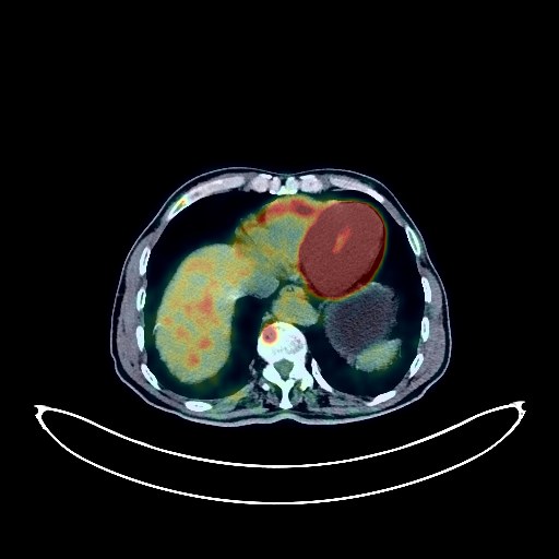
Lung Cancer PET/CT (case 983824-000015 from PETWB-REP)
7 views10 days agoWhole-body 18F-FDG PET/CT scan in a patient with Lung Cancer taken from the PETWB-REP dataset. The following English report (translated from original Chinese) is taken verbatim from the public dataset and has not been modified or otherwise checked for accuracy (see the end for citation). Impression a. A mass in the posterior segment of the right upper lobe, with elevated FDG metabolism, suggestive of peripheral lung cancer. Multiple bone metastases throughout the body. Possible metastases to the right hilar and mediastinal lymph nodes. b. Several small chronic inflammatory nodules in both lungs are highly probable; follow-up CT scan is recommended to rule out other possibilities. A few chronic inflammations and old lesions in both lungs. Some arterial wall calcifications. c. Bilateral adrenal hyperplasia; follow-up CT scan is recommended to rule out metastasis. An irregular mass in the lower pole of the left kidney, with heterogeneous elevated FDG metabolism, suggestive of malignancy, most likely renal cell carcinoma; please combine clinicopathological examination and contrast-enhanced MRI. Small liver cyst. Left kidney cyst. Right kidney calcification. Duodenal diverticulum. Chronic antral gastritis. Spinal degeneration. L4/5, L5/S1 intervertebral disc bulge. A few deep ischemic lesions in both lobes of the brain, suggestive of age-related brain disorders; follow-up MRI is recommended. If the thyroid gland has uneven density and increased FDG uptake, ultrasound and thyroid function tests are recommended. This case is from PETWB-REP, a curated dataset of whole-body 18F-FDG PET/CT scans and corresponding radiology reports from 490 patients with a broad spectrum of malignancies. The data were retrospectively collected from patients who underwent clinically indicated whole-body 18F-FDG PET/CT scans at the Shanghai Universal Medical Imaging Diagnostic Center between 2021 and 2024. License: Creative Commons Attribution 4.0 International (CC BY 4.0) Citation: Xue, L., Feng, G., Wenbo, Z., Zhang, Y., Li, L., Wang, S., Peng, L., Peng, S., & Gao, X. (2026). PETWB-REP: A Multi-Cancer Whole-Body FDG PET/CT Dataset with Corresponding Radiology Reports [Data set]. Zenodo. https://doi.org/10.5281/zenodo.18670487
Whole BodyPET/CT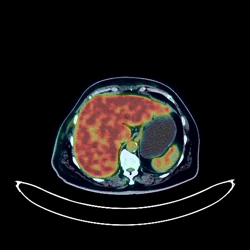
Breast Cancer PET/CT (case 983824-000029 from PETWB-REP)
8 views10 days agoWhole-body 18F-FDG PET/CT scan in a patient with Breast Cancer taken from the PETWB-REP dataset. The following English report (translated from original Chinese) is taken verbatim from the public dataset and has not been modified or otherwise checked for accuracy (see the end for citation). Impression a. Multiple lesions in the right breast with increased FDG metabolism, consistent with breast cancer. b. Multiple lymph node metastases in the right axilla and right internal mammary chain. Reactive hyperplasia of lymph nodes in the left axilla. Several small chronic inflammatory nodules (solid) in both lungs. Scattered chronic inflammation and old lesions in both lungs. Reactive hyperplasia of lymph nodes in the hilar and mediastinal regions of both lungs. Calcification of some arterial walls (including coronary arteries). Calcification in the left lobe of the liver. Diverticulum in the descending part of the duodenum. Continuous increased FDG metabolism in the colon and rectum, considered to be inflammatory or physiological uptake. Possible uterine fibroids; Nabothian cysts of the cervix. Degenerative changes in the spine. L4/5 and L5/S1 intervertebral disc bulges. Bilateral frozen shoulder. A few ischemic lesions in the deep bilateral brain regions, age-related brain changes. This case is from PETWB-REP, a curated dataset of whole-body 18F-FDG PET/CT scans and corresponding radiology reports from 490 patients with a broad spectrum of malignancies. The data were retrospectively collected from patients who underwent clinically indicated whole-body 18F-FDG PET/CT scans at the Shanghai Universal Medical Imaging Diagnostic Center between 2021 and 2024. License: Creative Commons Attribution 4.0 International (CC BY 4.0) Citation: Xue, L., Feng, G., Wenbo, Z., Zhang, Y., Li, L., Wang, S., Peng, L., Peng, S., & Gao, X. (2026). PETWB-REP: A Multi-Cancer Whole-Body FDG PET/CT Dataset with Corresponding Radiology Reports [Data set]. Zenodo. https://doi.org/10.5281/zenodo.18670487
Whole BodyPET/CT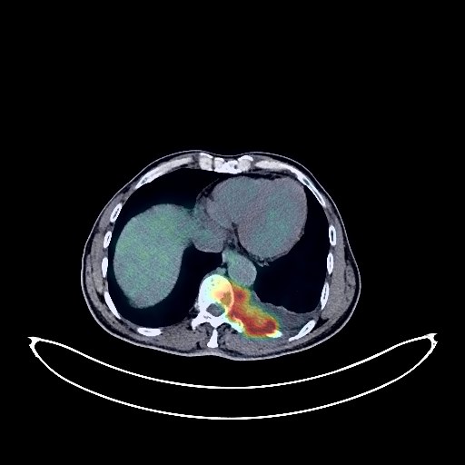
Lung Cancer PET/CT (case 983824-000002 from PETWB-REP)
10 views10 days agoWhole-body 18F-FDG PET/CT scan in a patient with Lung Cancer taken from the PETWB-REP dataset. The following English report (translated from original Chinese) is taken verbatim from the public dataset and has not been modified or otherwise checked for accuracy (see the end for citation). Impression a. A nodule at the left apex of the lung, highly suggestive of primary lung cancer; please correlate with clinicopathology. Enlarged hilar and mediastinal lymph nodes bilaterally with mildly elevated FDG metabolism suggest metastasis; follow-up examination after treatment is recommended. b. Left pleural effusion, multiple thickenings of the left pleura with elevated FDG metabolism suggest multiple pleural metastases. c. Destruction of the left portion of the T10 vertebral body and the left 10th and 11th posterior ribs with soft tissue mass, elevated FDG metabolism, suggest metastasis. Scattered emphysema and bullae in both lungs; cystic dilatation of some bronchi in the right middle and lower lobes. Mixed ground-glass nodules in the posterior segment of the right lower lobe and the anterior-medial basal segment of the left lower lobe, with no elevated FDG metabolism, suggestive of atypical adenomatous hyperplasia or chronic inflammatory nodules; multiple chronic inflammations and old lesions in both lungs (possible old pulmonary tuberculosis in the right upper lobe). Localized thickening of the pleura in both oblique fissures. Calcification of some arterial walls (including coronary arteries). Localized soft tissue nodules in the mid-sigmoid colon with increased FDG metabolism, suggestive of a polyp with potential malignancy; please confirm with colonoscopy pathology. Inflammatory changes in the lower sigmoid colon and rectum, reactive hyperplasia of lymph nodes at the root of the mesocolic colon. Small cyst in the left lobe of the liver. Prostatic calcification. Calcification of the tunica vaginalis on the left side of the testis, bilateral hydrocele. Spinal osteophyte formation. Lacunar ischemic foci in the bilateral basal ganglia. Age-related brain changes. Chronic inflammation of the nasopharynx. Minor inflammation of the right maxillary sinus. This case is from PETWB-REP, a curated dataset of whole-body 18F-FDG PET/CT scans and corresponding radiology reports from 490 patients with a broad spectrum of malignancies. The data were retrospectively collected from patients who underwent clinically indicated whole-body 18F-FDG PET/CT scans at the Shanghai Universal Medical Imaging Diagnostic Center between 2021 and 2024. License: Creative Commons Attribution 4.0 International (CC BY 4.0) Citation: Xue, L., Feng, G., Wenbo, Z., Zhang, Y., Li, L., Wang, S., Peng, L., Peng, S., & Gao, X. (2026). PETWB-REP: A Multi-Cancer Whole-Body FDG PET/CT Dataset with Corresponding Radiology Reports [Data set]. Zenodo. https://doi.org/10.5281/zenodo.18670487
Whole BodyPET/CT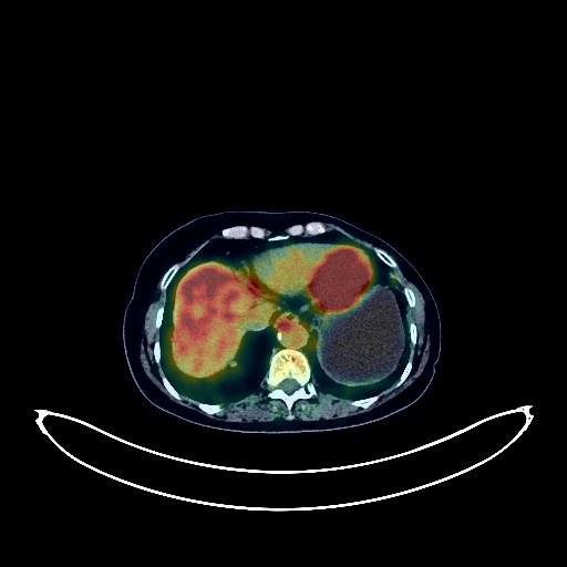
Pancreatic Cancer PET/CT (case 983824-000087 from PETWB-REP)
2 views10 days agoWhole-body 18F-FDG PET/CT scan in a patient with Pancreatic Cancer taken from the PETWB-REP dataset. The following English report (translated from original Chinese) is taken verbatim from the public dataset and has not been modified or otherwise checked for accuracy (see the end for citation). Impression A mass in the body and tail of the pancreas with increased FDG metabolism suggests a malignant tumor (possibly pancreatic cancer). Please correlate with clinicopathology. Multiple inflammatory foci in both lungs; multiple masses and nodules in both lungs with increased FDG metabolism suggest a high probability of metastatic tumors. Please monitor clinical progress. Reactive hyperplasia of the hilar and mediastinal lymph nodes is possible, but metastasis cannot be ruled out. Postoperative left breast cancer surgery; no signs of tumor recurrence were observed in the surgical area. Enlarged cardiac silhouette; calcification of some arterial walls (including coronary arteries). Further specialist clinical examination is recommended. Chronic cholecystitis; gallstones. Degenerative changes in the spine. L4/5 and L5/S1 intervertebral disc bulges. No abnormalities were found on cranial scintigraphy. Chronic inflammation of the left maxillary sinus. This case is from PETWB-REP, a curated dataset of whole-body 18F-FDG PET/CT scans and corresponding radiology reports from 490 patients with a broad spectrum of malignancies. The data were retrospectively collected from patients who underwent clinically indicated whole-body 18F-FDG PET/CT scans at the Shanghai Universal Medical Imaging Diagnostic Center between 2021 and 2024. License: Creative Commons Attribution 4.0 International (CC BY 4.0) Citation: Xue, L., Feng, G., Wenbo, Z., Zhang, Y., Li, L., Wang, S., Peng, L., Peng, S., & Gao, X. (2026). PETWB-REP: A Multi-Cancer Whole-Body FDG PET/CT Dataset with Corresponding Radiology Reports [Data set]. Zenodo. https://doi.org/10.5281/zenodo.18670487
Whole BodyPET/CT
Colon Cancer PET/CT (case 983824-000054 from PETWB-REP)
3 views10 days agoWhole-body 18F-FDG PET/CT scan in a patient with Colon Cancer taken from the PETWB-REP dataset. The following English report (translated from original Chinese) is taken verbatim from the public dataset and has not been modified or otherwise checked for accuracy (see the end for citation). Impression a. Thickening and narrowing of the descending colon wall with elevated FDG metabolism suggest colon cancer with surrounding infiltration; please confirm with colonoscopy and pathology. b. Bilateral pelvic lesions with elevated FDG metabolism suggest bilateral ovarian metastases, more likely than primary ovarian cancer; please consider clinical tumor markers and medical history. Metastasis to the peritoneum and rectouterine pouch. Significant abdominal and pelvic effusions. c. Elevated FDG metabolism in the right humeral head; metastasis to be ruled out; MRI is recommended. Possible multiple uterine fibroids; please follow up with ultrasound. Possible chronic inflammatory nodules in the left lower lobe and right upper lobe; please follow up with CT. Small amount of pleural effusion on the left, large amount on the right. Anemia. Partial vertebral osteophyte formation. L4/5 disc bulge. No obvious abnormalities seen on cranial scintigraphy. Bilateral ethmoid sinusitis. This case is from PETWB-REP, a curated dataset of whole-body 18F-FDG PET/CT scans and corresponding radiology reports from 490 patients with a broad spectrum of malignancies. The data were retrospectively collected from patients who underwent clinically indicated whole-body 18F-FDG PET/CT scans at the Shanghai Universal Medical Imaging Diagnostic Center between 2021 and 2024. License: Creative Commons Attribution 4.0 International (CC BY 4.0) Citation: Xue, L., Feng, G., Wenbo, Z., Zhang, Y., Li, L., Wang, S., Peng, L., Peng, S., & Gao, X. (2026). PETWB-REP: A Multi-Cancer Whole-Body FDG PET/CT Dataset with Corresponding Radiology Reports [Data set]. Zenodo. https://doi.org/10.5281/zenodo.18670487
Whole BodyPET/CT