Loading...
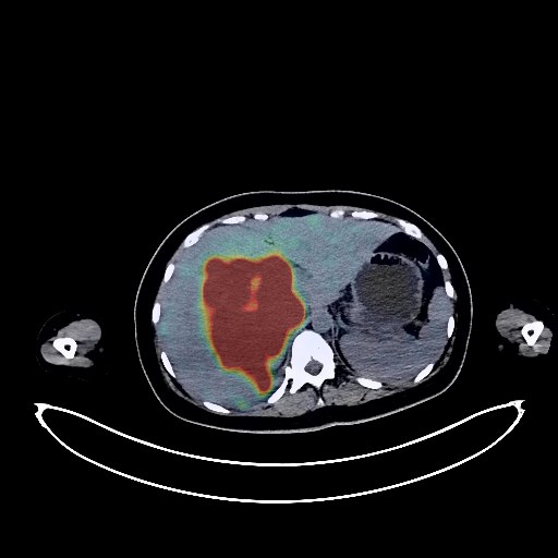
Renal Cancer PET/CT (case 983824-000143 from PETWB-REP)
2 views10 days agoWhole-body 18F-FDG PET/CT scan in a patient with Renal Cancer taken from the PETWB-REP dataset. The following English report (translated from original Chinese) is taken verbatim from the public dataset and has not been modified or otherwise checked for accuracy (see the end for citation). Impression a. Right renal malignancy followed by treatment, with a mass in the right kidney accompanied by increased FDG metabolism, suggesting high tumor activity. b. Peritoneal seeding metastasis, pelvic effusion. Multiple retroperitoneal lymph node metastases. c. Right lower lobe lung metastasis, with a high probability of right hilar lymph node metastasis. d. Multiple bone metastases throughout the body (see description for details). a. Several ground-glass nodules in both lungs, with normal FDG metabolism, suggesting possible inflammation; follow-up CT scan recommended. b. Chronic inflammatory micronodules in both lungs. A few post-inflammatory remnants in both lungs. Right pleural thickening. Anemia changes; right anterior chest wall port inserted. Bilateral breast hyperplasia. Small liver cysts. Accessory spleen. Chronic inflammatory changes or physiological uptake in the entire esophagus and part of the intestine; please follow up with endoscopy. No obvious abnormalities were found on cranial scintigraphy. This case is from PETWB-REP, a curated dataset of whole-body 18F-FDG PET/CT scans and corresponding radiology reports from 490 patients with a broad spectrum of malignancies. The data were retrospectively collected from patients who underwent clinically indicated whole-body 18F-FDG PET/CT scans at the Shanghai Universal Medical Imaging Diagnostic Center between 2021 and 2024. License: Creative Commons Attribution 4.0 International (CC BY 4.0) Citation: Xue, L., Feng, G., Wenbo, Z., Zhang, Y., Li, L., Wang, S., Peng, L., Peng, S., & Gao, X. (2026). PETWB-REP: A Multi-Cancer Whole-Body FDG PET/CT Dataset with Corresponding Radiology Reports [Data set]. Zenodo. https://doi.org/10.5281/zenodo.18670487
Whole BodyPET/CT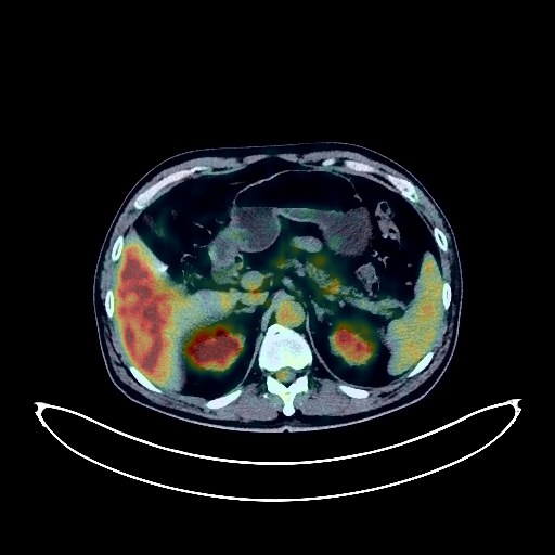
Lung Cancer PET/CT (case 983824-000049 from PETWB-REP)
7 views10 days agoWhole-body 18F-FDG PET/CT scan in a patient with Lung Cancer taken from the PETWB-REP dataset. The following English report (translated from original Chinese) is taken verbatim from the public dataset and has not been modified or otherwise checked for accuracy (see the end for citation). Impression a. Mass in the lower lobe of the left lung, with increased FDG metabolism, consistent with lung cancer. b. Multiple lymph nodes in the right hilum and mediastinum with some showing increased FDG metabolism, suggesting possible reactive lymph node hyperplasia; partial metastasis is pending. Please compare with previous scans for follow-up. c. Interstitial lung inflammation in both lungs. Bullae in the lower lobes of both lungs. Pleural thickening bilaterally. Multiple calcifications in the right hilum. Calcification of some arterial walls. Small kidney stone in the right kidney. Degenerative changes in the spine. No obvious abnormalities seen on cranial scintigraphy. Bilateral maxillary sinusitis. This case is from PETWB-REP, a curated dataset of whole-body 18F-FDG PET/CT scans and corresponding radiology reports from 490 patients with a broad spectrum of malignancies. The data were retrospectively collected from patients who underwent clinically indicated whole-body 18F-FDG PET/CT scans at the Shanghai Universal Medical Imaging Diagnostic Center between 2021 and 2024. License: Creative Commons Attribution 4.0 International (CC BY 4.0) Citation: Xue, L., Feng, G., Wenbo, Z., Zhang, Y., Li, L., Wang, S., Peng, L., Peng, S., & Gao, X. (2026). PETWB-REP: A Multi-Cancer Whole-Body FDG PET/CT Dataset with Corresponding Radiology Reports [Data set]. Zenodo. https://doi.org/10.5281/zenodo.18670487
Whole BodyPET/CT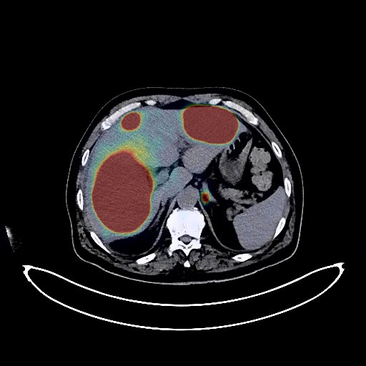
Lymphoma PET/CT (case 983824-000141 from PETWB-REP)
2 views10 days agoWhole-body 18F-FDG PET/CT scan in a patient with Lymphoma taken from the PETWB-REP dataset. The following English report (translated from original Chinese) is taken verbatim from the public dataset and has not been modified or otherwise checked for accuracy (see the end for citation). Impression a. Multiple lesions in the liver with increased FDG metabolism suggestive of malignancy, most likely lymphoma, with metastases to be ruled out; small amount of perihepatic effusion. b. Multiple enlarged lymph nodes in the hepatic hilum, intercaval space, hepatogastric space, perigastric region, right cardiophrenic angle, mesentery, retroperitoneum, and right pelvic wall with increased FDG uptake suggestive of lymphoma infiltration, with metastases to be ruled out. A biopsy is recommended for the above to clarify the pathology. Slight thickening of the lower thoracic esophageal wall with increased FDG metabolism suggests possible inflammation or lymphoma infiltration; endoscopy is recommended. Increased FDG metabolism in the bilateral peripheral zone of the prostate suggests possible inflammation; tumor to be ruled out; PSA analysis is recommended. a. Multiple chronic inflammatory micronodules in both lungs, calcification in the left lung, and a few post-inflammatory remnants in both lungs. b. Small diverticulum beside the right trachea. Bilateral pleural thickening. Slight pericardial thickening. Mild bilateral gynecomastia. Small cyst in the left medial lobe of the liver. Gallstones and chronic cholecystitis. Accessory spleen. Right renal cyst. Small amount of hydrocele in both testes. Diverticulum in the horizontal part of the duodenum. Focal increase in FDG uptake in the anal area, suggestive of hemorrhoids or physiological uptake; please correlate with clinical findings. Uneven thyroid density, with mild FDG uptake in some areas, suggestive of nodular goiter; ultrasound and thyroid function tests are recommended. Degenerative changes in the spine, with slight posterior slippage of the L3 vertebral body and slight anterior slippage of the L4 vertebral body. L4/5 and L5/S1 intervertebral disc bulges. Age-related brain changes. This case is from PETWB-REP, a curated dataset of whole-body 18F-FDG PET/CT scans and corresponding radiology reports from 490 patients with a broad spectrum of malignancies. The data were retrospectively collected from patients who underwent clinically indicated whole-body 18F-FDG PET/CT scans at the Shanghai Universal Medical Imaging Diagnostic Center between 2021 and 2024. License: Creative Commons Attribution 4.0 International (CC BY 4.0) Citation: Xue, L., Feng, G., Wenbo, Z., Zhang, Y., Li, L., Wang, S., Peng, L., Peng, S., & Gao, X. (2026). PETWB-REP: A Multi-Cancer Whole-Body FDG PET/CT Dataset with Corresponding Radiology Reports [Data set]. Zenodo. https://doi.org/10.5281/zenodo.18670487
Whole BodyPET/CT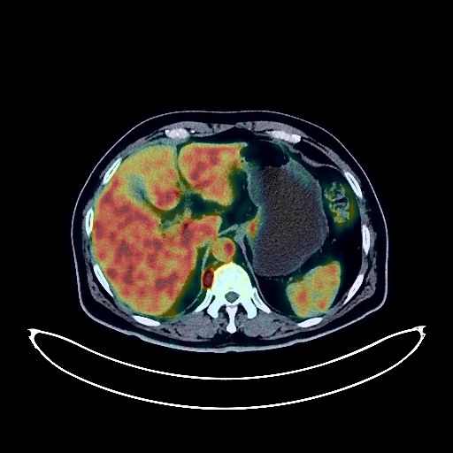
Gastric Cancer PET/CT (case 983824-000091 from PETWB-REP)
1 views10 days agoWhole-body 18F-FDG PET/CT scan in a patient with Gastric Cancer taken from the PETWB-REP dataset. The following English report (translated from original Chinese) is taken verbatim from the public dataset and has not been modified or otherwise checked for accuracy (see the end for citation). Impression a. Gastric antrum mass with increased FDG metabolism, consistent with gastric cancer. b. Multiple lymph node metastases in the left supraclavicular fossa and thoracic and abdominal regions (see description for details). a. Ground-glass nodule in the lingular segment of the left upper lobe, FDG metabolism normal, suggestive of inflammatory nodule or atypical adenomatous hyperplasia; annual HRCT follow-up recommended. b. Several small chronic inflammatory nodules (solid) in both lungs. A few chronic inflammations and old lesions in both lungs. Emphysema in both lungs. Bilateral renal cysts. Pancreatic fatty infiltration. Benign prostatic hyperplasia. Continuous increased FDG metabolism in the descending colon, sigmoid colon, and rectum, suggestive of inflammatory or physiological uptake; colonoscopy follow-up recommended. Degenerative changes in the spine. L4/5 and L5/S1 disc bulges. Minor ischemic lesions in the deep bilateral brain regions, indicative of age-related cerebral insufficiency. Chronic inflammation of the left maxillary sinus. This case is from PETWB-REP, a curated dataset of whole-body 18F-FDG PET/CT scans and corresponding radiology reports from 490 patients with a broad spectrum of malignancies. The data were retrospectively collected from patients who underwent clinically indicated whole-body 18F-FDG PET/CT scans at the Shanghai Universal Medical Imaging Diagnostic Center between 2021 and 2024. License: Creative Commons Attribution 4.0 International (CC BY 4.0) Citation: Xue, L., Feng, G., Wenbo, Z., Zhang, Y., Li, L., Wang, S., Peng, L., Peng, S., & Gao, X. (2026). PETWB-REP: A Multi-Cancer Whole-Body FDG PET/CT Dataset with Corresponding Radiology Reports [Data set]. Zenodo. https://doi.org/10.5281/zenodo.18670487
Whole BodyPET/CT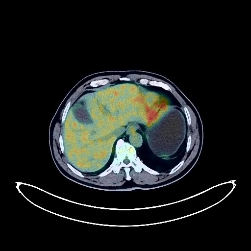
Esophageal Cancer PET/CT (case 983824-000188 from PETWB-REP)
2 views10 days agoWhole-body 18F-FDG PET/CT scan in a patient with Esophageal Cancer taken from the PETWB-REP dataset. The following English report (translated from original Chinese) is taken verbatim from the public dataset and has not been modified or otherwise checked for accuracy (see the end for citation). Impression a. Mass in the lower esophagus-cardia region, with increased FDG uptake, suggestive of cancer; please confirm with pathology. b. Chronic gastritis; endoscopic follow-up recommended. Reactive hyperplasia of lesser omental sac lymph nodes. a. Pure ground-glass nodules in the posterior segment of the left upper lobe and the apical segment of the right upper lobe, with no abnormal FDG uptake, suggestive of inflammatory nodules or atypical adenomatous hyperplasia; annual HRCT recommended. b. Multiple chronic inflammatory micronodules (solid) in both lungs. Partial calcification of the aorta and coronary artery walls. Accessory spleen. Small cyst in the right kidney. Small amount of hydrocele in both testes. Scoliosis with degenerative changes. Mild anterior slippage of the L4 vertebral body. L4/5 and L5/S1 intervertebral disc bulges. Right femoral head herniation fossa. Bilateral deep lacunar infarcts, age-related brain changes. Minor inflammation of the right maxillary sinus. This case is from PETWB-REP, a curated dataset of whole-body 18F-FDG PET/CT scans and corresponding radiology reports from 490 patients with a broad spectrum of malignancies. The data were retrospectively collected from patients who underwent clinically indicated whole-body 18F-FDG PET/CT scans at the Shanghai Universal Medical Imaging Diagnostic Center between 2021 and 2024. License: Creative Commons Attribution 4.0 International (CC BY 4.0) Citation: Xue, L., Feng, G., Wenbo, Z., Zhang, Y., Li, L., Wang, S., Peng, L., Peng, S., & Gao, X. (2026). PETWB-REP: A Multi-Cancer Whole-Body FDG PET/CT Dataset with Corresponding Radiology Reports [Data set]. Zenodo. https://doi.org/10.5281/zenodo.18670487
Whole BodyPET/CT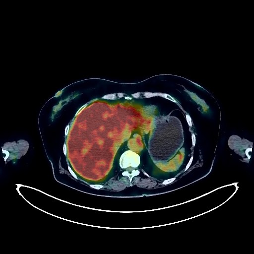
Glioma PET/CT (case 983824-000160 from PETWB-REP)
2 views10 days agoWhole-body 18F-FDG PET/CT scan in a patient with Glioma taken from the PETWB-REP dataset. The following English report (translated from original Chinese) is taken verbatim from the public dataset and has not been modified or otherwise checked for accuracy (see the end for citation). Impression a. A mass in the left basal ganglia with increased FDG metabolism, highly suggestive of a glioma; please confirm with pathology. b. Small ischemic foci in the remaining brain parenchyma, mild senile cerebral changes. Scattered chronic inflammation and remnants in both lungs. Mediastinal calcification. Trend towards cirrhosis. Increased FDG metabolism in the sigmoid colon, suggestive of physiological or inflammatory uptake; colonoscopy is recommended to rule out other possibilities. Partial vertebral osteophyte formation. Schmorl's node formation in the T9 vertebral body. L4/5 and L5/S1 intervertebral disc bulges. This case is from PETWB-REP, a curated dataset of whole-body 18F-FDG PET/CT scans and corresponding radiology reports from 490 patients with a broad spectrum of malignancies. The data were retrospectively collected from patients who underwent clinically indicated whole-body 18F-FDG PET/CT scans at the Shanghai Universal Medical Imaging Diagnostic Center between 2021 and 2024. License: Creative Commons Attribution 4.0 International (CC BY 4.0) Citation: Xue, L., Feng, G., Wenbo, Z., Zhang, Y., Li, L., Wang, S., Peng, L., Peng, S., & Gao, X. (2026). PETWB-REP: A Multi-Cancer Whole-Body FDG PET/CT Dataset with Corresponding Radiology Reports [Data set]. Zenodo. https://doi.org/10.5281/zenodo.18670487
Whole BodyPET/CT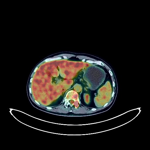
Lung Cancer PET/CT (case 983824-000159 from PETWB-REP)
2 views10 days agoWhole-body 18F-FDG PET/CT scan in a patient with Lung Cancer taken from the PETWB-REP dataset. The following English report (translated from original Chinese) is taken verbatim from the public dataset and has not been modified or otherwise checked for accuracy (see the end for citation). Impression a. A mass in the posterior segment of the left upper lobe, with elevated FDG metabolism, suggestive of lung cancer with surrounding obstructive inflammation; multiple lymph node metastases in the left hilum, mediastinum, left internal mammary chain, and left supraclavicular fossa; widespread bone metastases throughout the body. b. A patchy shadow in the anterior segment of the left upper lobe, with elevated FDG metabolism, highly suggestive of chronic inflammation. Multiple small (solid) chronic inflammatory nodules in the remaining lungs are highly probable; close CT observation is recommended to rule out mixed metastases. A few chronic inflammations and old lesions in both lungs. c. Slight thickening of the pleura bilaterally. Small amount of effusion in the superior pericardial recess. Calcification of some arterial walls. A few ischemic lesions in the deep bilateral brain regions, with mild age-related brain changes. A few chronic inflammations in the bilateral maxillary sinuses. Small renal calculus in the left kidney. Left renal cyst. Mild prostatic hyperplasia. Degenerative changes in the spine. L4/5 and L5/S1 intervertebral disc bulge. This case is from PETWB-REP, a curated dataset of whole-body 18F-FDG PET/CT scans and corresponding radiology reports from 490 patients with a broad spectrum of malignancies. The data were retrospectively collected from patients who underwent clinically indicated whole-body 18F-FDG PET/CT scans at the Shanghai Universal Medical Imaging Diagnostic Center between 2021 and 2024. License: Creative Commons Attribution 4.0 International (CC BY 4.0) Citation: Xue, L., Feng, G., Wenbo, Z., Zhang, Y., Li, L., Wang, S., Peng, L., Peng, S., & Gao, X. (2026). PETWB-REP: A Multi-Cancer Whole-Body FDG PET/CT Dataset with Corresponding Radiology Reports [Data set]. Zenodo. https://doi.org/10.5281/zenodo.18670487
Whole BodyPET/CT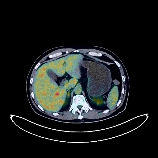
Lung Cancer PET/CT (case 983824-000016 from PETWB-REP)
7 views10 days agoWhole-body 18F-FDG PET/CT scan in a patient with Lung Cancer taken from the PETWB-REP dataset. The following English report (translated from original Chinese) is taken verbatim from the public dataset and has not been modified or otherwise checked for accuracy (see the end for citation). Impression a. A soft tissue mass with elevated FDG metabolism in the lateral segment of the right middle lobe of the lung, suggestive of malignancy, most likely small cell lung cancer, but metastasis cannot be ruled out; multiple lymph node metastases throughout the body (right hilum, mediastinum, left occipital region, bilateral supraclavicular regions, bilateral thoracic inlets, peripancreatic region, right retrorenal space, part of the superior mesenteric region, bilateral iliac vessels), some of which are fused. b. Right adrenal metastasis. Multiple bone metastases (see above for details). Peritoneal seeding metastasis. Subcutaneous metastatic nodules on the left anterior chest wall. c. Malignant tumor in the pancreatic head region, possibly originating from the pancreatic head, with enlarged and fused lymph nodes not ruled out; please combine with enhanced MRI for comprehensive analysis. Irregular shape of the pancreatic body and tail, elevated FDG metabolism, suggesting possible secondary inflammatory changes. Several nodules in the left lower lobe of the lung with mildly elevated FDG metabolism, suggestive of chronic inflammatory nodules, metastasis to be ruled out; close CT observation is recommended. Chronic inflammation and remnants in both lungs. Calcification of some arterial walls (including coronary arteries). Left renal cyst. Right testicular hydrocele. Partial vertebral osteophyte formation. L4/5 and L5/S1 intervertebral disc bulge. Sellar region mass, possibly pituitary tumor; age-related brain changes. Further enhanced MRI is recommended. This case is from PETWB-REP, a curated dataset of whole-body 18F-FDG PET/CT scans and corresponding radiology reports from 490 patients with a broad spectrum of malignancies. The data were retrospectively collected from patients who underwent clinically indicated whole-body 18F-FDG PET/CT scans at the Shanghai Universal Medical Imaging Diagnostic Center between 2021 and 2024. License: Creative Commons Attribution 4.0 International (CC BY 4.0) Citation: Xue, L., Feng, G., Wenbo, Z., Zhang, Y., Li, L., Wang, S., Peng, L., Peng, S., & Gao, X. (2026). PETWB-REP: A Multi-Cancer Whole-Body FDG PET/CT Dataset with Corresponding Radiology Reports [Data set]. Zenodo. https://doi.org/10.5281/zenodo.18670487
Whole BodyPET/CT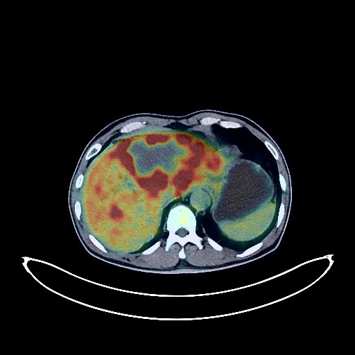
Liver Cancer PET/CT (case 983824-000083 from PETWB-REP)
2 views10 days agoWhole-body 18F-FDG PET/CT scan in a patient with Liver Cancer taken from the PETWB-REP dataset. The following English report (translated from original Chinese) is taken verbatim from the public dataset and has not been modified or otherwise checked for accuracy (see the end for citation). Impression a. Multiple intrahepatic lesions, the largest located in the left lobe with internal necrosis and increased FDG metabolism, suggestive of primary hepatocellular carcinoma with intrahepatic dissemination. b. Multiple lymph node metastases in the hepatogastric space, hilar space, and retroperitoneum. Bilateral adrenal metastases are highly probable. c. Left femoral neck metastasis to be ruled out; please confirm with MRI. Widespread mild dilatation of intrahepatic bile ducts. Post-cholecystectomy changes. Right renal cyst. Small amount of pelvic effusion. Manifestations of chronic gastritis; endoscopic re-examination necessary. A few chronic pulmonary lesions (including miliary foci) and remnants, lower lobular hypostatic effect, paraseptal emphysema in the right lower lobe, reactive hyperplasia of small hilar lymph nodes in both lungs. Degenerative changes in the spine, lumbar vertebral instability, L3/4 and L4/5 disc bulges, and L5/S1 disc herniation. No obvious abnormalities were found on cranial scintigraphy. Chronic inflammation of the right frontal sinus. This case is from PETWB-REP, a curated dataset of whole-body 18F-FDG PET/CT scans and corresponding radiology reports from 490 patients with a broad spectrum of malignancies. The data were retrospectively collected from patients who underwent clinically indicated whole-body 18F-FDG PET/CT scans at the Shanghai Universal Medical Imaging Diagnostic Center between 2021 and 2024. License: Creative Commons Attribution 4.0 International (CC BY 4.0) Citation: Xue, L., Feng, G., Wenbo, Z., Zhang, Y., Li, L., Wang, S., Peng, L., Peng, S., & Gao, X. (2026). PETWB-REP: A Multi-Cancer Whole-Body FDG PET/CT Dataset with Corresponding Radiology Reports [Data set]. Zenodo. https://doi.org/10.5281/zenodo.18670487
Whole BodyPET/CT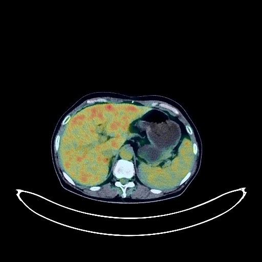
Lung Cancer PET/CT (case 983824-000007 from PETWB-REP)
8 views10 days agoWhole-body 18F-FDG PET/CT scan in a patient with Lung Cancer taken from the PETWB-REP dataset. The following English report (translated from original Chinese) is taken verbatim from the public dataset and has not been modified or otherwise checked for accuracy (see the end for citation). Impression a. Soft tissue mass in the left upper lobe of the lung with increased FDG metabolism, consistent with lung cancer based on pathology. b. Solid nodule in the posterior segment of the left upper lobe with increased FDG metabolism, suggestive of metastasis. c. Left hilar lymph node metastasis. Multiple bone metastases throughout the body (see description for details). d. Multiple small solid nodules in the remaining lungs, with no significant FDG uptake, highly suggestive of chronic inflammatory nodules; follow-up is recommended. Interstitial fibrosis in both lungs with scattered chronic inflammation and remnants. Bilateral pleural thickening. Bilateral incomplete breast regression; follow-up with ultrasound is recommended. Postoperative changes in the pancreatic mass; no abnormalities were found in FDG metabolism at the surgical site; follow-up is recommended. Highly suggestive of a liver cyst. Thickening of the left perirenal fascia, possible complex cyst in the left kidney; calcification of the left renal papillae; follow-up with ultrasound is recommended. a. Post-gastric surgery changes: increased FDG metabolism in part of the anastomotic wall, possibly due to physiological uptake or chronic inflammation. Follow-up with clinical findings and gastroscopy is recommended. b. Increased FDG metabolism in part of the intestinal tract, possibly due to physiological uptake or chronic inflammation. Further colonoscopy is recommended to rule out other possibilities. Osteoporosis. Spinal degeneration. L2/3, L3/4, L4/5 disc bulging, L5/S1 disc herniation. Right external oblique muscle lipoma. Bilateral deep lacunar infarcts, age-related brain abnormalities. MRI is recommended. Unevenly decreased density in the left and right lobes of the thyroid gland, with no abnormalities in FDG metabolism. Follow-up with ultrasound is recommended. This case is from PETWB-REP, a curated dataset of whole-body 18F-FDG PET/CT scans and corresponding radiology reports from 490 patients with a broad spectrum of malignancies. The data were retrospectively collected from patients who underwent clinically indicated whole-body 18F-FDG PET/CT scans at the Shanghai Universal Medical Imaging Diagnostic Center between 2021 and 2024. License: Creative Commons Attribution 4.0 International (CC BY 4.0) Citation: Xue, L., Feng, G., Wenbo, Z., Zhang, Y., Li, L., Wang, S., Peng, L., Peng, S., & Gao, X. (2026). PETWB-REP: A Multi-Cancer Whole-Body FDG PET/CT Dataset with Corresponding Radiology Reports [Data set]. Zenodo. https://doi.org/10.5281/zenodo.18670487
Whole BodyPET/CT