Loading...
Lung Cancer PET/CT (case 983824-000059 from PETWB-REP)
1 views10 days agoWhole-body 18F-FDG PET/CT scan in a patient with Lung Cancer taken from the PETWB-REP dataset. The following English report (translated from original Chinese) is taken verbatim from the public dataset and has not been modified or otherwise checked for accuracy (see the end for citation). Impression a. A mass in the lower lobe of the left lung with increased FDG metabolism, suggestive of lung cancer, with a high probability of invasion of adjacent pleura and interlobar pleural metastasis. Pathological examination is recommended. Multiple lymph node metastases in the bilateral hilar, mediastinal, and bilateral supraclavicular fossae. Multiple bone metastases throughout the body. b. A ground-glass nodule in the apical-posterior segment of the left upper lobe, with normal FDG metabolism, suggestive of an inflammatory nodule or atypical adenomatous hyperplasia. Annual HRCT follow-up is recommended. Several small, solid, chronic inflammatory nodules in both lungs. A few chronic inflammations and old lesions in both lungs. c. A soft tissue nodule in the right adrenal gland with basal FDG uptake, suggestive of adenoma, metastasis to be ruled out. Follow-up CT examination is recommended. Bilateral breast proliferative changes. Thyroid gland with uneven density and increased FDG metabolism, suggestive of chronic inflammation. Ultrasound and laboratory tests are recommended. Calcification in the right lobe of the thyroid gland. Slight thickening of the gastric body and antrum walls, with mildly increased FDG uptake, suggestive of chronic gastritis; increased FDG metabolism in some intestinal segments, suggestive of inflammatory or physiological uptake. Follow-up gastroscopy and colonoscopy are recommended. Uterine fibroids. Cystic lesions in both adnexa, most likely ovarian cysts; the right lesion is a cystadenoma or other possibilities to be ruled out. Please combine clinical findings with MRI examination. Mild osteophyte formation in the cervical, thoracic, and lumbar spine. L4/5 disc bulge with pneumothorax and degeneration; L5/S1 disc herniation. No abnormalities seen on cranial scintigraphy; please follow up with MRI. A small amount of chronic inflammation in the right maxillary sinus. This case is from PETWB-REP, a curated dataset of whole-body 18F-FDG PET/CT scans and corresponding radiology reports from 490 patients with a broad spectrum of malignancies. The data were retrospectively collected from patients who underwent clinically indicated whole-body 18F-FDG PET/CT scans at the Shanghai Universal Medical Imaging Diagnostic Center between 2021 and 2024. License: Creative Commons Attribution 4.0 International (CC BY 4.0) Citation: Xue, L., Feng, G., Wenbo, Z., Zhang, Y., Li, L., Wang, S., Peng, L., Peng, S., & Gao, X. (2026). PETWB-REP: A Multi-Cancer Whole-Body FDG PET/CT Dataset with Corresponding Radiology Reports [Data set]. Zenodo. https://doi.org/10.5281/zenodo.18670487
Whole BodyPET/CT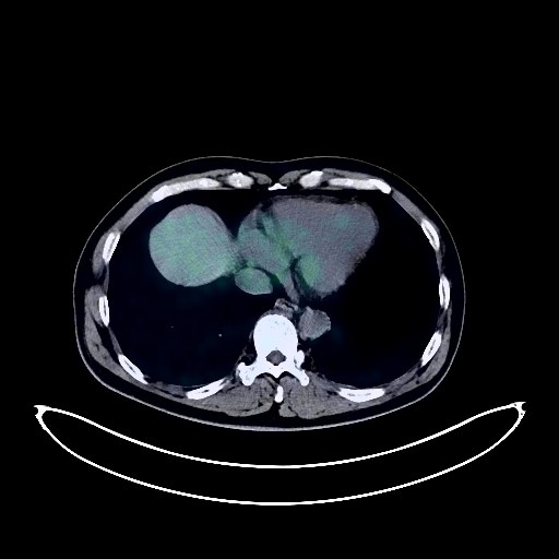
Lung Cancer PET/CT (case 983824-000173 from PETWB-REP)
1 views10 days agoWhole-body 18F-FDG PET/CT scan in a patient with Lung Cancer taken from the PETWB-REP dataset. The following English report (translated from original Chinese) is taken verbatim from the public dataset and has not been modified or otherwise checked for accuracy (see the end for citation). Impression a. A mass in the anterior segment of the right upper lobe, with increased FDG metabolism, suggestive of lung cancer. Multiple lymph node metastases in the right hilum and mediastinum. b. A mixed ground-glass opacity in the apical segment of the right upper lobe, with mildly increased FDG metabolism, suggestive of atypical adenomatous hyperplasia. Early lung cancer needs to be ruled out. Further comparison with older CT scans is recommended, and clinicopathological examination may be necessary. Several solid micronodules of chronic inflammation in both lungs. Calcification in the posterior segment of the left upper lobe. A few scattered chronic inflammatory and old lesions in both lungs. A few ischemic lesions in the deep bilateral brain regions; MRI is recommended. Liver cyst. Left kidney cyst. Benign prostatic hyperplasia with calcifications. Bilateral inguinal canal dilation. Increased FDG metabolism in parts of the intestine, likely due to inflammatory uptake; colonoscopy is recommended. Scoliosis with degenerative changes, sacralization into lumbar spine, instability of L2 and L4 vertebral bodies. L3/4 vertebral endplate inflammation. L4/5 intervertebral disc bulge, L5/S1 intervertebral disc herniation. Uneven thyroid density, increased FDG metabolism, suggesting a high probability of inflammatory changes; ultrasound and thyroid function tests are recommended. This case is from PETWB-REP, a curated dataset of whole-body 18F-FDG PET/CT scans and corresponding radiology reports from 490 patients with a broad spectrum of malignancies. The data were retrospectively collected from patients who underwent clinically indicated whole-body 18F-FDG PET/CT scans at the Shanghai Universal Medical Imaging Diagnostic Center between 2021 and 2024. License: Creative Commons Attribution 4.0 International (CC BY 4.0) Citation: Xue, L., Feng, G., Wenbo, Z., Zhang, Y., Li, L., Wang, S., Peng, L., Peng, S., & Gao, X. (2026). PETWB-REP: A Multi-Cancer Whole-Body FDG PET/CT Dataset with Corresponding Radiology Reports [Data set]. Zenodo. https://doi.org/10.5281/zenodo.18670487
Whole BodyPET/CT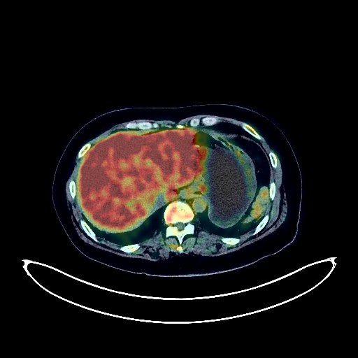
Lung Cancer PET/CT (case 983824-000193 from PETWB-REP)
2 views10 days agoWhole-body 18F-FDG PET/CT scan in a patient with Lung Cancer taken from the PETWB-REP dataset. The following English report (translated from original Chinese) is taken verbatim from the public dataset and has not been modified or otherwise checked for accuracy (see the end for citation). Impression a. A mass near the hilum in the right upper lobe of the lung, with increased FDG metabolism, consistent with lung cancer and surrounding obstructive changes. b. Multiple chronic inflammatory nodules in both lungs, possibly old tuberculosis in the left upper lobe. Reactive hyperplasia of mediastinal lymph nodes; follow-up is recommended. Accessory spleen. Uterine fibroids. Spinal osteophyte formation. L3/4 and L4/5 intervertebral disc bulge. Bilateral sacroiliac joint condensation osteitis. No obvious abnormalities were seen on cranial imaging; MRI is recommended. Left mastoiditis. This case is from PETWB-REP, a curated dataset of whole-body 18F-FDG PET/CT scans and corresponding radiology reports from 490 patients with a broad spectrum of malignancies. The data were retrospectively collected from patients who underwent clinically indicated whole-body 18F-FDG PET/CT scans at the Shanghai Universal Medical Imaging Diagnostic Center between 2021 and 2024. License: Creative Commons Attribution 4.0 International (CC BY 4.0) Citation: Xue, L., Feng, G., Wenbo, Z., Zhang, Y., Li, L., Wang, S., Peng, L., Peng, S., & Gao, X. (2026). PETWB-REP: A Multi-Cancer Whole-Body FDG PET/CT Dataset with Corresponding Radiology Reports [Data set]. Zenodo. https://doi.org/10.5281/zenodo.18670487
Whole BodyPET/CT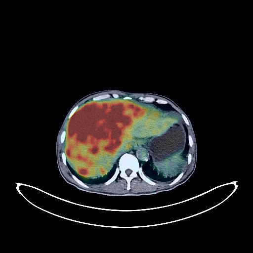
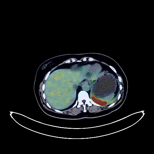
Liver Cancer PET/CT (case 983824-000050 from PETWB-REP)
10 views10 days agoWhole-body 18F-FDG PET/CT scan in a patient with Liver Cancer taken from the PETWB-REP dataset. The following English report (translated from original Chinese) is taken verbatim from the public dataset and has not been modified or otherwise checked for accuracy (see the end for citation). Impression a. Liver cirrhosis; multiple intrahepatic lesions with increased FDG uptake, suggestive of malignancy, with a high probability of hepatocellular carcinoma with intrahepatic dissemination; portal vein tumor thrombus formation is also possible. Comprehensive analysis with contrast-enhanced MRI is recommended. b. Abdominal and pelvic effusion; peritoneal seeding metastasis to be ruled out, follow-up is recommended. Slightly thickened gastric antrum wall, increased FDG metabolism, suggestive of inflammation; endoscopic follow-up is recommended to rule out other possibilities. a. One ground-glass nodule each in the left upper lobe and right lower lobe posterior segment, FDG metabolism normal, suggestive of atypical adenomatous hyperplasia or inflammatory nodules; annual HRCT follow-up is recommended. b. Multiple chronic inflammatory nodules and calcifications in both lungs. Emphysema and fibrosis in both lungs. c. Partial calcification of the aorta and coronary artery walls. Chronic cholecystitis. Small renal cyst in the right kidney. Spinal osteophyte formation, L4/5 and L5/S1 intervertebral disc bulge. White matter degeneration. Senile cerebral changes. This case is from PETWB-REP, a curated dataset of whole-body 18F-FDG PET/CT scans and corresponding radiology reports from 490 patients with a broad spectrum of malignancies. The data were retrospectively collected from patients who underwent clinically indicated whole-body 18F-FDG PET/CT scans at the Shanghai Universal Medical Imaging Diagnostic Center between 2021 and 2024. License: Creative Commons Attribution 4.0 International (CC BY 4.0) Citation: Xue, L., Feng, G., Wenbo, Z., Zhang, Y., Li, L., Wang, S., Peng, L., Peng, S., & Gao, X. (2026). PETWB-REP: A Multi-Cancer Whole-Body FDG PET/CT Dataset with Corresponding Radiology Reports [Data set]. Zenodo. https://doi.org/10.5281/zenodo.18670487
Whole BodyPET/CT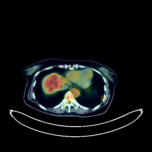
Cervical Cancer PET/CT (case 983824-000121 from PETWB-REP)
1 views10 days agoWhole-body 18F-FDG PET/CT scan in a patient with Cervical Cancer taken from the PETWB-REP dataset. The following English report (translated from original Chinese) is taken verbatim from the public dataset and has not been modified or otherwise checked for accuracy (see the end for citation). Impression Post-comprehensive treatment for cervical cancer: No clear signs of tumor recurrence were observed in the surgical area. Enlarged lymph nodes in the left groin with increased FDG metabolism suggest reactive hyperplasia; metastasis is not ruled out. Close follow-up is recommended. Small amount of pelvic effusion. a. Irregular nodules with increased FDG metabolism in the anterior medial basal segment of the left lower lobe, newly added compared to the previous image, are highly suggestive of metastasis. Please follow up with CT scans and perform a biopsy if necessary. b. Multiple lymph nodes in the bilateral hilar and mediastinal regions and bilateral supraclavicular fossae show increased FDG metabolism, newly added compared to the previous image, suggesting lymph node metastasis. c. A slightly enlarged air-filled cavity in the left lower lobe, with normal FDG metabolism, suggests possible bronchial cystic dilatation. Please follow up regularly with CT scans. A few fibrotic lesions in both lungs. A cyst in the superior pericardial recess. Bilateral breast hyperplasia. A slightly low-density nodule in the upper segment of the left lateral lobe of the liver, with decreased FDG metabolism, roughly similar to the previous scan, suggestive of a hemangioma; please confirm with contrast-enhanced MRI. Liver cyst. Possible mild hyperplasia of the left adrenal gland. Increased FDG metabolism in part of the gastric wall, suggestive of physiological or inflammatory changes; please confirm with gastroscopy. No obvious space-occupying lesions in the intestines; please confirm with colonoscopy follow-up. Osteophyte formation in some vertebral bodies of the spine; persistent epiphysis of the L4 vertebral body. L4/5 intervertebral disc herniation. No obvious abnormalities seen on cranial scintigraphy. This case is from PETWB-REP, a curated dataset of whole-body 18F-FDG PET/CT scans and corresponding radiology reports from 490 patients with a broad spectrum of malignancies. The data were retrospectively collected from patients who underwent clinically indicated whole-body 18F-FDG PET/CT scans at the Shanghai Universal Medical Imaging Diagnostic Center between 2021 and 2024. License: Creative Commons Attribution 4.0 International (CC BY 4.0) Citation: Xue, L., Feng, G., Wenbo, Z., Zhang, Y., Li, L., Wang, S., Peng, L., Peng, S., & Gao, X. (2026). PETWB-REP: A Multi-Cancer Whole-Body FDG PET/CT Dataset with Corresponding Radiology Reports [Data set]. Zenodo. https://doi.org/10.5281/zenodo.18670487
Whole BodyPET/CT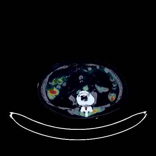
Rectal Cancer PET/CT (case 983824-000202 from PETWB-REP)
1 views10 days agoWhole-body 18F-FDG PET/CT scan in a patient with Rectal Cancer taken from the PETWB-REP dataset. The following English report (translated from original Chinese) is taken verbatim from the public dataset and has not been modified or otherwise checked for accuracy (see the end for citation). Impression A mass in the mid-rectum with elevated FDG metabolism, suggestive of colorectal cancer involving the adventitia; please confirm with pathology. Presacral lymph node metastasis is pending; follow-up is recommended. Chronic inflammatory micronodules in both lungs; CT follow-up is recommended. Chronic inflammation and sequelae in both lungs. Right-sided pleural effusion. Reactive hyperplasia of hilar and mediastinal lymph nodes in both lungs. Mild anemia; partial calcification of arterial walls (including coronary arteries). Postoperative changes after gallbladder and uterine surgery. Accessory spleen. Left adrenal hyperplasia. Bilateral renal cysts. Chronic inflammatory changes in the gastric antrum; please confirm with endoscopy. Osteoporosis, scoliosis, degenerative changes in the spine, multiple lumbar disc bulges with pneumoconiosis, mild posterior slippage of the L4 vertebra. Postoperative changes after cervical spine surgery. Bilateral knee joint degeneration with inflammation. Calcification in the left lobe of the thyroid gland; ultrasound follow-up is recommended. Age-related brain changes; deep lacunar infarcts; MRI follow-up is recommended. This case is from PETWB-REP, a curated dataset of whole-body 18F-FDG PET/CT scans and corresponding radiology reports from 490 patients with a broad spectrum of malignancies. The data were retrospectively collected from patients who underwent clinically indicated whole-body 18F-FDG PET/CT scans at the Shanghai Universal Medical Imaging Diagnostic Center between 2021 and 2024. License: Creative Commons Attribution 4.0 International (CC BY 4.0) Citation: Xue, L., Feng, G., Wenbo, Z., Zhang, Y., Li, L., Wang, S., Peng, L., Peng, S., & Gao, X. (2026). PETWB-REP: A Multi-Cancer Whole-Body FDG PET/CT Dataset with Corresponding Radiology Reports [Data set]. Zenodo. https://doi.org/10.5281/zenodo.18670487
Whole BodyPET/CT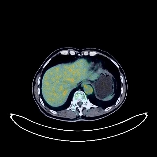
Bladder Cancer PET/CT (case 983824-000074 from PETWB-REP)
2 views10 days agoWhole-body 18F-FDG PET/CT scan in a patient with Bladder Cancer taken from the PETWB-REP dataset. The following English report (translated from original Chinese) is taken verbatim from the public dataset and has not been modified or otherwise checked for accuracy (see the end for citation). Impression a. A slightly high-density nodule on the left side of the lower bladder with increased FDG metabolism, suggestive of a malignant tumor, most likely bladder cancer. Please confirm with pathology. b. Small bladder stone. c. Postoperative left femoral head fracture surgery, unevenly decreased bone density in the left femoral head, slight FDG metabolism, suggesting postoperative changes. Please correlate with clinical findings and follow up. a. Postoperative changes after cerebral hemangioblastoma, no significant abnormalities in FDG uptake in the surgical area. b. A few lacunar lesions in the deep bilateral brain, mild age-related brain changes. Poor pneumatization of the left mastoid air cells. Chronic inflammatory nodules and nodule-like lesions in the right lung. Please follow up with CT scan. A few post-inflammatory remnants in the left lower lobe. Postoperative changes after coronary artery stenting; partial calcification of the arterial wall. Nodular goiter is highly probable. Calcifications are present in the left lobe of the thyroid gland. Please repeat ultrasound and thyroid function tests. Cystic mass in the tail of the pancreas. Enhanced MRI is recommended. Calcifications are present in the lower segment of the right posterior lobe of the liver. Benign prostatic hyperplasia. Mildly increased FDG metabolism in some gastric wall areas, possibly due to physiological uptake or chronic inflammation. Spinal degenerative changes. Bone islands in the right 7th and 8th posterior ribs. This case is from PETWB-REP, a curated dataset of whole-body 18F-FDG PET/CT scans and corresponding radiology reports from 490 patients with a broad spectrum of malignancies. The data were retrospectively collected from patients who underwent clinically indicated whole-body 18F-FDG PET/CT scans at the Shanghai Universal Medical Imaging Diagnostic Center between 2021 and 2024. License: Creative Commons Attribution 4.0 International (CC BY 4.0) Citation: Xue, L., Feng, G., Wenbo, Z., Zhang, Y., Li, L., Wang, S., Peng, L., Peng, S., & Gao, X. (2026). PETWB-REP: A Multi-Cancer Whole-Body FDG PET/CT Dataset with Corresponding Radiology Reports [Data set]. Zenodo. https://doi.org/10.5281/zenodo.18670487
Whole BodyPET/CT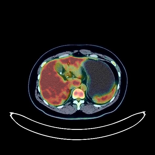
Lung Cancer PET/CT (case 983824-000081 from PETWB-REP)
4 views10 days agoWhole-body 18F-FDG PET/CT scan in a patient with Lung Cancer taken from the PETWB-REP dataset. The following English report (translated from original Chinese) is taken verbatim from the public dataset and has not been modified or otherwise checked for accuracy (see the end for citation). Impression a. A space-occupying lesion in the posterior segment of the left lower lobe bronchus with increased FDG metabolism, consistent with carcinoid features based on pathology. b. Several solid micronodules in the lower lobes of both lungs and the left interlobar fissure, with normal FDG uptake, suggestive of chronic inflammatory nodules, metastasis to be ruled out; please review with CT for comparison. T10-T11 vertebral endplate inflammation. T10/11 intervertebral disc pneumothorax. Gallstones. Accessory spleen. Prostatic calcification. Chronic inflammation of both palatine tonsils. No obvious abnormalities were found on cranial scintigraphy. This case is from PETWB-REP, a curated dataset of whole-body 18F-FDG PET/CT scans and corresponding radiology reports from 490 patients with a broad spectrum of malignancies. The data were retrospectively collected from patients who underwent clinically indicated whole-body 18F-FDG PET/CT scans at the Shanghai Universal Medical Imaging Diagnostic Center between 2021 and 2024. License: Creative Commons Attribution 4.0 International (CC BY 4.0) Citation: Xue, L., Feng, G., Wenbo, Z., Zhang, Y., Li, L., Wang, S., Peng, L., Peng, S., & Gao, X. (2026). PETWB-REP: A Multi-Cancer Whole-Body FDG PET/CT Dataset with Corresponding Radiology Reports [Data set]. Zenodo. https://doi.org/10.5281/zenodo.18670487
Whole BodyPET/CT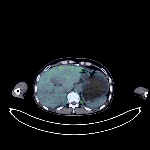
Renal Cancer PET/CT (case 983824-000147 from PETWB-REP)
2 views10 days agoWhole-body 18F-FDG PET/CT scan in a patient with Renal Cancer taken from the PETWB-REP dataset. The following English report (translated from original Chinese) is taken verbatim from the public dataset and has not been modified or otherwise checked for accuracy (see the end for citation). Impression a. Multiple enlarged lymph nodes throughout the body with increased FDG metabolism (see description for details), consistent with metastatic lesions based on pathology. b. Focal increased FDG metabolism in the right part of the L5 vertebral body, highly suggestive of metastatic lesions; close observation is recommended. Increased FDG metabolism in the upper pole of the left kidney; slightly increased density on the same CT scan, suggestive of a space-occupying lesion; further enhanced MRI of both kidneys is recommended. Slightly low-density lesion in the middle of the right kidney; no abnormal FDG metabolism; highly suggestive of a cyst. Calcification in the upper lobe of the left lung. A few post-inflammatory lesions in both lungs. Bilateral pleural thickening. Anemia changes; partial calcification of arterial walls (including coronary arteries). Bilateral breast hyperplasia; calcification in the right breast. Schistosomiasis intestinal changes; possible chronic inflammatory changes in part of the gastric wall and intestinal tract; endoscopic examination is recommended. Small amount of pelvic effusion. Degenerative changes in the spine, multiple bulging lumbar intervertebral discs. Subcutaneous edema in the abdomen and buttocks. Age-related brain conditions, deep lacunar infarcts. This case is from PETWB-REP, a curated dataset of whole-body 18F-FDG PET/CT scans and corresponding radiology reports from 490 patients with a broad spectrum of malignancies. The data were retrospectively collected from patients who underwent clinically indicated whole-body 18F-FDG PET/CT scans at the Shanghai Universal Medical Imaging Diagnostic Center between 2021 and 2024. License: Creative Commons Attribution 4.0 International (CC BY 4.0) Citation: Xue, L., Feng, G., Wenbo, Z., Zhang, Y., Li, L., Wang, S., Peng, L., Peng, S., & Gao, X. (2026). PETWB-REP: A Multi-Cancer Whole-Body FDG PET/CT Dataset with Corresponding Radiology Reports [Data set]. Zenodo. https://doi.org/10.5281/zenodo.18670487
Whole BodyPET/CT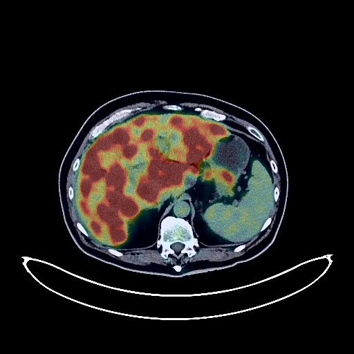
Pancreatic Cancer PET/CT (case 983824-000072 from PETWB-REP)
2 views10 days agoWhole-body 18F-FDG PET/CT scan in a patient with Pancreatic Cancer taken from the PETWB-REP dataset. The following English report (translated from original Chinese) is taken verbatim from the public dataset and has not been modified or otherwise checked for accuracy (see the end for citation). Impression a. A mass in the body and tail of the pancreas with elevated FDG metabolism, suggestive of pancreatic cancer; please correlate with clinicopathology. b. Diffuse liver metastases. Multiple metastases in the greater omentum and mesentery of the abdominopelvic cavity, with abdominopelvic effusion. c. Multiple bone metastases in the L4 vertebral body, left L5 adnexa, left sacrum, and left iliac bone. Intramuscular metastases in the left paramuscular region of the L4 and L5 spinous processes. Multiple solid nodules in both lungs, some with slightly elevated FDG metabolism, suggestive of inflammatory nodules; CT follow-up is recommended to rule out other possibilities. Scattered post-inflammatory lesions in both lungs. Small amount of pleural effusion bilaterally. Reactive hyperplasia of mediastinal lymph nodes. Calcification of some arterial walls (including coronary arteries). Liver cysts. Chronic cholecystitis, gallstones. Accessory spleen. Prostatic calcifications. Chronic inflammatory changes in part of the gastric wall; please follow up with endoscopy. Degenerative changes in the spine, with L4/5 and L5/S1 intervertebral disc bulges. Subcutaneous calcifications in the left buttock. No obvious abnormalities were found on cranial scintigraphy. This case is from PETWB-REP, a curated dataset of whole-body 18F-FDG PET/CT scans and corresponding radiology reports from 490 patients with a broad spectrum of malignancies. The data were retrospectively collected from patients who underwent clinically indicated whole-body 18F-FDG PET/CT scans at the Shanghai Universal Medical Imaging Diagnostic Center between 2021 and 2024. License: Creative Commons Attribution 4.0 International (CC BY 4.0) Citation: Xue, L., Feng, G., Wenbo, Z., Zhang, Y., Li, L., Wang, S., Peng, L., Peng, S., & Gao, X. (2026). PETWB-REP: A Multi-Cancer Whole-Body FDG PET/CT Dataset with Corresponding Radiology Reports [Data set]. Zenodo. https://doi.org/10.5281/zenodo.18670487
Whole BodyPET/CT