Loading...
Nasopharyngeal Cancer PET/CT (case 983824-000169 from PETWB-REP)
0 views10 days agoWhole-body 18F-FDG PET/CT scan in a patient with Nasopharyngeal Cancer taken from the PETWB-REP dataset. The following English report (translated from original Chinese) is taken verbatim from the public dataset and has not been modified or otherwise checked for accuracy (see the end for citation). Impression a. Nasopharyngeal mass with elevated FDG metabolism, consistent with nasopharyngeal carcinoma with skull base bone destruction and extensive surrounding invasion (as described above), metastasis to right retropharyngeal and right deep cervical lymph nodes. Reactive hyperplasia of small lymph nodes in the bilateral deep cervical spaces, submandibular, and submental regions. b. Right maxillary sinus submucosal cyst. Bilateral mastoid incomplete pneumatization and bilateral chronic mastoiditis. Chronic inflammatory nodule in the posterior segment of the right upper lobe of the lung. Scattered chronic inflammation and remnants in both lungs. Mild pleural thickening bilaterally. a. Uneven FDG metabolism in the liver; MRI follow-up is recommended to rule out other possibilities. Multiple liver cysts. b. Possible gallstones. Prostatic calcification. Degenerative changes in the spine. L4/5 and L5/S1 intervertebral disc bulge. L2 vertebral body instability. No obvious abnormalities were found on cranial scintigraphy. This case is from PETWB-REP, a curated dataset of whole-body 18F-FDG PET/CT scans and corresponding radiology reports from 490 patients with a broad spectrum of malignancies. The data were retrospectively collected from patients who underwent clinically indicated whole-body 18F-FDG PET/CT scans at the Shanghai Universal Medical Imaging Diagnostic Center between 2021 and 2024. License: Creative Commons Attribution 4.0 International (CC BY 4.0) Citation: Xue, L., Feng, G., Wenbo, Z., Zhang, Y., Li, L., Wang, S., Peng, L., Peng, S., & Gao, X. (2026). PETWB-REP: A Multi-Cancer Whole-Body FDG PET/CT Dataset with Corresponding Radiology Reports [Data set]. Zenodo. https://doi.org/10.5281/zenodo.18670487
Whole BodyPET/CT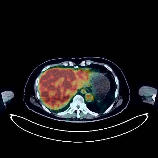
Renal Cancer PET/CT (case 983824-000057 from PETWB-REP)
0 views10 days agoWhole-body 18F-FDG PET/CT scan in a patient with Renal Cancer taken from the PETWB-REP dataset. The following English report (translated from original Chinese) is taken verbatim from the public dataset and has not been modified or otherwise checked for accuracy (see the end for citation). Impression a. Left kidney mass with increased FDG metabolism, suggestive of renal cell carcinoma; please correlate with clinicopathology. b. Multiple renal cysts in both kidneys. Renal stones in both kidneys. Several small, solid, chronic inflammatory nodules in both lungs; regular CT scans are recommended to rule out other involvement. Minor chronic inflammation and old lesions in both lungs. Calcification of some arterial walls (including coronary arteries). Liver cysts. Fatty infiltration of the pancreas. Prostatic calcification. Degenerative changes in the spine. L4/5 and L5/S1 intervertebral disc bulges. A few ischemic lesions in the deep bilateral brain regions, suggestive of senile encephalopathy. This case is from PETWB-REP, a curated dataset of whole-body 18F-FDG PET/CT scans and corresponding radiology reports from 490 patients with a broad spectrum of malignancies. The data were retrospectively collected from patients who underwent clinically indicated whole-body 18F-FDG PET/CT scans at the Shanghai Universal Medical Imaging Diagnostic Center between 2021 and 2024. License: Creative Commons Attribution 4.0 International (CC BY 4.0) Citation: Xue, L., Feng, G., Wenbo, Z., Zhang, Y., Li, L., Wang, S., Peng, L., Peng, S., & Gao, X. (2026). PETWB-REP: A Multi-Cancer Whole-Body FDG PET/CT Dataset with Corresponding Radiology Reports [Data set]. Zenodo. https://doi.org/10.5281/zenodo.18670487
Whole BodyPET/CT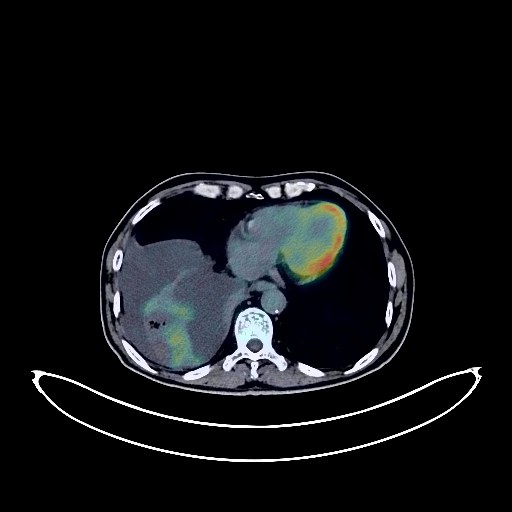
Lung Cancer PET/CT (case 983824-000199 from PETWB-REP)
0 views10 days agoWhole-body 18F-FDG PET/CT scan in a patient with Lung Cancer taken from the PETWB-REP dataset. The following English report (translated from original Chinese) is taken verbatim from the public dataset and has not been modified or otherwise checked for accuracy (see the end for citation). Impression a. A mass near the right hilum at the bronchial opening in the right lower lobe, encircling adjacent blood vessels, with increased FDG metabolism, suggestive of central lung cancer with obstructive inflammation. Right-sided pleural effusion. b. Lymph node metastases in the right hilum, mediastinum, right supraclavicular fossa, and below the pancreatic head. c. Multiple liver metastases. A left adrenal metastasis is highly probable. Bone metastases in the T2 spinous process, T12 vertebral body, left sacrum, and right pubic tubercle. d. Several small, solid, chronic inflammatory nodules in both lungs; please follow up with CT scans. A few post-inflammatory remnants and calcifications in both lungs. Paraseptal emphysema in both upper lobes. Partial arteriosclerosis (including coronary arteries). Right adrenal hyperplasia. Bilateral hydrocele. Increased FDG metabolism in parts of the colon and rectum, possibly due to physiological uptake or chronic inflammation; please follow up with endoscopy. Degenerative changes in the spine. Sacral canal cyst, L4/5 and L5/S1 disc bulge. Age-related brain changes; MRI recommended. Deep lacunar infarcts. Bilateral mastoid hypopneumatization. Likely a mixed tumor or adenolymphoma in the deep lobe of the left parotid gland; ultrasound follow-up recommended. This case is from PETWB-REP, a curated dataset of whole-body 18F-FDG PET/CT scans and corresponding radiology reports from 490 patients with a broad spectrum of malignancies. The data were retrospectively collected from patients who underwent clinically indicated whole-body 18F-FDG PET/CT scans at the Shanghai Universal Medical Imaging Diagnostic Center between 2021 and 2024. License: Creative Commons Attribution 4.0 International (CC BY 4.0) Citation: Xue, L., Feng, G., Wenbo, Z., Zhang, Y., Li, L., Wang, S., Peng, L., Peng, S., & Gao, X. (2026). PETWB-REP: A Multi-Cancer Whole-Body FDG PET/CT Dataset with Corresponding Radiology Reports [Data set]. Zenodo. https://doi.org/10.5281/zenodo.18670487
Whole BodyPET/CT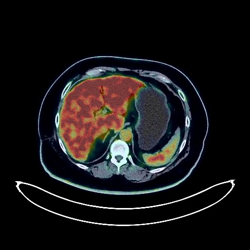
Lung Cancer PET/CT (case 983824-000145 from PETWB-REP)
0 views10 days agoWhole-body 18F-FDG PET/CT scan in a patient with Lung Cancer taken from the PETWB-REP dataset. The following English report (translated from original Chinese) is taken verbatim from the public dataset and has not been modified or otherwise checked for accuracy (see the end for citation). Impression a. No obvious signs of tumor recurrence in the right lung surgical area. b. Post-operatively, no abnormal density shadows or abnormal FDG metabolic foci were observed in the surgical area following a right 3rd rib lesion. Changes were observed after a fracture of the right 4th rib. c. Multiple nodules in both lungs, more numerous and larger than before, with no abnormal FDG uptake, suggesting a high probability of metastatic tumors. Regular follow-up based on clinical findings and CT scans is recommended. d. Left supraclavicular fossa lymph nodes showed mild FDG uptake and slight enlargement, suggesting a possible metastatic tumor. Regular follow-up based on clinical findings is recommended. Fibrotic lesions in both lungs, mild thickening of the right pleura. Partial calcification of the aorta and coronary artery walls. Fatty liver. Post-operative absence of the gallbladder. Small renal calculus in the right kidney. Uterine fibroids. Scoliosis with degenerative changes. Multiple lumbar vertebral instability. L3/4, L4/5, and L5/S1 intervertebral disc bulging with pneumoconiosis and degeneration. Bilateral femoral head insular cranial fossa. Bilateral frozen shoulder. Bilateral deep lacunar infarcts in the brain, age-related brain changes. This case is from PETWB-REP, a curated dataset of whole-body 18F-FDG PET/CT scans and corresponding radiology reports from 490 patients with a broad spectrum of malignancies. The data were retrospectively collected from patients who underwent clinically indicated whole-body 18F-FDG PET/CT scans at the Shanghai Universal Medical Imaging Diagnostic Center between 2021 and 2024. License: Creative Commons Attribution 4.0 International (CC BY 4.0) Citation: Xue, L., Feng, G., Wenbo, Z., Zhang, Y., Li, L., Wang, S., Peng, L., Peng, S., & Gao, X. (2026). PETWB-REP: A Multi-Cancer Whole-Body FDG PET/CT Dataset with Corresponding Radiology Reports [Data set]. Zenodo. https://doi.org/10.5281/zenodo.18670487
Whole BodyPET/CT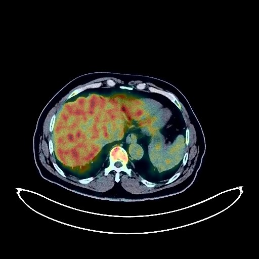
Bladder Cancer PET/CT (case 983824-000099 from PETWB-REP)
0 views10 days agoWhole-body 18F-FDG PET/CT scan in a patient with Bladder Cancer taken from the PETWB-REP dataset. The following English report (translated from original Chinese) is taken verbatim from the public dataset and has not been modified or otherwise checked for accuracy (see the end for citation). Impression a. Post-bladder cancer surgery, no clear signs of tumor recurrence were observed; please follow up with ultrasound. b. A mass in the left lateral lobe of the liver, with increased FDG metabolism, suggests a malignant tumor, more likely primary than metastatic. A cyst on the upper margin of the pancreatic body is highly probable; MRI follow-up is recommended. Liver cyst. Reactive hyperplasia of lymph nodes in both inguinal regions. Bilateral renal cysts, complex cyst in the right kidney, bilateral renal calcifications, small stone in the left kidney. Prostatic calcifications. Stones in the right vas deferens and seminal vesicle. Changes after partial gastrectomy, physiological or inflammatory uptake at the gastrojejunostomy, and physiological uptake of part of the intestine. a. Chronic inflammatory solid nodules in the lower lobe of the right lung, roughly similar to the CT scan from our center on October 19 The previously observed ground-glass opacity in the posterior segment of the lower lobe of the left lung was not clearly visualized this time. b. Pulmonary emphysema with bullae in both lungs, a few chronic inflammations and sequelae in both lungs. Partial arteriosclerosis. c. Benign patchy shadows in the aortic window, similar to the previous one. Degenerative changes in the spine, cervical lordosis reversal, L5/S1 endplate inflammation, bilateral vertebral arch collapse of the L5 vertebral body. Deep lacunar ischemic lesions in the brain, senile encephalopathy. A few chronic inflammations in multiple paranasal sinuses. This case is from PETWB-REP, a curated dataset of whole-body 18F-FDG PET/CT scans and corresponding radiology reports from 490 patients with a broad spectrum of malignancies. The data were retrospectively collected from patients who underwent clinically indicated whole-body 18F-FDG PET/CT scans at the Shanghai Universal Medical Imaging Diagnostic Center between 2021 and 2024. License: Creative Commons Attribution 4.0 International (CC BY 4.0) Citation: Xue, L., Feng, G., Wenbo, Z., Zhang, Y., Li, L., Wang, S., Peng, L., Peng, S., & Gao, X. (2026). PETWB-REP: A Multi-Cancer Whole-Body FDG PET/CT Dataset with Corresponding Radiology Reports [Data set]. Zenodo. https://doi.org/10.5281/zenodo.18670487
Whole BodyPET/CT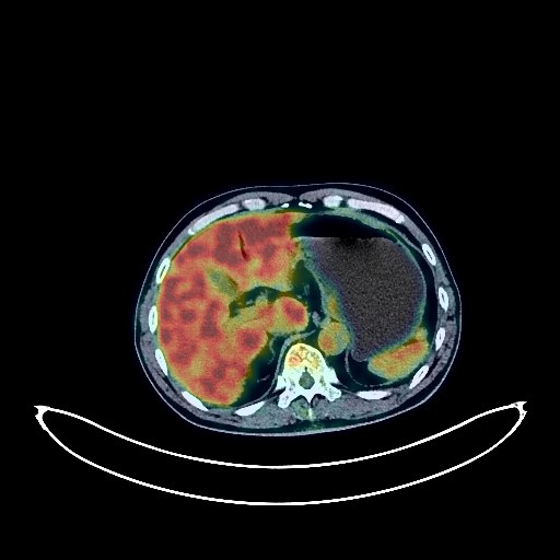
Prostate Cancer PET/CT (case 983824-000155 from PETWB-REP)
2 views10 days agoWhole-body 18F-FDG PET/CT scan in a patient with Prostate Cancer taken from the PETWB-REP dataset. The following English report (translated from original Chinese) is taken verbatim from the public dataset and has not been modified or otherwise checked for accuracy (see the end for citation). Impression Benign prostatic hyperplasia with calcification; prostatic mass with increased FDG metabolism, consistent with prostate cancer, involving the posterior wall of the bladder and seminal vesicles, bladder mass to be ruled out. Reactive hyperplasia of bilateral inguinal lymph nodes. Chronic inflammatory micronodules (solid) in both lungs. Cystic cavity in the posterior wall of the posterior basal segment of the left lower lobe, cystic lung cancer should be suspected, HRCT follow-up is recommended in 3-6 months. Scattered post-inflammatory lesions in both lungs. Calcification of some arterial walls (including coronary arteries). Pancreatic calcification. Left renal cyst. Chronic inflammatory changes in the antrum of the stomach, please follow up with endoscopy. Degenerative changes in the spine, slight posterior slippage of the L4 vertebral body, L4/5 intervertebral disc bulge. Age-related brain, deep lacunar ischemic lesions in the brain. Chronic inflammation of both ethmoid sinuses and both maxillary sinuses. Inflammation of the base of the tongue and both palatine tonsils. Nodular goiter; ultrasound follow-up is recommended. This case is from PETWB-REP, a curated dataset of whole-body 18F-FDG PET/CT scans and corresponding radiology reports from 490 patients with a broad spectrum of malignancies. The data were retrospectively collected from patients who underwent clinically indicated whole-body 18F-FDG PET/CT scans at the Shanghai Universal Medical Imaging Diagnostic Center between 2021 and 2024. License: Creative Commons Attribution 4.0 International (CC BY 4.0) Citation: Xue, L., Feng, G., Wenbo, Z., Zhang, Y., Li, L., Wang, S., Peng, L., Peng, S., & Gao, X. (2026). PETWB-REP: A Multi-Cancer Whole-Body FDG PET/CT Dataset with Corresponding Radiology Reports [Data set]. Zenodo. https://doi.org/10.5281/zenodo.18670487
Whole BodyPET/CT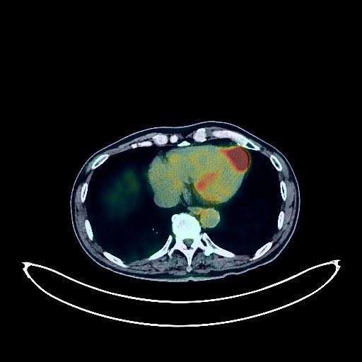
Lung Cancer PET/CT (case 983824-000131 from PETWB-REP)
2 views10 days agoWhole-body 18F-FDG PET/CT scan in a patient with Lung Cancer taken from the PETWB-REP dataset. The following English report (translated from original Chinese) is taken verbatim from the public dataset and has not been modified or otherwise checked for accuracy (see the end for citation). Impression a. A mass in the right middle lobe of the lung with increased FDG metabolism, highly suggestive of lung cancer with atelectasis; bronchoscopy recommended. b. Reactive hyperplasia of the right hilar and mediastinal lymph nodes; CT follow-up recommended to rule out other possibilities. Partial calcification of the aorta and coronary artery walls. c. Emphysema in the upper lobes of both lungs, multiple chronic inflammatory micronodules in both lungs, and multiple calcifications in both lungs. A slightly low-density nodule in the left adrenal gland, with no abnormal FDG uptake, highly suggestive of an adenoma; contrast-enhanced MRI recommended. Right renal cyst. Small crystals in the left kidney. Prostatic calcification. Thickening of the gastric fundus and body mucosa with increased FDG metabolism, suggestive of inflammation or physiological uptake; clinical correlation recommended. Increased FDG metabolism in the nasopharynx, suggestive of inflammation or physiological uptake; please correlate with clinical findings and, if necessary, undergo specialist examination. Spinal osteophyte formation, mild wedge-shaped deformity of the L1 vertebral body. L3/4, L4/5, and L5/S1 intervertebral disc bulges. Age-related brain changes. This case is from PETWB-REP, a curated dataset of whole-body 18F-FDG PET/CT scans and corresponding radiology reports from 490 patients with a broad spectrum of malignancies. The data were retrospectively collected from patients who underwent clinically indicated whole-body 18F-FDG PET/CT scans at the Shanghai Universal Medical Imaging Diagnostic Center between 2021 and 2024. License: Creative Commons Attribution 4.0 International (CC BY 4.0) Citation: Xue, L., Feng, G., Wenbo, Z., Zhang, Y., Li, L., Wang, S., Peng, L., Peng, S., & Gao, X. (2026). PETWB-REP: A Multi-Cancer Whole-Body FDG PET/CT Dataset with Corresponding Radiology Reports [Data set]. Zenodo. https://doi.org/10.5281/zenodo.18670487
Whole BodyPET/CT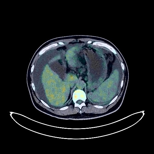
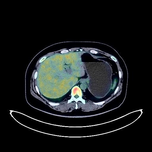
Liver Cancer PET/CT (case 983824-000069 from PETWB-REP)
1 views10 days agoWhole-body 18F-FDG PET/CT scan in a patient with Liver Cancer taken from the PETWB-REP dataset. The following English report (translated from original Chinese) is taken verbatim from the public dataset and has not been modified or otherwise checked for accuracy (see the end for citation). Impression a. Uneven liver density and FDG metabolism; multiple lesions in the liver indicated by an external hospital were not clearly visualized on PET/CT; enhanced MRI is recommended for comprehensive analysis. b. Liver cirrhosis, calcification in the right lobe of the liver. Splenomegaly. Massive ascites. Gallbladder poorly visualized. c. Slight thickening of the peritoneum and mesentery in the abdomen and pelvis; FDG metabolism normal, likely reactive hyperplasia; reactive hyperplasia of small lymph nodes in the porta hepatis, hilar space, retroperitoneum, and part of the mesenteric region. Follow-up is recommended to rule out other complications. a. Chronic inflammatory micronodules in the right lung; follow-up with CT is recommended. A few chronic inflammations and sequelae in both lungs. Bilateral pleural effusion; partial atelectasis in the lower lobes of both lungs. b. Reactive hyperplasia of small mediastinal lymph nodes. Calcification of some arterial walls (including coronary arteries). Possible small kidney stone in the left kidney. Increased FDG metabolism in parts of the stomach wall and intestines, possibly due to physiological uptake or chronic inflammation; please follow up with endoscopy. Osteophyte formation in some vertebral bodies of the spine, straightening of the cervical curvature. L5/S1 disc bulge. No obvious abnormalities seen on cranial scintigraphy. Chronic inflammation of both palatine tonsils. This case is from PETWB-REP, a curated dataset of whole-body 18F-FDG PET/CT scans and corresponding radiology reports from 490 patients with a broad spectrum of malignancies. The data were retrospectively collected from patients who underwent clinically indicated whole-body 18F-FDG PET/CT scans at the Shanghai Universal Medical Imaging Diagnostic Center between 2021 and 2024. License: Creative Commons Attribution 4.0 International (CC BY 4.0) Citation: Xue, L., Feng, G., Wenbo, Z., Zhang, Y., Li, L., Wang, S., Peng, L., Peng, S., & Gao, X. (2026). PETWB-REP: A Multi-Cancer Whole-Body FDG PET/CT Dataset with Corresponding Radiology Reports [Data set]. Zenodo. https://doi.org/10.5281/zenodo.18670487
Whole BodyPET/CT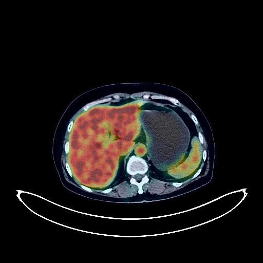
Pancreatic Cancer PET/CT (case 983824-000061 from PETWB-REP)
2 views10 days agoWhole-body 18F-FDG PET/CT scan in a patient with Pancreatic Cancer taken from the PETWB-REP dataset. The following English report (translated from original Chinese) is taken verbatim from the public dataset and has not been modified or otherwise checked for accuracy (see the end for citation). Impression A mass in the head of the pancreas with increased FDG metabolism and pancreatic duct dilation suggests pancreatic head cancer complicated by pancreatitis; please correlate with clinicopathology. Reactive hyperplasia of the peripancreatic, retroperitoneal, left posterior diaphragmatic crura, and mesenteric lymph nodes is possible; follow-up is recommended to rule out other possibilities. Chronic inflammatory micronodules in the upper lobes of both lungs. A few post-inflammatory lesions in both lungs. Calcification of some arterial walls (including coronary arteries). Multiple liver cysts. Small renal cysts. Post-hysterectomy changes. Chronic inflammatory changes in the antrum of the stomach and part of the intestine; please follow up with endoscopy. Degenerative changes in the spine, L4/5 and L5/S1 intervertebral disc bulges. Subcutaneous calcification in the left buttock. Inflammation of the right shoulder. No obvious abnormalities were found on cranial scintigraphy. This case is from PETWB-REP, a curated dataset of whole-body 18F-FDG PET/CT scans and corresponding radiology reports from 490 patients with a broad spectrum of malignancies. The data were retrospectively collected from patients who underwent clinically indicated whole-body 18F-FDG PET/CT scans at the Shanghai Universal Medical Imaging Diagnostic Center between 2021 and 2024. License: Creative Commons Attribution 4.0 International (CC BY 4.0) Citation: Xue, L., Feng, G., Wenbo, Z., Zhang, Y., Li, L., Wang, S., Peng, L., Peng, S., & Gao, X. (2026). PETWB-REP: A Multi-Cancer Whole-Body FDG PET/CT Dataset with Corresponding Radiology Reports [Data set]. Zenodo. https://doi.org/10.5281/zenodo.18670487
Whole BodyPET/CT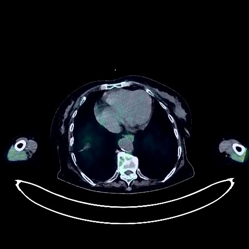
Lung Cancer PET/CT (case 983824-000165 from PETWB-REP)
2 views10 days agoWhole-body 18F-FDG PET/CT scan in a patient with Lung Cancer taken from the PETWB-REP dataset. The following English report (translated from original Chinese) is taken verbatim from the public dataset and has not been modified or otherwise checked for accuracy (see the end for citation). Impression A mass near the hilum of the right lung with increased FDG metabolism; a soft tissue nodule in the right pleura with increased FDG metabolism; multiple soft tissue nodules and masses with increased FDG metabolism in the right axilla, right supraclavicular fossa, right hilum, mediastinum, hepatic hilum, mesentery in the abdominoperineal cavity, bilateral iliac vessels, bilateral pelvic walls, left iliac vessels, left groin, and bilateral adrenal regions. All of these are considered malignant tumors, possibly right lung cancer with multiple metastases. A biopsy is recommended to confirm the pathology and rule out other possibilities. Old lesions in the upper lobes of both lungs. Scattered post-inflammatory lesions in both lungs. Anemia changes, partial calcification of arterial walls (including coronary arteries). Calcified lesions in the liver. Concentrated bile or sludge-like stones in the gallbladder. Accessory spleen. Left ovarian cyst. Chronic inflammatory changes or physiological uptake in parts of the stomach wall and intestines; please follow up with endoscopy. Osteoporosis, degenerative changes in the spine, bilateral L4 spondylolysis with anterior vertebral slippage. Multiple intervertebral disc bulges, multiple vertebral wedging deformities in the thoracolumbar spine. Right hip periarthritis. Umbilical hernia. Benign subcutaneous lesion in the left anterior chest wall, suggestive of a cyst. Low-density thyroid nodule, normal FDG metabolism, suggestive of nodular goiter; please combine with ultrasound examination. Age-related brain, deep lacunar infarcts. This case is from PETWB-REP, a curated dataset of whole-body 18F-FDG PET/CT scans and corresponding radiology reports from 490 patients with a broad spectrum of malignancies. The data were retrospectively collected from patients who underwent clinically indicated whole-body 18F-FDG PET/CT scans at the Shanghai Universal Medical Imaging Diagnostic Center between 2021 and 2024. License: Creative Commons Attribution 4.0 International (CC BY 4.0) Citation: Xue, L., Feng, G., Wenbo, Z., Zhang, Y., Li, L., Wang, S., Peng, L., Peng, S., & Gao, X. (2026). PETWB-REP: A Multi-Cancer Whole-Body FDG PET/CT Dataset with Corresponding Radiology Reports [Data set]. Zenodo. https://doi.org/10.5281/zenodo.18670487
Whole BodyPET/CT