Loading...
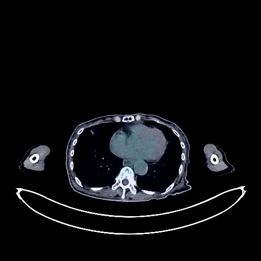
Gastric Cancer PET/CT (case 983824-000161 from PETWB-REP)
0 views10 days agoWhole-body 18F-FDG PET/CT scan in a patient with Gastric Cancer taken from the PETWB-REP dataset. The following English report (translated from original Chinese) is taken verbatim from the public dataset and has not been modified or otherwise checked for accuracy (see the end for citation). Impression a. Postoperative changes after gastric cancer surgery, no signs of tumor recurrence in the surgical area, no obvious space-occupying lesions in the abdominopelvic cavity; fluid retention in the upper and middle esophagus; increased FDG uptake in some intestinal segments, considered inflammatory or physiological uptake. Endoscopic follow-up is recommended. b. Small amount of pelvic effusion. Postoperative changes in the anterior abdominal wall. Nodular goiter, ultrasound follow-up is recommended. Multiple solid micronodules and ground-glass opacities in both lungs, considered chronic inflammatory nodules or atypical adenomatous hyperplasia, annual HRCT follow-up is recommended. Scattered chronic inflammation and sequelae in both lungs. Partial arteriosclerosis (including coronary arteries). Small amount of pericardial effusion. Intrahepatic calcifications. Concentrated bile in the gallbladder. Bilateral renal cysts, small stone in the left kidney. Bilateral adrenal hyperplasia is highly probable; a follow-up MRI with contrast is recommended to rule out other possibilities. Residual contrast agent in the bladder. Scoliosis, degenerative changes in the spine. Sacral canal cyst, L4/5 and L5/S1 intervertebral disc bulge. Age-related brain changes, deep lacunar infarcts in the brain. This case is from PETWB-REP, a curated dataset of whole-body 18F-FDG PET/CT scans and corresponding radiology reports from 490 patients with a broad spectrum of malignancies. The data were retrospectively collected from patients who underwent clinically indicated whole-body 18F-FDG PET/CT scans at the Shanghai Universal Medical Imaging Diagnostic Center between 2021 and 2024. License: Creative Commons Attribution 4.0 International (CC BY 4.0) Citation: Xue, L., Feng, G., Wenbo, Z., Zhang, Y., Li, L., Wang, S., Peng, L., Peng, S., & Gao, X. (2026). PETWB-REP: A Multi-Cancer Whole-Body FDG PET/CT Dataset with Corresponding Radiology Reports [Data set]. Zenodo. https://doi.org/10.5281/zenodo.18670487
Whole BodyPET/CT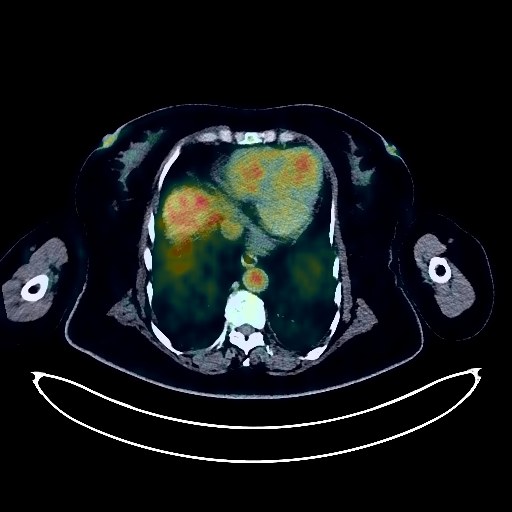
Renal Cancer PET/CT (case 983824-000134 from PETWB-REP)
0 views10 days agoWhole-body 18F-FDG PET/CT scan in a patient with Renal Cancer taken from the PETWB-REP dataset. The following English report (translated from original Chinese) is taken verbatim from the public dataset and has not been modified or otherwise checked for accuracy (see the end for citation). Impression a. Left renal mass with increased FDG metabolism, consistent with renal cell carcinoma, involving the spleen. Reactive hyperplasia of small retroperitoneal lymph nodes. b. Multiple metastatic tumors in both lungs. Scattered post-inflammatory lesions in both lungs. Anemia changes, partial calcification of arterial walls (including coronary arteries). Bilateral breast hyperplasia, calcification in the right breast. Accessory spleen. Small cyst in the right kidney. Post-uterine surgery changes. Partial chronic inflammatory changes in the gastric wall. Osteoporosis, degenerative changes in the spine, L4/5 and L5/S1 intervertebral disc bulge. Bilateral frozen shoulder. No obvious abnormalities seen on cranial scintigraphy. This case is from PETWB-REP, a curated dataset of whole-body 18F-FDG PET/CT scans and corresponding radiology reports from 490 patients with a broad spectrum of malignancies. The data were retrospectively collected from patients who underwent clinically indicated whole-body 18F-FDG PET/CT scans at the Shanghai Universal Medical Imaging Diagnostic Center between 2021 and 2024. License: Creative Commons Attribution 4.0 International (CC BY 4.0) Citation: Xue, L., Feng, G., Wenbo, Z., Zhang, Y., Li, L., Wang, S., Peng, L., Peng, S., & Gao, X. (2026). PETWB-REP: A Multi-Cancer Whole-Body FDG PET/CT Dataset with Corresponding Radiology Reports [Data set]. Zenodo. https://doi.org/10.5281/zenodo.18670487
Whole BodyPET/CT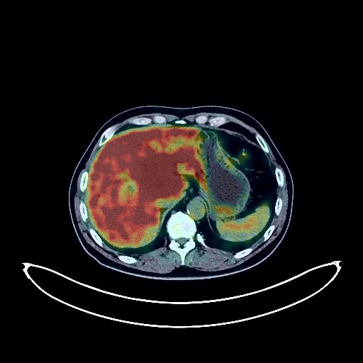
Cholangiocarcinoma PET/CT (case 983824-000207 from PETWB-REP)
1 views10 days agoWhole-body 18F-FDG PET/CT scan in a patient with Cholangiocarcinoma taken from the PETWB-REP dataset. The following English report (translated from original Chinese) is taken verbatim from the public dataset and has not been modified or otherwise checked for accuracy (see the end for citation). Impression Mass in the hepatic hilum, with increased FDG uptake and intrahepatic bile duct dilation, suggestive of cholangiocarcinoma; intrahepatic metastasis; hilar lymph node metastasis. Multiple chronic inflammatory micronodules in both lungs, with a few post-inflammatory lesions in the lower lobes of both lungs. Calcification of some arterial walls (including coronary arteries). Accessory spleen. Left renal cyst. Prostatic calcification. Postoperative changes after right inguinal hernia surgery. Slight thickening of the rectosigmoid junction wall with increased FDG uptake, suggestive of possible inflammation or physiological changes; endoscopic re-examination is recommended to rule out other possibilities. Spinal osteophyte formation. L4/5 and L5/S1 intervertebral disc bulge, L5/S1 disc pneumatosis and degeneration. Mild age-related brain changes. This case is from PETWB-REP, a curated dataset of whole-body 18F-FDG PET/CT scans and corresponding radiology reports from 490 patients with a broad spectrum of malignancies. The data were retrospectively collected from patients who underwent clinically indicated whole-body 18F-FDG PET/CT scans at the Shanghai Universal Medical Imaging Diagnostic Center between 2021 and 2024. License: Creative Commons Attribution 4.0 International (CC BY 4.0) Citation: Xue, L., Feng, G., Wenbo, Z., Zhang, Y., Li, L., Wang, S., Peng, L., Peng, S., & Gao, X. (2026). PETWB-REP: A Multi-Cancer Whole-Body FDG PET/CT Dataset with Corresponding Radiology Reports [Data set]. Zenodo. https://doi.org/10.5281/zenodo.18670487
Whole BodyPET/CT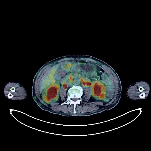
Lymphoma PET/CT (case 983824-000070 from PETWB-REP)
2 views10 days agoWhole-body 18F-FDG PET/CT scan in a patient with Lymphoma taken from the PETWB-REP dataset. The following English report (translated from original Chinese) is taken verbatim from the public dataset and has not been modified or otherwise checked for accuracy (see the end for citation). Impression a. Multiple lymphadenopathy throughout the body with increased FDG metabolism (see description for details). b. Extensive thickening of soft tissue in the left ethmoid sinus and bilateral nasal cavities with increased FDG metabolism; nodular thickening of the left pleura with increased FDG metabolism. c. Slightly low-density lesion in the left lobe of the liver with increased FDG metabolism; space-occupying lesion in the bilateral adrenal regions with increased FDG metabolism. d. Multiple areas of bone with increased FDG metabolism throughout the body, with partial bone destruction (see description for details); multiple subcutaneous soft tissue nodules with increased FDG metabolism throughout the body. All of the above suggest multisystemic lymphoma infiltration; follow-up examination after treatment is recommended. a. Bronchiectasis with infection in the left lower lobe of the lung; follow-up CT scan after treatment is recommended. b. Emphysema in both lungs; bullae in the right upper lobe of the lung. Calcification in the lower lobe of the right lung, scattered post-inflammatory lesions in both lungs. ? c. Thickening and adhesions of the left pleura. Enlarged cardiac silhouette, thickened pericardium with a small amount of effusion, anemic changes, calcification of some arterial walls (including coronary arteries). Chronic cholecystitis, gallstones. Left renal cyst, bilateral renal pelvis and calyces dilation with effusion. Benign prostatic hyperplasia with calcification. Chronic inflammatory changes in part of the gastric wall. Slight scoliosis, degenerative changes in the spine, L4/5 intervertebral disc bulge with pneumoconiosis. Senile brain, deep lacunar infarcts. Chronic inflammation of both maxillary sinuses. This case is from PETWB-REP, a curated dataset of whole-body 18F-FDG PET/CT scans and corresponding radiology reports from 490 patients with a broad spectrum of malignancies. The data were retrospectively collected from patients who underwent clinically indicated whole-body 18F-FDG PET/CT scans at the Shanghai Universal Medical Imaging Diagnostic Center between 2021 and 2024. License: Creative Commons Attribution 4.0 International (CC BY 4.0) Citation: Xue, L., Feng, G., Wenbo, Z., Zhang, Y., Li, L., Wang, S., Peng, L., Peng, S., & Gao, X. (2026). PETWB-REP: A Multi-Cancer Whole-Body FDG PET/CT Dataset with Corresponding Radiology Reports [Data set]. Zenodo. https://doi.org/10.5281/zenodo.18670487
Whole BodyPET/CT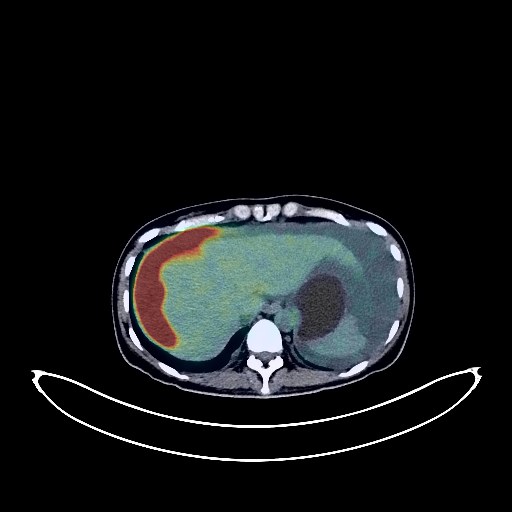
Ovarian Cancer PET/CT (case 983824-000052 from PETWB-REP)
4 views10 days agoWhole-body 18F-FDG PET/CT scan in a patient with Ovarian Cancer taken from the PETWB-REP dataset. The following English report (translated from original Chinese) is taken verbatim from the public dataset and has not been modified or otherwise checked for accuracy (see the end for citation). Impression Bilateral adnexal region cystic-solid lesions with increased FDG metabolism in the solid component; nodular FDG hypermetabolic foci within the uterus; multiple lymph nodes in the retroperitoneum, mesentery, and bilateral prediaphragmatic groups showing some with increased FDG metabolism; thickening of the subdiaphragmatic, abdominopelvic peritoneum, and pelvic floor fascia with increased FDG metabolism. Considering all of the above, this is likely a malignant tumor, possibly originating from the ovary with multiple metastases. Metastatic lesions in the adnexa cannot be ruled out. Please combine clinical findings with enhanced MRI for comprehensive analysis. Abdominal and pelvic effusion. Chronic inflammatory nodule in the right upper lobe of the lung. Calcification in the left upper lobe of the lung, fibrosis in both lower lobes of the lungs. Small amount of bilateral pleural effusion. Increased FDG metabolism in parts of the gastric wall and intestines, considered physiological uptake or chronic inflammation. High-density nodule at approximately the T11 vertebral level within the spinal canal with increased FDG metabolism, considered a possible meningioma. Enhanced MRI follow-up is recommended. Mild osteophyte formation in some vertebral bodies. No obvious abnormalities were found on cranial imaging. This case is from PETWB-REP, a curated dataset of whole-body 18F-FDG PET/CT scans and corresponding radiology reports from 490 patients with a broad spectrum of malignancies. The data were retrospectively collected from patients who underwent clinically indicated whole-body 18F-FDG PET/CT scans at the Shanghai Universal Medical Imaging Diagnostic Center between 2021 and 2024. License: Creative Commons Attribution 4.0 International (CC BY 4.0) Citation: Xue, L., Feng, G., Wenbo, Z., Zhang, Y., Li, L., Wang, S., Peng, L., Peng, S., & Gao, X. (2026). PETWB-REP: A Multi-Cancer Whole-Body FDG PET/CT Dataset with Corresponding Radiology Reports [Data set]. Zenodo. https://doi.org/10.5281/zenodo.18670487
Whole BodyPET/CT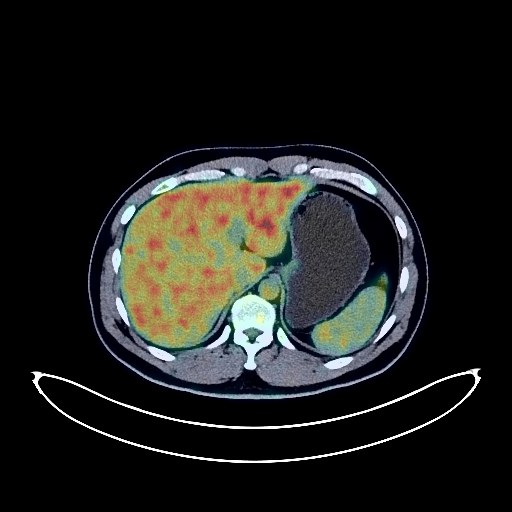
Nasopharyngeal Cancer PET/CT (case 983824-000027 from PETWB-REP)
9 views10 days agoWhole-body 18F-FDG PET/CT scan in a patient with Nasopharyngeal Cancer taken from the PETWB-REP dataset. The following English report (translated from original Chinese) is taken verbatim from the public dataset and has not been modified or otherwise checked for accuracy (see the end for citation). Impression A mass in the nasopharynx with elevated FDG metabolism, consistent with nasopharyngeal carcinoma, invading the posterior nasal aperture, bilateral pterygopalatine fossa, and adjacent skull base; multiple lymph node metastases in the bilateral retropharyngeal and deep cervical spaces. Several solid, chronic inflammatory micronodules in both lungs; follow-up is recommended to rule out other confounding lesions. Minor chronic inflammation and old lesions in both lungs. Mild fatty liver. Partial vertebral osteophyte formation and Schmorl's nodes. No abnormalities seen on cranial scintigraphy. Minor chronic inflammation in the right maxillary sinus. This case is from PETWB-REP, a curated dataset of whole-body 18F-FDG PET/CT scans and corresponding radiology reports from 490 patients with a broad spectrum of malignancies. The data were retrospectively collected from patients who underwent clinically indicated whole-body 18F-FDG PET/CT scans at the Shanghai Universal Medical Imaging Diagnostic Center between 2021 and 2024. License: Creative Commons Attribution 4.0 International (CC BY 4.0) Citation: Xue, L., Feng, G., Wenbo, Z., Zhang, Y., Li, L., Wang, S., Peng, L., Peng, S., & Gao, X. (2026). PETWB-REP: A Multi-Cancer Whole-Body FDG PET/CT Dataset with Corresponding Radiology Reports [Data set]. Zenodo. https://doi.org/10.5281/zenodo.18670487
Whole BodyPET/CT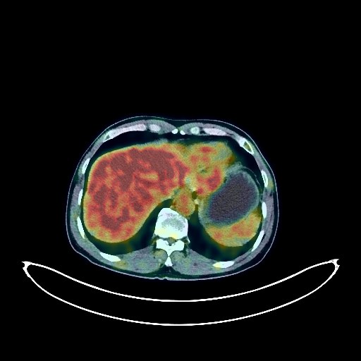
Lung Cancer PET/CT (case 983824-000048 from PETWB-REP)
6 views10 days agoWhole-body 18F-FDG PET/CT scan in a patient with Lung Cancer taken from the PETWB-REP dataset. The following English report (translated from original Chinese) is taken verbatim from the public dataset and has not been modified or otherwise checked for accuracy (see the end for citation). Impression a. A mass near the hilum in the right upper lobe of the lung, with increased FDG metabolism, suggestive of central lung cancer with obstructive inflammation or airway dissemination. Right hilar lymph node metastasis. Reactive hyperplasia of the left hilar and mediastinal lymph nodes is highly probable; follow-up is recommended. b. Scattered inflammation in the remaining lungs. Calcification of some arterial walls (including coronary arteries). Left renal cyst. Slight thickening of the walls of part of the gastric body and antrum, with mildly increased FDG uptake, suggestive of chronic gastritis; follow-up with gastroscopy is recommended. Degenerative changes in the spine. L4/5 and L5/S1 intervertebral disc bulge. Right shoulder periarthritis. Multiple nodules in both parotid glands, with increased FDG metabolism, suggestive of adenolymphoma; follow-up with MRI is recommended. A few ischemic lesions in the deep bilateral brain regions, suggestive of senile encephalopathy. Chronic inflammation of the left maxillary sinus. This case is from PETWB-REP, a curated dataset of whole-body 18F-FDG PET/CT scans and corresponding radiology reports from 490 patients with a broad spectrum of malignancies. The data were retrospectively collected from patients who underwent clinically indicated whole-body 18F-FDG PET/CT scans at the Shanghai Universal Medical Imaging Diagnostic Center between 2021 and 2024. License: Creative Commons Attribution 4.0 International (CC BY 4.0) Citation: Xue, L., Feng, G., Wenbo, Z., Zhang, Y., Li, L., Wang, S., Peng, L., Peng, S., & Gao, X. (2026). PETWB-REP: A Multi-Cancer Whole-Body FDG PET/CT Dataset with Corresponding Radiology Reports [Data set]. Zenodo. https://doi.org/10.5281/zenodo.18670487
Whole BodyPET/CT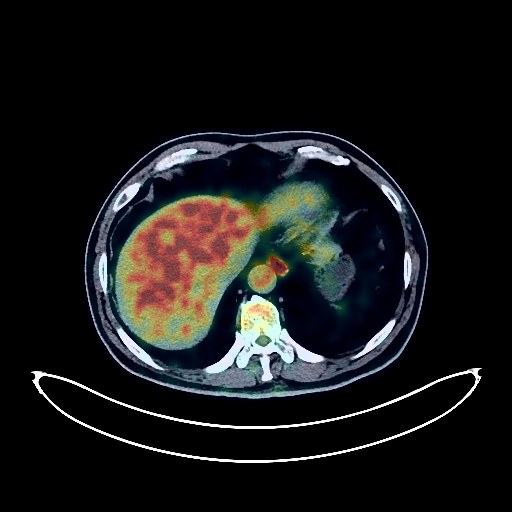
Lung Cancer PET/CT (case 983824-000123 from PETWB-REP)
0 views10 days agoWhole-body 18F-FDG PET/CT scan in a patient with Lung Cancer taken from the PETWB-REP dataset. The following English report (translated from original Chinese) is taken verbatim from the public dataset and has not been modified or otherwise checked for accuracy (see the end for citation). Impression a. A mass in the posterior segment of the left upper lobe with increased FDG metabolism, suggestive of lung cancer with obstructive inflammation; left hilar and mediastinal lymph node metastasis. b. Bilateral emphysema. Several chronic inflammatory micronodules in the right lung. Chronic inflammation in the lingular segment of the left upper lobe. Left pleural thickening. Calcification of some arterial walls (including coronary arteries). a. Benign prostatic hyperplasia with calcification, uneven FDG metabolism; please follow up with PSA and MRI to rule out prostate cancer. b. Mild fatty liver. Small liver cysts. Possible adenomyosis of the gallbladder floor. Accessory splenic nodules. Right renal cyst. Inflammation in the middle and lower esophagus; inflammatory or physiological uptake in the gastric antrum. Endoscopic follow-up is recommended for all of the above. Spinal degeneration, L5 vertebral endplate inflammation. L4/5 and L5/S1 disc herniation. Age-related brain disease, bilateral deep lacunar infarcts. This case is from PETWB-REP, a curated dataset of whole-body 18F-FDG PET/CT scans and corresponding radiology reports from 490 patients with a broad spectrum of malignancies. The data were retrospectively collected from patients who underwent clinically indicated whole-body 18F-FDG PET/CT scans at the Shanghai Universal Medical Imaging Diagnostic Center between 2021 and 2024. License: Creative Commons Attribution 4.0 International (CC BY 4.0) Citation: Xue, L., Feng, G., Wenbo, Z., Zhang, Y., Li, L., Wang, S., Peng, L., Peng, S., & Gao, X. (2026). PETWB-REP: A Multi-Cancer Whole-Body FDG PET/CT Dataset with Corresponding Radiology Reports [Data set]. Zenodo. https://doi.org/10.5281/zenodo.18670487
Whole BodyPET/CT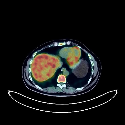
Nasopharyngeal Cancer PET/CT (case 983824-000175 from PETWB-REP)
0 views10 days agoWhole-body 18F-FDG PET/CT scan in a patient with Nasopharyngeal Cancer taken from the PETWB-REP dataset. The following English report (translated from original Chinese) is taken verbatim from the public dataset and has not been modified or otherwise checked for accuracy (see the end for citation). Impression a. No obvious space-occupying lesions were observed in the nasopharynx, and FDG metabolism was normal, suggesting that tumor activity was largely suppressed after treatment. b. Bilateral cervical lymph nodes showed a significant reduction in size compared to before, with most disappearing, and FDG metabolism slightly increased, suggesting that tumor activity was largely suppressed after treatment. c. Multiple bone metastases throughout the body (see description for details) showed a reduction in lesion size compared to before, and decreased FDG metabolism, suggesting that most tumor activity was suppressed, with some tumors still showing activity. Pathological fractures of the T11 and L2 vertebral bodies were observed. The original right erector spinae muscle lesion was not visualized. a. Pure ground-glass nodules were found in the apical segment of the right upper lobe and the anterior segment of the left upper lobe, with no abnormal FDG uptake, similar to before, suggesting atypical adenomatous hyperplasia or chronic inflammatory nodules. Annual HRCT follow-up is recommended. ? b. Chronic inflammatory micronodules (solid) in the posterior basal segment of the left lower lobe; a few fibrotic foci in the medial segment of the right middle lobe and the lingular segment of the left upper lobe. Anemia changes, PICC line inserted. Benign prostatic hyperplasia with calcification. Scoliosis with cervical recurvature. Right shoulder periarthritis. No obvious abnormalities seen on cranial imaging. Left maxillary sinusitis. This case is from PETWB-REP, a curated dataset of whole-body 18F-FDG PET/CT scans and corresponding radiology reports from 490 patients with a broad spectrum of malignancies. The data were retrospectively collected from patients who underwent clinically indicated whole-body 18F-FDG PET/CT scans at the Shanghai Universal Medical Imaging Diagnostic Center between 2021 and 2024. License: Creative Commons Attribution 4.0 International (CC BY 4.0) Citation: Xue, L., Feng, G., Wenbo, Z., Zhang, Y., Li, L., Wang, S., Peng, L., Peng, S., & Gao, X. (2026). PETWB-REP: A Multi-Cancer Whole-Body FDG PET/CT Dataset with Corresponding Radiology Reports [Data set]. Zenodo. https://doi.org/10.5281/zenodo.18670487
Whole BodyPET/CT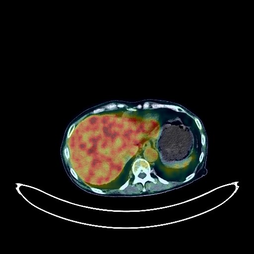
Renal Cancer PET/CT (case 983824-000158 from PETWB-REP)
0 views10 days agoWhole-body 18F-FDG PET/CT scan in a patient with Renal Cancer taken from the PETWB-REP dataset. The following English report (translated from original Chinese) is taken verbatim from the public dataset and has not been modified or otherwise checked for accuracy (see the end for citation). Impression a. Right renal mass with increased FDG metabolism, suggestive of renal cell carcinoma; reactive hyperplasia of retroperitoneal lymph nodes, follow-up recommended to rule out mixed metastases. b. Multiple lung metastases. Liver metastases are highly probable; enhanced MRI analysis recommended. c. No clear tumor thrombus findings in the right renal vein and inferior vena cava; enhanced CT scan recommended. Scattered chronic inflammation and remnants in both lungs. Bilateral pleural thickening. Pericardial thickening with a small amount of effusion, anemia changes, and calcification of some arterial walls (including coronary arteries). Bilateral breast hyperplasia; fibroadenoma possible in the left breast, calcification in the right breast; ultrasound examination recommended. Accessory spleen. Left adrenal hyperplasia. Multiple cysts in the left kidney. Osteoporosis, degenerative changes in the spine, multiple lumbar disc herniations. Sacral canal cyst. Subcutaneous calcification in the right buttock. Age-related brain conditions, deep lacunar infarcts. Submucosal cyst in the left maxillary sinus. This case is from PETWB-REP, a curated dataset of whole-body 18F-FDG PET/CT scans and corresponding radiology reports from 490 patients with a broad spectrum of malignancies. The data were retrospectively collected from patients who underwent clinically indicated whole-body 18F-FDG PET/CT scans at the Shanghai Universal Medical Imaging Diagnostic Center between 2021 and 2024. License: Creative Commons Attribution 4.0 International (CC BY 4.0) Citation: Xue, L., Feng, G., Wenbo, Z., Zhang, Y., Li, L., Wang, S., Peng, L., Peng, S., & Gao, X. (2026). PETWB-REP: A Multi-Cancer Whole-Body FDG PET/CT Dataset with Corresponding Radiology Reports [Data set]. Zenodo. https://doi.org/10.5281/zenodo.18670487
Whole BodyPET/CT