Loading...
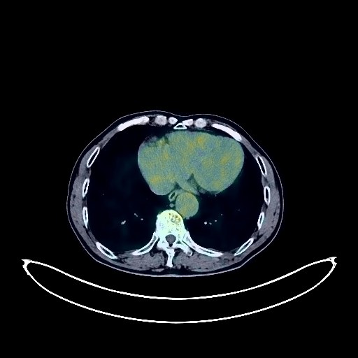
Lung Cancer PET/CT (case 983824-000213 from PETWB-REP)
2 views10 days agoWhole-body 18F-FDG PET/CT scan in a patient with Lung Cancer taken from the PETWB-REP dataset. The following English report (translated from original Chinese) is taken verbatim from the public dataset and has not been modified or otherwise checked for accuracy (see the end for citation). Impression a. A mass in the left upper lobe, lingular segment, closely adhering to the oblique fissure pleura, with increased FDG metabolism, highly suggestive of peripheral lung cancer; please correlate with clinicopathology. b. Multiple small, solid, chronic inflammatory nodules in both lungs; please follow up with CT scans. A few post-inflammatory lesions in both lungs. Reactive hyperplasia of bilateral hilar and mediastinal lymph nodes; please follow up. c. Slightly enlarged cardiac silhouette, with partial arteriosclerosis (including coronary arteries); specialist follow-up is recommended. A nodule with increased FDG metabolism in the deep lobe of the right parotid gland, highly suggestive of a mixed parotid tumor; specialist follow-up is recommended. Hepatic parenchyma with uneven density, small cyst in the left lobe of the liver, and possible gallbladder polyps; ultrasound follow-up is recommended for all of the above. Benign prostatic hyperplasia with calcification. Bilateral hydrocele. Increased FDG metabolism in parts of the colon and rectum, possibly due to physiological uptake or chronic inflammation; please follow up with endoscopy. Osteoporosis, degenerative changes in the spine. L4/5 and L5/S1 disc bulges. Right-sided frozen shoulder. Age-related brain changes, deep lacunar infarcts; MRI is recommended. This case is from PETWB-REP, a curated dataset of whole-body 18F-FDG PET/CT scans and corresponding radiology reports from 490 patients with a broad spectrum of malignancies. The data were retrospectively collected from patients who underwent clinically indicated whole-body 18F-FDG PET/CT scans at the Shanghai Universal Medical Imaging Diagnostic Center between 2021 and 2024. License: Creative Commons Attribution 4.0 International (CC BY 4.0) Citation: Xue, L., Feng, G., Wenbo, Z., Zhang, Y., Li, L., Wang, S., Peng, L., Peng, S., & Gao, X. (2026). PETWB-REP: A Multi-Cancer Whole-Body FDG PET/CT Dataset with Corresponding Radiology Reports [Data set]. Zenodo. https://doi.org/10.5281/zenodo.18670487
Whole BodyPET/CT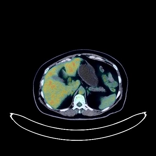
Ovarian Cancer PET/CT (case 983824-000060 from PETWB-REP)
3 views10 days agoWhole-body 18F-FDG PET/CT scan in a patient with Ovarian Cancer taken from the PETWB-REP dataset. The following English report (translated from original Chinese) is taken verbatim from the public dataset and has not been modified or otherwise checked for accuracy (see the end for citation). Impression A large cystic-solid mass in the abdominopelvic cavity with significantly increased FDG metabolism in the solid portion, suggestive of malignancy, most likely ovarian cancer; please confirm with pathology. a. Postoperative changes in the right lung; no signs of tumor recurrence were observed in the surgical area; please compare with old films and follow up with CT. b. Chronic inflammatory ground-glass nodules or atypical adenomatous hyperplasia in the lower lobes of both lungs; chronic inflammatory solid micronodules in both lungs; please have an annual HRCT follow-up. c. A few chronic inflammations and remnants in the remaining lungs. Hyperplastic changes in both breasts. Chronic inflammatory changes in the lower esophagus and stomach; some intestinal physiological uptake. Small liver cysts. Punctate calcifications in the right kidney. Degenerative changes in the spine; significant calcification of the posterior longitudinal ligament at the C5/6 level. No obvious abnormalities were seen on cranial scintigraphy. A few chronic inflammations in the right maxillary sinus. A nodular lesion posterior to the right lobe of the thyroid gland (density similar to the thyroid gland); no increased FDG metabolism was observed; suggestive of a posterior thyroid nodule; please confirm with ultrasound. This case is from PETWB-REP, a curated dataset of whole-body 18F-FDG PET/CT scans and corresponding radiology reports from 490 patients with a broad spectrum of malignancies. The data were retrospectively collected from patients who underwent clinically indicated whole-body 18F-FDG PET/CT scans at the Shanghai Universal Medical Imaging Diagnostic Center between 2021 and 2024. License: Creative Commons Attribution 4.0 International (CC BY 4.0) Citation: Xue, L., Feng, G., Wenbo, Z., Zhang, Y., Li, L., Wang, S., Peng, L., Peng, S., & Gao, X. (2026). PETWB-REP: A Multi-Cancer Whole-Body FDG PET/CT Dataset with Corresponding Radiology Reports [Data set]. Zenodo. https://doi.org/10.5281/zenodo.18670487
Whole BodyPET/CT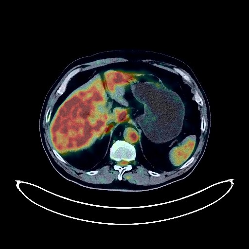
Lung Cancer PET/CT (case 983824-000209 from PETWB-REP)
2 views10 days agoWhole-body 18F-FDG PET/CT scan in a patient with Lung Cancer taken from the PETWB-REP dataset. The following English report (translated from original Chinese) is taken verbatim from the public dataset and has not been modified or otherwise checked for accuracy (see the end for citation). Impression a. A mass in the posterior segment of the left upper lobe, with increased FDG metabolism, suggestive of peripheral lung cancer. Metastasis to the hilar and mediastinal lymph nodes is highly probable. Please correlate with clinicopathology. b. Scattered chronic inflammatory nodules (solid and calcified) in both lungs. Please correlate with CT follow-up. A few post-inflammatory lesions in both lungs. Emphysema in both lungs. Mild pleural thickening bilaterally. Partial arteriosclerosis. Benign prostatic hyperplasia, with uneven FDG metabolism, suggestive of inflammatory or physiological uptake. PSA and ultrasound follow-up are recommended. A biopsy may be necessary. Uneven thyroid density, with unevenly increased FDG metabolism, suggestive of thyroiditis. Please correlate with ultrasound and thyroid function tests. Small liver cysts. Absence after gallbladder surgery. Cysts in the left kidney and left renal pelvis. Chronic antral gastritis. Degenerative changes in the spine. L4/5 and L5/S1 intervertebral disc bulge. Right-sided frozen shoulder. Age-related brain changes, deep lacunar infarcts in the brain; MRI recommended. Minor inflammation of the right maxillary sinus. This case is from PETWB-REP, a curated dataset of whole-body 18F-FDG PET/CT scans and corresponding radiology reports from 490 patients with a broad spectrum of malignancies. The data were retrospectively collected from patients who underwent clinically indicated whole-body 18F-FDG PET/CT scans at the Shanghai Universal Medical Imaging Diagnostic Center between 2021 and 2024. License: Creative Commons Attribution 4.0 International (CC BY 4.0) Citation: Xue, L., Feng, G., Wenbo, Z., Zhang, Y., Li, L., Wang, S., Peng, L., Peng, S., & Gao, X. (2026). PETWB-REP: A Multi-Cancer Whole-Body FDG PET/CT Dataset with Corresponding Radiology Reports [Data set]. Zenodo. https://doi.org/10.5281/zenodo.18670487
Whole BodyPET/CT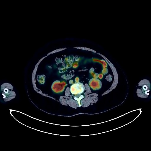
Lymphoma PET/CT (case 983824-000040 from PETWB-REP)
8 views10 days agoWhole-body 18F-FDG PET/CT scan in a patient with Lymphoma taken from the PETWB-REP dataset. The following English report (translated from original Chinese) is taken verbatim from the public dataset and has not been modified or otherwise checked for accuracy (see the end for citation). Impression a. Space-occupying lesions in the left nasal cavity, left maxillary sinus, and left ethmoid sinus, with increased FDG metabolism, consistent with lymphoma, involving the left orbit, with exophthalmos. b. Hypermetabolic lesions in the spleen, suggestive of lymphoma infiltration. c. Possible infection in the left lower lobe of the lung, lymphoma infiltration to be ruled out; follow-up examination after treatment is recommended for comparison. d. Multiple lymph nodes in both sides of the neck, both hilum, and mediastinum, with increased FDG metabolism, highly suggestive of reactive lymph node hyperplasia; follow-up is recommended to rule out lymphoma infiltration. e. Possible reactive hyperplasia of the entire bone marrow; follow-up is recommended to rule out lymphoma infiltration. A few chronic inflammations and old lesions in both lungs. Calcification of some arterial walls (including coronary arteries). Increased FDG metabolism in the terminal ileum and part of the colon, suggestive of inflammatory or physiological uptake. Duodenal diverticulum in the descending part. Possible uterine cavity effusion, cystic lesions cannot be ruled out; further ultrasound examination is recommended. Degenerative changes in the spine. L4/5 and L5/S1 intervertebral disc bulge. No abnormalities were found on cranial scintigraphy. This case is from PETWB-REP, a curated dataset of whole-body 18F-FDG PET/CT scans and corresponding radiology reports from 490 patients with a broad spectrum of malignancies. The data were retrospectively collected from patients who underwent clinically indicated whole-body 18F-FDG PET/CT scans at the Shanghai Universal Medical Imaging Diagnostic Center between 2021 and 2024. License: Creative Commons Attribution 4.0 International (CC BY 4.0) Citation: Xue, L., Feng, G., Wenbo, Z., Zhang, Y., Li, L., Wang, S., Peng, L., Peng, S., & Gao, X. (2026). PETWB-REP: A Multi-Cancer Whole-Body FDG PET/CT Dataset with Corresponding Radiology Reports [Data set]. Zenodo. https://doi.org/10.5281/zenodo.18670487
Whole BodyPET/CT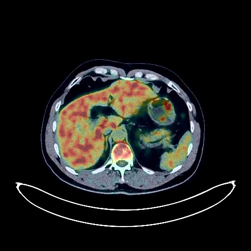
Renal Cancer PET/CT (case 983824-000011 from PETWB-REP)
7 views10 days agoWhole-body 18F-FDG PET/CT scan in a patient with Renal Cancer taken from the PETWB-REP dataset. The following English report (translated from original Chinese) is taken verbatim from the public dataset and has not been modified or otherwise checked for accuracy (see the end for citation). Impression a. A slightly low-density soft tissue lesion in the left kidney, with background uptake on FDG, suggestive of a renal space-occupying lesion, possibly malignant. Please confirm with contrast-enhanced MRI and clinicopathological examination. Small renal stone in the left kidney. Cyst in the right kidney. b. A soft tissue lesion in the right anterior lobe of the liver, with background uptake on FDG, highly suggestive of a neoplastic lesion. Chronic inflammatory nodules in the posterior segment of the left upper lobe and the apical segment of the right upper lobe. Please confirm with CT follow-up. Soft tissue nodules in the anterior mediastinum, with absent FDG metabolism, suggestive of a thymic cyst or thymoma. Please confirm with CT follow-up. Calcification of some arterial walls. Benign prostatic hyperplasia. Degenerative changes in the spine. Wedge-shaped L2 vertebral body. Intervertebral disc bulging at L3/4, L4/5, and L5/S1. No obvious abnormalities were found on cranial scintigraphy. This case is from PETWB-REP, a curated dataset of whole-body 18F-FDG PET/CT scans and corresponding radiology reports from 490 patients with a broad spectrum of malignancies. The data were retrospectively collected from patients who underwent clinically indicated whole-body 18F-FDG PET/CT scans at the Shanghai Universal Medical Imaging Diagnostic Center between 2021 and 2024. License: Creative Commons Attribution 4.0 International (CC BY 4.0) Citation: Xue, L., Feng, G., Wenbo, Z., Zhang, Y., Li, L., Wang, S., Peng, L., Peng, S., & Gao, X. (2026). PETWB-REP: A Multi-Cancer Whole-Body FDG PET/CT Dataset with Corresponding Radiology Reports [Data set]. Zenodo. https://doi.org/10.5281/zenodo.18670487
Whole BodyPET/CT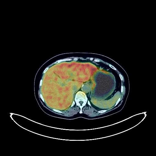
Cervical Cancer PET/CT (case 983824-000171 from PETWB-REP)
2 views10 days agoWhole-body 18F-FDG PET/CT scan in a patient with Cervical Cancer taken from the PETWB-REP dataset. The following English report (translated from original Chinese) is taken verbatim from the public dataset and has not been modified or otherwise checked for accuracy (see the end for citation). Impression Cervical mass with elevated FDG metabolism, consistent with cervical cancer; reactive hyperplasia of small lymph nodes in the left pelvic wall, left iliac vessels, and bilateral inguinal regions. Chronic inflammatory micronodules in both lungs. Chronic inflammation in the right upper lobe, with a few post-inflammatory lesions in both lungs. Mild anemia, slight arteriosclerosis in some arteries. Calcification in the right breast. Calcification in the liver, cyst in the right lobe of the liver. Chronic cholecystitis. Chronic inflammatory changes in part of the gastric wall; please follow up with endoscopy. Degenerative changes in the spine, L4/5 disc herniation. No obvious abnormalities on cranial scintigraphy. Chronic inflammation of the right frontal sinus, right ethmoid sinus, and right maxillary sinus. This case is from PETWB-REP, a curated dataset of whole-body 18F-FDG PET/CT scans and corresponding radiology reports from 490 patients with a broad spectrum of malignancies. The data were retrospectively collected from patients who underwent clinically indicated whole-body 18F-FDG PET/CT scans at the Shanghai Universal Medical Imaging Diagnostic Center between 2021 and 2024. License: Creative Commons Attribution 4.0 International (CC BY 4.0) Citation: Xue, L., Feng, G., Wenbo, Z., Zhang, Y., Li, L., Wang, S., Peng, L., Peng, S., & Gao, X. (2026). PETWB-REP: A Multi-Cancer Whole-Body FDG PET/CT Dataset with Corresponding Radiology Reports [Data set]. Zenodo. https://doi.org/10.5281/zenodo.18670487
Whole BodyPET/CT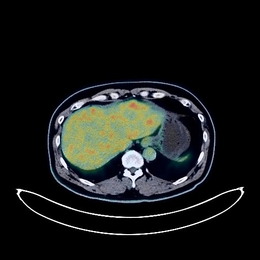
Glioma PET/CT (case 983824-000020 from PETWB-REP)
7 views10 days agoWhole-body 18F-FDG PET/CT scan in a patient with Glioma taken from the PETWB-REP dataset. The following English report (translated from original Chinese) is taken verbatim from the public dataset and has not been modified or otherwise checked for accuracy (see the end for citation). Impression a. A mass in the left basal ganglia region with increased FDG metabolism, suggesting a high probability of a primary malignant tumor, such as glioblastoma. Further examination with contrast-enhanced MRI is recommended. b. Senile brain changes. Cavity septum pellucidum formation. a. Ground-glass nodule in the lower lingular segment of the left upper lobe, with no increased FDG metabolism, suggesting a chronic inflammatory nodule or atypical adenomatous hyperplasia. Annual HRCT follow-up is recommended. Chronic inflammatory micronodule (solid) in the lower lingular segment of the left upper lobe. b. Slight thickening of the pleura on both sides. Reactive hyperplasia of the lymph nodes in the deep cervical space and axilla on both sides. Calcification of the arterial walls on both sides. Small cyst in the left lobe of the liver. Localized calcium salt deposition or contrast agent residue in the left kidney. Small vascular leiomyolipomas of the left kidney. Calcification of the prostate. Possible hemorrhoids. Osteophyte formation in the vertebrae on both sides. L4/5 and L5/S1 intervertebral disc bulge. Reactive hyperplasia of small lymph nodes in the parotid glands on both sides. This case is from PETWB-REP, a curated dataset of whole-body 18F-FDG PET/CT scans and corresponding radiology reports from 490 patients with a broad spectrum of malignancies. The data were retrospectively collected from patients who underwent clinically indicated whole-body 18F-FDG PET/CT scans at the Shanghai Universal Medical Imaging Diagnostic Center between 2021 and 2024. License: Creative Commons Attribution 4.0 International (CC BY 4.0) Citation: Xue, L., Feng, G., Wenbo, Z., Zhang, Y., Li, L., Wang, S., Peng, L., Peng, S., & Gao, X. (2026). PETWB-REP: A Multi-Cancer Whole-Body FDG PET/CT Dataset with Corresponding Radiology Reports [Data set]. Zenodo. https://doi.org/10.5281/zenodo.18670487
Whole BodyPET/CT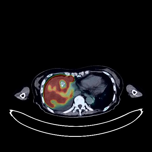
Colon Cancer PET/CT (case 983824-000067 from PETWB-REP)
0 views10 days agoWhole-body 18F-FDG PET/CT scan in a patient with Colon Cancer taken from the PETWB-REP dataset. The following English report (translated from original Chinese) is taken verbatim from the public dataset and has not been modified or otherwise checked for accuracy (see the end for citation). Impression Changes after left hemicolectomy and transverse colostomy: a. Multiple peritoneal seeding metastases (including around the anastomosis site of the descending colon stump). Multiple lymph node metastases throughout the body. Multiple liver metastases. Multiple lung metastases. Multiple bone metastases throughout the body. b. A cystic-solid mass in the pelvis with significantly increased FDG metabolism in the solid portion, suggesting possible malignancy; primary or metastatic origin in the adnexa is possible. Possible uterine metastases. Manifestations of chronic gastritis. Chronic inflammatory changes in part of the right colon wall. Chronic cholecystitis with cholestasis of the gallbladder. Left renal cyst. Bilateral pleural effusion, more pronounced on the right side, with partial atelectasis of the right lower lobe. Bilateral breast hyperplasia. Degenerative changes in the spine, L4 vertebral body grade I anterior slippage. Possible falx cerebri meningioma. Chronic inflammation of the right maxillary sinus. This case is from PETWB-REP, a curated dataset of whole-body 18F-FDG PET/CT scans and corresponding radiology reports from 490 patients with a broad spectrum of malignancies. The data were retrospectively collected from patients who underwent clinically indicated whole-body 18F-FDG PET/CT scans at the Shanghai Universal Medical Imaging Diagnostic Center between 2021 and 2024. License: Creative Commons Attribution 4.0 International (CC BY 4.0) Citation: Xue, L., Feng, G., Wenbo, Z., Zhang, Y., Li, L., Wang, S., Peng, L., Peng, S., & Gao, X. (2026). PETWB-REP: A Multi-Cancer Whole-Body FDG PET/CT Dataset with Corresponding Radiology Reports [Data set]. Zenodo. https://doi.org/10.5281/zenodo.18670487
Whole BodyPET/CT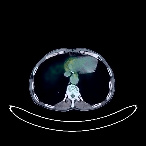
Lung Cancer PET/CT (case 983824-000192 from PETWB-REP)
0 views10 days agoWhole-body 18F-FDG PET/CT scan in a patient with Lung Cancer taken from the PETWB-REP dataset. The following English report (translated from original Chinese) is taken verbatim from the public dataset and has not been modified or otherwise checked for accuracy (see the end for citation). Impression a. A mass near the bronchial opening in the lingular segment of the left upper lobe at the left upper hilum, with elevated FDG metabolism, consistent with lung cancer, and left hilar lymph node metastasis. A few atelectasis or inflammations in the lingular segment of the left upper lobe. b. Scattered chronic inflammatory micronodules (solid and calcified) in both lungs; please follow up with CT scans. A few post-inflammatory lesions in both lungs. Partial arteriosclerosis (including coronary arteries). Left kidney stone. Degenerative changes in the spine. L4/5 and L5/S1 intervertebral disc bulges. Schmorl's nodes at the upper margin of the S1 vertebral body. Age-related brain changes; deep lacunar infarcts in the brain; MRI is recommended. A few ethmoid sinuses bilaterally. Poor pneumatization of the mastoid process bilaterally. This case is from PETWB-REP, a curated dataset of whole-body 18F-FDG PET/CT scans and corresponding radiology reports from 490 patients with a broad spectrum of malignancies. The data were retrospectively collected from patients who underwent clinically indicated whole-body 18F-FDG PET/CT scans at the Shanghai Universal Medical Imaging Diagnostic Center between 2021 and 2024. License: Creative Commons Attribution 4.0 International (CC BY 4.0) Citation: Xue, L., Feng, G., Wenbo, Z., Zhang, Y., Li, L., Wang, S., Peng, L., Peng, S., & Gao, X. (2026). PETWB-REP: A Multi-Cancer Whole-Body FDG PET/CT Dataset with Corresponding Radiology Reports [Data set]. Zenodo. https://doi.org/10.5281/zenodo.18670487
Whole BodyPET/CT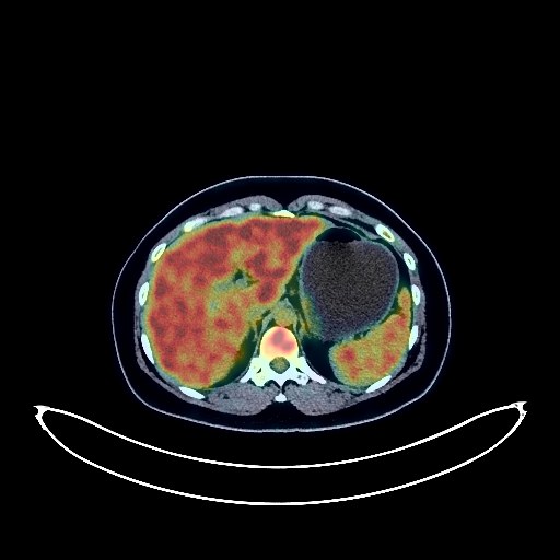
Cervical Cancer PET/CT (case 983824-000077 from PETWB-REP)
0 views10 days agoWhole-body 18F-FDG PET/CT scan in a patient with Cervical Cancer taken from the PETWB-REP dataset. The following English report (translated from original Chinese) is taken verbatim from the public dataset and has not been modified or otherwise checked for accuracy (see the end for citation). Impression a. Cervical mass with increased FDG metabolism, consistent with cervical cancer; reactive hyperplasia of small lymph nodes beside bilateral iliac vessels. b. Slightly low-density lesion in the right adnexal region with increased FDG metabolism, likely due to physiological uptake by the ovary; ultrasound follow-up is recommended. Chronic inflammatory micronodules in both lungs. Small amount of pleural effusion bilaterally. Mild anemia. Slight incomplete thymic regression. Left renal calculus. Right renal cyst. Chronic inflammatory changes or physiological uptake in some intestinal segments; endoscopic follow-up is recommended. No obvious abnormalities seen on cranial scintigraphy. Chronic inflammation of bilateral ethmoid sinuses and bilateral maxillary sinuses. This case is from PETWB-REP, a curated dataset of whole-body 18F-FDG PET/CT scans and corresponding radiology reports from 490 patients with a broad spectrum of malignancies. The data were retrospectively collected from patients who underwent clinically indicated whole-body 18F-FDG PET/CT scans at the Shanghai Universal Medical Imaging Diagnostic Center between 2021 and 2024. License: Creative Commons Attribution 4.0 International (CC BY 4.0) Citation: Xue, L., Feng, G., Wenbo, Z., Zhang, Y., Li, L., Wang, S., Peng, L., Peng, S., & Gao, X. (2026). PETWB-REP: A Multi-Cancer Whole-Body FDG PET/CT Dataset with Corresponding Radiology Reports [Data set]. Zenodo. https://doi.org/10.5281/zenodo.18670487
Whole BodyPET/CT