Loading...
Liver Cancer PET/CT (case 984005-000017 from PETWB-REP)
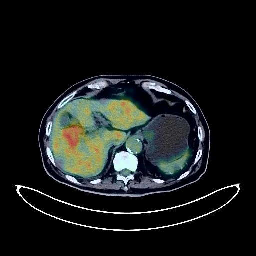
2 views9 days agoWhole-body 18F-FDG PET/CT scan in a patient with Liver Cancer taken from the PETWB-REP dataset. The following English report (translated from original Chinese) is taken verbatim from the public dataset and has not been modified or otherwise checked for accuracy (see the end for citation). Impression Soft tissue mass in the lower right lobe of the liver with increased FDG metabolism, suggestive of hepatocellular carcinoma. Liver cirrhosis. Multiple liver cysts. Postoperative changes after right lung cancer surgery, no obvious signs of tumor recurrence in the surgical area. Multiple ground-glass nodules in both lungs, FDG metabolism normal, suggestive of chronic inflammatory nodules or atypical adenomatous hyperplasia. Multiple solid chronic inflammatory micronodules in both lungs. Emphysema, sequelae of pneumonia in both lungs. Calcification of some arterial walls (including coronary arteries). Localized FDG metabolism increase in the left ventricle, physiological metabolism is the primary consideration, pathological changes to be ruled out, echocardiography/enhanced CT follow-up recommended. Multiple cysts in both kidneys, complex cyst in the left kidney. Kidney stones in both kidneys. Spinal degenerative changes. Pneumothorax and degeneration of the L4/5 and L5/S1 intervertebral discs. Bone island in the right iliac bone. Multiple ischemic lesions in the brain, white matter degeneration, age-related brain changes, MRI follow-up recommended. Slight chronic inflammation of the right maxillary sinus. This case is from PETWB-REP, a curated dataset of whole-body 18F-FDG PET/CT scans and corresponding radiology reports from 490 patients with a broad spectrum of malignancies. The data were retrospectively collected from patients who underwent clinically indicated whole-body 18F-FDG PET/CT scans at the Shanghai Universal Medical Imaging Diagnostic Center between 2021 and 2024. License: Creative Commons Attribution 4.0 International (CC BY 4.0) Citation: Xue, L., Feng, G., Wenbo, Z., Zhang, Y., Li, L., Wang, S., Peng, L., Peng, S., & Gao, X. (2026). PETWB-REP: A Multi-Cancer Whole-Body FDG PET/CT Dataset with Corresponding Radiology Reports [Data set]. Zenodo. https://doi.org/10.5281/zenodo.18670487
Whole BodyPET/CT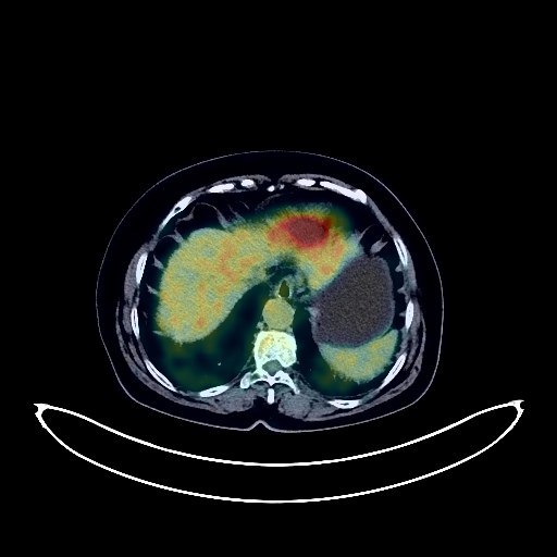
Liver Cancer PET/CT (case 984005-000016 from PETWB-REP)
2 views9 days agoWhole-body 18F-FDG PET/CT scan in a patient with Liver Cancer taken from the PETWB-REP dataset. The following English report (translated from original Chinese) is taken verbatim from the public dataset and has not been modified or otherwise checked for accuracy (see the end for citation). Impression a. A slightly low-density mass in the left inner lobe of the liver with increased FDG metabolism, strongly suggestive of hepatocellular carcinoma; please confirm with pathology. b. Liver cirrhosis. Accessory spleen. Post-cholecystectomy. Multiple chronic inflammatory lesions and sequelae in both lungs. Reactive hyperplasia of mediastinal lymph nodes. Calcification of some arterial walls (including coronary arteries). Left renal calculi. Spinal osteophyte formation. L4/5 and L5/S1 intervertebral disc bulge. Bilateral frozen shoulder. Right iliac bone island. A few ischemic lesions deep in the brain. Senile cerebral changes. Left maxillary sinusitis. This case is from PETWB-REP, a curated dataset of whole-body 18F-FDG PET/CT scans and corresponding radiology reports from 490 patients with a broad spectrum of malignancies. The data were retrospectively collected from patients who underwent clinically indicated whole-body 18F-FDG PET/CT scans at the Shanghai Universal Medical Imaging Diagnostic Center between 2021 and 2024. License: Creative Commons Attribution 4.0 International (CC BY 4.0) Citation: Xue, L., Feng, G., Wenbo, Z., Zhang, Y., Li, L., Wang, S., Peng, L., Peng, S., & Gao, X. (2026). PETWB-REP: A Multi-Cancer Whole-Body FDG PET/CT Dataset with Corresponding Radiology Reports [Data set]. Zenodo. https://doi.org/10.5281/zenodo.18670487
Whole BodyPET/CT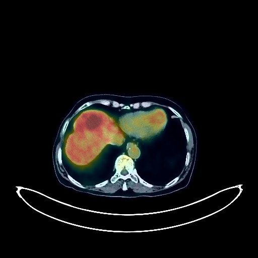
Liver Cancer PET/CT (case 984005-000015 from PETWB-REP)
3 views9 days agoWhole-body 18F-FDG PET/CT scan in a patient with Liver Cancer taken from the PETWB-REP dataset. The following English report (translated from original Chinese) is taken verbatim from the public dataset and has not been modified or otherwise checked for accuracy (see the end for citation). Impression Two liver lesions with elevated FDG metabolism, suggestive of malignancy, hepatocellular carcinoma is the primary consideration, cholangiocarcinoma to be ruled out. Please combine tumor markers and enhanced MRI for comprehensive analysis. Metastasis to the right para-inferior vena cava lymph nodes in the upper abdomen. Reactive hyperplasia of the remaining retroperitoneal lymph nodes. a. Several ground-glass nodules in both lungs, FDG metabolism normal, suggest inflammation or atypical adenomatous hyperplasia, CT follow-up recommended. b. Chronic inflammatory micronodules (solid) in both lungs, CT follow-up recommended. Slight bronchial dilatation with chronic inflammation in the lower lingular segment of the left upper lobe, a few post-inflammatory remnants in both lungs. Partial pleural thickening bilaterally. Tracheal diverticulum. Mild anemia changes, partial calcification of arterial walls (including coronary arteries). Small nodules in both breasts, FDG metabolism normal, suggest fibroadenoma or hyperplastic nodules, calcification in the right breast, ultrasound follow-up recommended. Calcification in the right lobe of the liver. Left adrenal hyperplasia. Chronic inflammatory changes or physiological metabolic changes in parts of the stomach wall and intestines; please follow up with endoscopy. Osteoporosis, degenerative changes in the spine. L4/5 and L5/S1 intervertebral disc bulge with pneumoconiosis and degeneration. Age-related brain changes, deep lacunar infarcts in the brain, and formation of some small softening lesions. Bilateral chronic ethmoid sinusitis. This case is from PETWB-REP, a curated dataset of whole-body 18F-FDG PET/CT scans and corresponding radiology reports from 490 patients with a broad spectrum of malignancies. The data were retrospectively collected from patients who underwent clinically indicated whole-body 18F-FDG PET/CT scans at the Shanghai Universal Medical Imaging Diagnostic Center between 2021 and 2024. License: Creative Commons Attribution 4.0 International (CC BY 4.0) Citation: Xue, L., Feng, G., Wenbo, Z., Zhang, Y., Li, L., Wang, S., Peng, L., Peng, S., & Gao, X. (2026). PETWB-REP: A Multi-Cancer Whole-Body FDG PET/CT Dataset with Corresponding Radiology Reports [Data set]. Zenodo. https://doi.org/10.5281/zenodo.18670487
Whole BodyPET/CT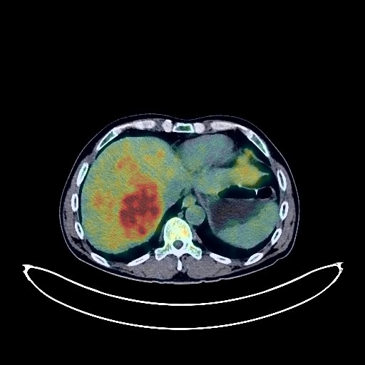
Liver Cancer PET/CT (case 984005-000014 from PETWB-REP)
3 views9 days agoWhole-body 18F-FDG PET/CT scan in a patient with Liver Cancer taken from the PETWB-REP dataset. The following English report (translated from original Chinese) is taken verbatim from the public dataset and has not been modified or otherwise checked for accuracy (see the end for citation). Impression a. Multiple slightly low-density nodules and masses in the liver parenchyma, with unevenly increased FDG metabolism, suggestive of malignancy, with hepatocellular carcinoma being the primary consideration. Please combine AFP and enhanced MRI for comprehensive analysis. b. Metastasis to lymph nodes in the porta hepatis, hepatogastric space, peripancreatic region, mesenteric root, and retroperitoneal perivascular region. A small amount of pelvic effusion. Chronic inflammatory micronodules in the apical-posterior segment of the left upper lobe of the lung. Please follow up with CT. A few post-inflammatory lesions in both lungs. Partial arteriosclerosis. Small liver cysts. Prostatic calcifications. Small amount of hydrocele bilaterally. Chronic gastritis; increased FDG metabolism in parts of the colon and rectum, suggestive of physiological metabolism or chronic inflammatory changes; small diverticulum in the hepatic flexure of the colon. Please follow up with endoscopy for the above. Degenerative changes in the spine. L4/5 and L5/S1 intervertebral disc bulges. Small bony island in the left iliac bone. No obvious abnormalities were found on cranial scintigraphy. There is slight inflammation of the right maxillary sinus. This case is from PETWB-REP, a curated dataset of whole-body 18F-FDG PET/CT scans and corresponding radiology reports from 490 patients with a broad spectrum of malignancies. The data were retrospectively collected from patients who underwent clinically indicated whole-body 18F-FDG PET/CT scans at the Shanghai Universal Medical Imaging Diagnostic Center between 2021 and 2024. License: Creative Commons Attribution 4.0 International (CC BY 4.0) Citation: Xue, L., Feng, G., Wenbo, Z., Zhang, Y., Li, L., Wang, S., Peng, L., Peng, S., & Gao, X. (2026). PETWB-REP: A Multi-Cancer Whole-Body FDG PET/CT Dataset with Corresponding Radiology Reports [Data set]. Zenodo. https://doi.org/10.5281/zenodo.18670487
Whole BodyPET/CT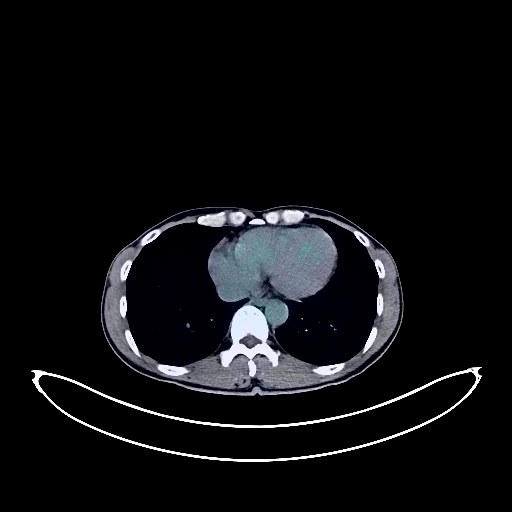
Liver Cancer PET/CT (case 984005-000013 from PETWB-REP)
1 views9 days agoWhole-body 18F-FDG PET/CT scan in a patient with Liver Cancer taken from the PETWB-REP dataset. The following English report (translated from original Chinese) is taken verbatim from the public dataset and has not been modified or otherwise checked for accuracy (see the end for citation). Impression a. Slightly low-density nodule with increased FDG metabolism in the lower right lobe of the liver, suggestive of hepatocellular carcinoma. b. Hemangioma in the upper left lateral lobe of the liver. Widening of the main portal vein. Slightly enlarged spleen. Cholestasis in the gallbladder. a. Small nodule in the left main bronchus, FDG metabolism normal, currently considered benign, follow-up CT recommended. b. Scattered chronic inflammatory micronodules (solid) in both lungs. Physiological changes or inflammatory lesions in the gastric cardia, follow-up gastroscopy recommended. Small amount of pelvic effusion. Degenerative changes in the spine. Calcification of the falx cerebri, cisterna magna and occipital region. Right maxillary sinusitis. This case is from PETWB-REP, a curated dataset of whole-body 18F-FDG PET/CT scans and corresponding radiology reports from 490 patients with a broad spectrum of malignancies. The data were retrospectively collected from patients who underwent clinically indicated whole-body 18F-FDG PET/CT scans at the Shanghai Universal Medical Imaging Diagnostic Center between 2021 and 2024. License: Creative Commons Attribution 4.0 International (CC BY 4.0) Citation: Xue, L., Feng, G., Wenbo, Z., Zhang, Y., Li, L., Wang, S., Peng, L., Peng, S., & Gao, X. (2026). PETWB-REP: A Multi-Cancer Whole-Body FDG PET/CT Dataset with Corresponding Radiology Reports [Data set]. Zenodo. https://doi.org/10.5281/zenodo.18670487
Whole BodyPET/CT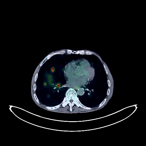
Liver Cancer PET/CT (case 984005-000012 from PETWB-REP)
1 views9 days agoWhole-body 18F-FDG PET/CT scan in a patient with Liver Cancer taken from the PETWB-REP dataset. The following English report (translated from original Chinese) is taken verbatim from the public dataset and has not been modified or otherwise checked for accuracy (see the end for citation). Impression a. Multiple fused nodules and masses in the liver with unevenly increased FDG metabolism suggestive of malignancy, with primary liver cancer being the first consideration. Please correlate with clinicopathology. Multiple lung metastases; left adrenal metastasis is highly probable. b. Possible lymph node metastases in the hepatic hilum, hilar space, retroperitoneum, bilateral hilar regions, and mediastinum. c. Liver cirrhosis; small amount of effusion in the abdomen and pelvis. Partial flocculent thickening of the greater omentum and mesentery, with normal FDG metabolism. Please confirm with CT follow-up to rule out metastasis. A few post-inflammatory lesions in both lungs, emphysema, and bullae in both lungs. Mild thickening of the pleura on both sides. Partial arteriosclerosis (including coronary arteries). Cholecystitis, gallstones. Schistosomiasis intestinal disease. Prostatic calcifications. Osteoporosis. Spinal degeneration. L4/5 and L5/S1 intervertebral disc herniation. Bilateral deep lacunar infarcts, age-related brain changes. Chronic inflammation of the paranasal sinuses. A slightly high-density nodule next to the left parotid gland with increased FDG metabolism is highly suggestive of a mixed tumor; please follow up with ultrasound. This case is from PETWB-REP, a curated dataset of whole-body 18F-FDG PET/CT scans and corresponding radiology reports from 490 patients with a broad spectrum of malignancies. The data were retrospectively collected from patients who underwent clinically indicated whole-body 18F-FDG PET/CT scans at the Shanghai Universal Medical Imaging Diagnostic Center between 2021 and 2024. License: Creative Commons Attribution 4.0 International (CC BY 4.0) Citation: Xue, L., Feng, G., Wenbo, Z., Zhang, Y., Li, L., Wang, S., Peng, L., Peng, S., & Gao, X. (2026). PETWB-REP: A Multi-Cancer Whole-Body FDG PET/CT Dataset with Corresponding Radiology Reports [Data set]. Zenodo. https://doi.org/10.5281/zenodo.18670487
Whole BodyPET/CT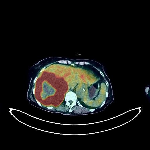
Liver Cancer PET/CT (case 984005-000011 from PETWB-REP)
1 views9 days agoWhole-body 18F-FDG PET/CT scan in a patient with Liver Cancer taken from the PETWB-REP dataset. The following English report (translated from original Chinese) is taken verbatim from the public dataset and has not been modified or otherwise checked for accuracy (see the end for citation). Impression a. Irregular mixed-density nodules and masses in the liver parenchyma, predominantly in the right lobe, with unevenly increased FDG metabolism, suggestive of malignancy. Hepatocellular carcinoma with intrahepatic metastasis is the primary consideration, but metastatic tumors should be ruled out. Please combine tumor markers for comprehensive analysis. b. Pelvic peritoneal seeding metastasis. Hilar lymph node metastasis to be ruled out; follow-up is recommended. a. Chronic inflammatory micronodules in the anterior segment of the left upper lobe; please follow up with CT. A few post-inflammatory lesions in both lungs. Bilateral pleural thickening. b. Cardiac enlargement, partial arteriosclerosis (including coronary arteries); abdominal aortic dilatation. Specialist follow-up is recommended. Mild hyperplasia or incomplete regression in both breasts, calcification in the right breast. Cholecystitis; ultrasound follow-up is recommended. Bilateral renal cysts. Left adrenal hyperplasia. High-density contrast agent residue in the urinary system. Chronic gastritis; please follow up with endoscopy. Increased FDG metabolism in a portion of the small intestine in the right lower pelvic cavity, suggesting physiological metabolism or chronic inflammatory changes; enhanced CT follow-up is recommended. Osteoporosis, degenerative changes in the spine. Old compressive changes in the T7 and L3 vertebral bodies, L4/5 and L5/S1 intervertebral disc bulges. Sacral canal cyst. Age-related brain changes, deep lacunar infarcts in the brain. Inflammation of the right ethmoid and maxillary sinuses. Reactive hyperplasia of bilateral deep cervical interspace, submandibular, and submental lymph nodes. Nodular goiter or thyroiditis; follow-up with ultrasound and thyroid function tests is recommended. This case is from PETWB-REP, a curated dataset of whole-body 18F-FDG PET/CT scans and corresponding radiology reports from 490 patients with a broad spectrum of malignancies. The data were retrospectively collected from patients who underwent clinically indicated whole-body 18F-FDG PET/CT scans at the Shanghai Universal Medical Imaging Diagnostic Center between 2021 and 2024. License: Creative Commons Attribution 4.0 International (CC BY 4.0) Citation: Xue, L., Feng, G., Wenbo, Z., Zhang, Y., Li, L., Wang, S., Peng, L., Peng, S., & Gao, X. (2026). PETWB-REP: A Multi-Cancer Whole-Body FDG PET/CT Dataset with Corresponding Radiology Reports [Data set]. Zenodo. https://doi.org/10.5281/zenodo.18670487
Whole BodyPET/CT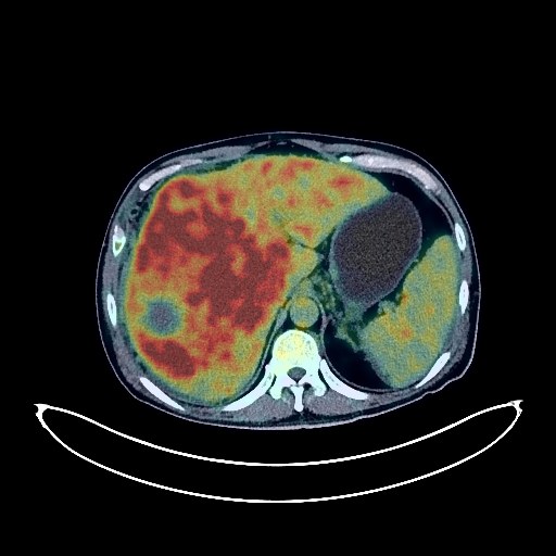
Liver Cancer PET/CT (case 984005-000010 from PETWB-REP)
2 views9 days agoWhole-body 18F-FDG PET/CT scan in a patient with Liver Cancer taken from the PETWB-REP dataset. The following English report (translated from original Chinese) is taken verbatim from the public dataset and has not been modified or otherwise checked for accuracy (see the end for citation). Impression a. A large, irregular mass in the right lobe of the liver and adjacent left medial lobe with increased FDG metabolism, suggestive of hepatocellular carcinoma; focal FDG metabolism increase in the left lobe of the liver suggests metastasis or daughter lesions; portal vein tumor thrombosis is the primary consideration. Please analyze the above in conjunction with enhanced MRI images. b. Liver cirrhosis, splenomegaly. Reactive hyperplasia of the hilar and retroperitoneal lymph nodes; follow-up is recommended to rule out other possibilities. Abdominal and pelvic effusion. c. Multiple metastatic tumors in both lungs. Scattered fibrotic lesions in both lungs. Small amount of pleural effusion bilaterally. Partial arteriosclerosis. Gallstones. Left adrenal hyperplasia. Right renal cyst. Prostatic hyperplasia with calcification. Postoperative changes after colon polypectomy, some chronic inflammatory changes or physiological metabolic changes in the gastric wall and intestinal tract; please follow up with endoscopy. Degenerative changes in the spine. L4/5 and L5/S1 intervertebral disc bulge. No obvious abnormalities were found on cranial scintigraphy. Chronic inflammation of the left maxillary sinus. This case is from PETWB-REP, a curated dataset of whole-body 18F-FDG PET/CT scans and corresponding radiology reports from 490 patients with a broad spectrum of malignancies. The data were retrospectively collected from patients who underwent clinically indicated whole-body 18F-FDG PET/CT scans at the Shanghai Universal Medical Imaging Diagnostic Center between 2021 and 2024. License: Creative Commons Attribution 4.0 International (CC BY 4.0) Citation: Xue, L., Feng, G., Wenbo, Z., Zhang, Y., Li, L., Wang, S., Peng, L., Peng, S., & Gao, X. (2026). PETWB-REP: A Multi-Cancer Whole-Body FDG PET/CT Dataset with Corresponding Radiology Reports [Data set]. Zenodo. https://doi.org/10.5281/zenodo.18670487
Whole BodyPET/CT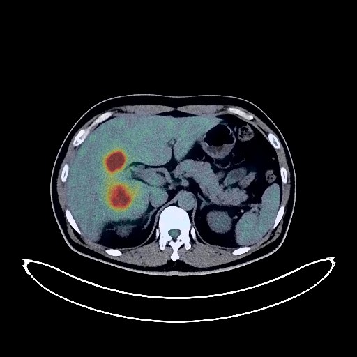
Liver Cancer PET/CT (case 984005-000009 from PETWB-REP)
2 views9 days agoWhole-body 18F-FDG PET/CT scan in a patient with Liver Cancer taken from the PETWB-REP dataset. The following English report (translated from original Chinese) is taken verbatim from the public dataset and has not been modified or otherwise checked for accuracy (see the end for citation). Impression a. Large, mixed-density mass in the liver, with increased FDG metabolism, suggestive of hepatocellular carcinoma. b. Portal vein visualization is indistinct. Multiple chronic inflammatory micronodules in both lungs, with a few post-inflammatory lesions in both lungs. Slight pleural thickening bilaterally. Cholestasis in the gallbladder; follow-up ultrasound is recommended. Multiple kidney stones in both kidneys, a stone in the upper left ureter with hydronephrosis of the ureter and renal pelvis above it. Calcification in the prostate. Slight scoliosis with osteophyte formation in the spine. L3/4 and L4/5 intervertebral disc bulges. No abnormalities were found on cranial imaging. This case is from PETWB-REP, a curated dataset of whole-body 18F-FDG PET/CT scans and corresponding radiology reports from 490 patients with a broad spectrum of malignancies. The data were retrospectively collected from patients who underwent clinically indicated whole-body 18F-FDG PET/CT scans at the Shanghai Universal Medical Imaging Diagnostic Center between 2021 and 2024. License: Creative Commons Attribution 4.0 International (CC BY 4.0) Citation: Xue, L., Feng, G., Wenbo, Z., Zhang, Y., Li, L., Wang, S., Peng, L., Peng, S., & Gao, X. (2026). PETWB-REP: A Multi-Cancer Whole-Body FDG PET/CT Dataset with Corresponding Radiology Reports [Data set]. Zenodo. https://doi.org/10.5281/zenodo.18670487
Whole BodyPET/CT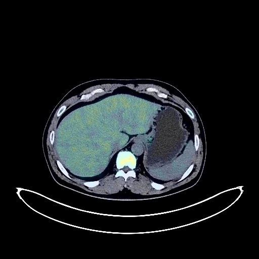
Liver Cancer PET/CT (case 984005-000008 from PETWB-REP)
1 views9 days agoWhole-body 18F-FDG PET/CT scan in a patient with Liver Cancer taken from the PETWB-REP dataset. The following English report (translated from original Chinese) is taken verbatim from the public dataset and has not been modified or otherwise checked for accuracy (see the end for citation). Impression a. A large mass with necrosis in the right lobe of the liver, with increased FDG metabolism, suggestive of hepatocellular carcinoma; please correlate with clinicopathology. b. A linear shadow in the right pelvic region, with some mild FDG metabolism, suggesting possible hematoma; implantation metastasis to be ruled out. c. Multiple reactive hyperplasia of lymph nodes in the pancreatic head region and mid-abdomen mesenteric region; follow-up is recommended. d. The right branch of the portal vein is not clearly visualized. Multiple chronic inflammatory micronodules in both lungs, with a few post-inflammatory remnants in both lungs. Slight thickening of the pleura bilaterally. Bilateral gynecomastia. Multiple gallstones and adenomyomatosis of the gallbladder. Small kidney stones bilaterally. Calcifications in the prostate. Spinal osteophyte formation. L3/4 and L4/5 intervertebral disc bulge. No obvious abnormalities were seen on cranial imaging. This case is from PETWB-REP, a curated dataset of whole-body 18F-FDG PET/CT scans and corresponding radiology reports from 490 patients with a broad spectrum of malignancies. The data were retrospectively collected from patients who underwent clinically indicated whole-body 18F-FDG PET/CT scans at the Shanghai Universal Medical Imaging Diagnostic Center between 2021 and 2024. License: Creative Commons Attribution 4.0 International (CC BY 4.0) Citation: Xue, L., Feng, G., Wenbo, Z., Zhang, Y., Li, L., Wang, S., Peng, L., Peng, S., & Gao, X. (2026). PETWB-REP: A Multi-Cancer Whole-Body FDG PET/CT Dataset with Corresponding Radiology Reports [Data set]. Zenodo. https://doi.org/10.5281/zenodo.18670487
Whole BodyPET/CT