Loading...
Nasopharyngeal Cancer PET/CT (case 983824-000103 from PETWB-REP)
0 views10 days agoWhole-body 18F-FDG PET/CT scan in a patient with Nasopharyngeal Cancer taken from the PETWB-REP dataset. The following English report (translated from original Chinese) is taken verbatim from the public dataset and has not been modified or otherwise checked for accuracy (see the end for citation). Impression a. Nasopharyngeal mass with increased FDG metabolism, consistent with nasopharyngeal carcinoma, involving the posterior nasal cavity, nasal septum, and adjacent skull base bone. b. High probability of left deep cervical lymph node metastasis. Right mastoiditis. Minor chronic inflammation and old lesions in both lungs. Slightly widened ascending aorta. Liver calcification. Small cyst in the left lobe of the liver. Post-tubal ligation changes. Mild degenerative changes in the spine. L4/5 and L5/S1 intervertebral disc bulge. Intramuscular lipoma in the upper right humerus. No abnormalities seen on cranial scintigraphy. Chronic inflammation of bilateral maxillary sinuses, bilateral ethmoid sinuses, and sphenoid sinuses. This case is from PETWB-REP, a curated dataset of whole-body 18F-FDG PET/CT scans and corresponding radiology reports from 490 patients with a broad spectrum of malignancies. The data were retrospectively collected from patients who underwent clinically indicated whole-body 18F-FDG PET/CT scans at the Shanghai Universal Medical Imaging Diagnostic Center between 2021 and 2024. License: Creative Commons Attribution 4.0 International (CC BY 4.0) Citation: Xue, L., Feng, G., Wenbo, Z., Zhang, Y., Li, L., Wang, S., Peng, L., Peng, S., & Gao, X. (2026). PETWB-REP: A Multi-Cancer Whole-Body FDG PET/CT Dataset with Corresponding Radiology Reports [Data set]. Zenodo. https://doi.org/10.5281/zenodo.18670487
Whole BodyPET/CT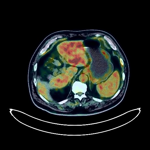
Lung Cancer PET/CT (case 983824-000086 from PETWB-REP)
2 views10 days agoWhole-body 18F-FDG PET/CT scan in a patient with Lung Cancer taken from the PETWB-REP dataset. The following English report (translated from original Chinese) is taken verbatim from the public dataset and has not been modified or otherwise checked for accuracy (see the end for citation). Impression Post-treatment PET/CT images of right lung cancer, compared with images from our center on February 7, 2023: a. A soft tissue mass in the posterior segment of the right upper lobe with increased FDG metabolism, significantly smaller than before, suggesting partial suppression of tumor activity after treatment, with some remaining; accompanied by minor distal obstructive changes. b. Right hilar and mediastinal lymph nodes, roughly the same size as before, with slightly decreased FDG metabolism; follow-up is recommended. c. Multiple small solid nodules in both lungs (some newly added), FDG metabolism normal, suggesting possible metastasis; regular CT follow-up is recommended. d. Ground-glass nodule in the posterior segment of the left lower lobe, FDG metabolism normal, roughly the same size as before; suggestive of chronic inflammatory nodules or atypical adenomatous hyperplasia; please combine with HRCT annual follow-up. Bilateral emphysema with bullae, bilateral pulmonary fibrosis. Slight thickening of the pleura in some areas. a. Thickening of the gastric angle and antrum walls with increased FDG metabolism, slightly relieved from before, suggesting possible inflammation; gastric cancer to be ruled out. Please combine with gastroscopy for comprehensive analysis. Lymph nodes in the hepatogastric space and lesser curvature of the stomach are roughly the same as before. b. Nodules in the lower rectal wall with increased FDG metabolism, suggesting possible inflammatory polyps or tubular adenomas. Colonoscopy follow-up is recommended to rule out other possibilities. c. Physiological or inflammatory uptake in the lower esophagus and part of the intestine. Duodenal diverticulum. Benign prostatic hyperplasia with calcification, uneven FDG metabolism; please follow up with PSA and MRI. Gallstones. Thickening of the perirenal septum in both kidneys, left renal cyst. Slightly enlarged heart. Partial arteriosclerosis (including coronary arteries). Degenerative changes in the spine, L1 vertebral hemangioma, L4/5 intervertebral disc bulge, partial cervical, thoracic and lumbar intervertebral disc degeneration with pneumoconiosis. Right-sided frozen shoulder. Left trapezius muscle lipoma. Bilateral deep lacunar infarcts, age-related cerebral encephalopathy. Minor chronic inflammation of the bilateral ethmoid sinuses and right maxillary sinus. This case is from PETWB-REP, a curated dataset of whole-body 18F-FDG PET/CT scans and corresponding radiology reports from 490 patients with a broad spectrum of malignancies. The data were retrospectively collected from patients who underwent clinically indicated whole-body 18F-FDG PET/CT scans at the Shanghai Universal Medical Imaging Diagnostic Center between 2021 and 2024. License: Creative Commons Attribution 4.0 International (CC BY 4.0) Citation: Xue, L., Feng, G., Wenbo, Z., Zhang, Y., Li, L., Wang, S., Peng, L., Peng, S., & Gao, X. (2026). PETWB-REP: A Multi-Cancer Whole-Body FDG PET/CT Dataset with Corresponding Radiology Reports [Data set]. Zenodo. https://doi.org/10.5281/zenodo.18670487
Whole BodyPET/CT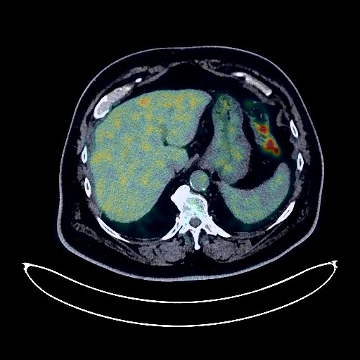
Colon Cancer PET/CT (case 983824-000005 from PETWB-REP)
8 views10 days agoWhole-body 18F-FDG PET/CT scan in a patient with Colon Cancer taken from the PETWB-REP dataset. The following English report (translated from original Chinese) is taken verbatim from the public dataset and has not been modified or otherwise checked for accuracy (see the end for citation). Impression Thickening of the ascending colon near the ileocecal junction with increased FDG uptake, thickening of the surrounding mesentery with several lymph nodes visible. Delayed scanning showed no significant change in the lesion near the ileocecal junction, but FDG uptake values in all lesions remained increased, suggesting possible colon cancer with surrounding mesenteric lymph node metastasis. A follow-up colonoscopy is recommended to rule out other possibilities; inflammatory uptake in the colon is also possible. Liver cirrhosis findings, gallstones in the gallbladder neck; no space-occupying lesions or foci of increased FDG uptake were observed in the liver, pancreas, or biliary system. Clinical and MRI findings are recommended. Thickening of the gastric wall with mildly increased FDG uptake is suggested, possibly due to inflammatory uptake. A follow-up gastroscopy is recommended. Multiple solid nodules in both lungs with increased FDG uptake are suggested, possibly due to inflammatory nodules. Clinical and old film comparison and follow-up CT scan are recommended. Interstitial pneumonia in the lower lobes of both lungs. Slight pleural thickening bilaterally. Reactive hyperplasia of hilar and mediastinal lymph nodes bilaterally. Calcification of some arterial walls (including coronary arteries). Bilateral renal atrophy, small renal stone in the left kidney. Benign prostatic hyperplasia with calcification, uneven FDG metabolism; please follow up with PSA levels. Bilateral hydrocele. Spinal degenerative changes. L4/5 and L5/S1 intervertebral disc bulge. Bilateral subcutaneous calcifications in the buttocks. Bilateral basal ganglia ischemia. Age-related brain changes. Bilateral ethmoid sinusitis and right maxillary sinusitis. This case is from PETWB-REP, a curated dataset of whole-body 18F-FDG PET/CT scans and corresponding radiology reports from 490 patients with a broad spectrum of malignancies. The data were retrospectively collected from patients who underwent clinically indicated whole-body 18F-FDG PET/CT scans at the Shanghai Universal Medical Imaging Diagnostic Center between 2021 and 2024. License: Creative Commons Attribution 4.0 International (CC BY 4.0) Citation: Xue, L., Feng, G., Wenbo, Z., Zhang, Y., Li, L., Wang, S., Peng, L., Peng, S., & Gao, X. (2026). PETWB-REP: A Multi-Cancer Whole-Body FDG PET/CT Dataset with Corresponding Radiology Reports [Data set]. Zenodo. https://doi.org/10.5281/zenodo.18670487
Whole BodyPET/CT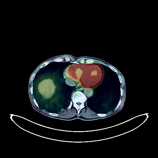
Lung Cancer PET/CT (case 983824-000191 from PETWB-REP)
0 views10 days agoWhole-body 18F-FDG PET/CT scan in a patient with Lung Cancer taken from the PETWB-REP dataset. The following English report (translated from original Chinese) is taken verbatim from the public dataset and has not been modified or otherwise checked for accuracy (see the end for citation). Impression a. A space-occupying lesion in the right lower lobe, paravertebral, with pleural infiltration and adhesion, and increased FDG metabolism, consistent with lung cancer; the tumor is still active. Metastasis to the right hilar and some mediastinal lymph nodes is highly probable. b. Uneven bone density in the right second rib and S2 region, with increased FDG metabolism, suggesting a high probability of bone metastasis; please compare with previous images. c. Chronic bronchitis and emphysema-like changes in both lungs, with bullae. A small amount of chronic inflammation in both lungs. Scattered chronic inflammatory micronodules (solid) in both lungs; please follow up with CT scans. Mild pleural thickening bilaterally. Partial arteriosclerosis (including coronary arteries). Chronic cholecystitis. Small kidney stone in the right kidney. Benign prostatic hyperplasia with calcification. Calcification lesion in the right testicular tunica vaginalis. Possible reactive hyperplasia of retroperitoneal lymph nodes; follow-up with CT or ultrasound is recommended. Chronic antral gastritis. Degenerative changes in the spine. L4/5 and L5/S1 intervertebral disc bulge. L5/S1 intervertebral disc pneumothorax and degeneration. Age-related brain changes, deep lacunar infarcts in the brain; MRI is recommended. Bilateral mastoid hypopneumatization. This case is from PETWB-REP, a curated dataset of whole-body 18F-FDG PET/CT scans and corresponding radiology reports from 490 patients with a broad spectrum of malignancies. The data were retrospectively collected from patients who underwent clinically indicated whole-body 18F-FDG PET/CT scans at the Shanghai Universal Medical Imaging Diagnostic Center between 2021 and 2024. License: Creative Commons Attribution 4.0 International (CC BY 4.0) Citation: Xue, L., Feng, G., Wenbo, Z., Zhang, Y., Li, L., Wang, S., Peng, L., Peng, S., & Gao, X. (2026). PETWB-REP: A Multi-Cancer Whole-Body FDG PET/CT Dataset with Corresponding Radiology Reports [Data set]. Zenodo. https://doi.org/10.5281/zenodo.18670487
Whole BodyPET/CT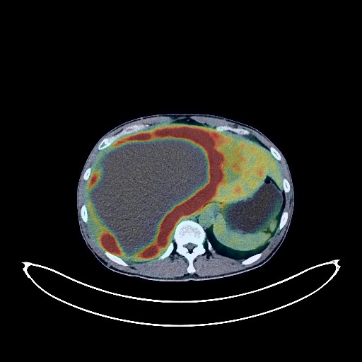
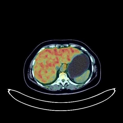
Pancreatic Cancer PET/CT (case 983824-000056 from PETWB-REP)
2 views10 days agoWhole-body 18F-FDG PET/CT scan in a patient with Pancreatic Cancer taken from the PETWB-REP dataset. The following English report (translated from original Chinese) is taken verbatim from the public dataset and has not been modified or otherwise checked for accuracy (see the end for citation). Impression a. Low-density mass in the tail of the pancreas with increased FDG metabolism; multiple low-density lesions in the liver parenchyma with increased FDG metabolism; enlarged retroperitoneal lymph nodes with increased FDG metabolism. All of these suggest malignancy, with a high probability of pancreatic cancer (possibly pancreatic carcinoma) accompanied by multiple metastases; metastases cannot be ruled out. b. A large, multilocular cystic lesion in the right lobe of the liver, with increased FDG metabolism in some septa and cyst walls, strongly suggests malignancy; enhanced MRI analysis is recommended. c. Multiple tortuous vessels in the abdominal cavity, indicating obstructed venous return. Small amount of effusion in the abdominopelvic cavity. d. Expansile changes in the sternum with mixed bone density and slightly increased FDG metabolism, suggesting possible fibrous dysplasia of bone; metastases need to be ruled out. Chronic miliary lesions in the upper lobe of the right lung; a small amount of chronic inflammation in the middle and lower lobes of the right lung. Possible physiological uptake at the gastric cardia; contracted state of the gastric antrum. Vertebral osteophyte formation in the spine. Internal fixation of the left clavicle with metal fixation underway. No obvious abnormalities were found on cranial scintigraphy. Reactive hyperplasia of bilateral cervical lymph nodes was observed. This case is from PETWB-REP, a curated dataset of whole-body 18F-FDG PET/CT scans and corresponding radiology reports from 490 patients with a broad spectrum of malignancies. The data were retrospectively collected from patients who underwent clinically indicated whole-body 18F-FDG PET/CT scans at the Shanghai Universal Medical Imaging Diagnostic Center between 2021 and 2024. License: Creative Commons Attribution 4.0 International (CC BY 4.0) Citation: Xue, L., Feng, G., Wenbo, Z., Zhang, Y., Li, L., Wang, S., Peng, L., Peng, S., & Gao, X. (2026). PETWB-REP: A Multi-Cancer Whole-Body FDG PET/CT Dataset with Corresponding Radiology Reports [Data set]. Zenodo. https://doi.org/10.5281/zenodo.18670487
Whole BodyPET/CT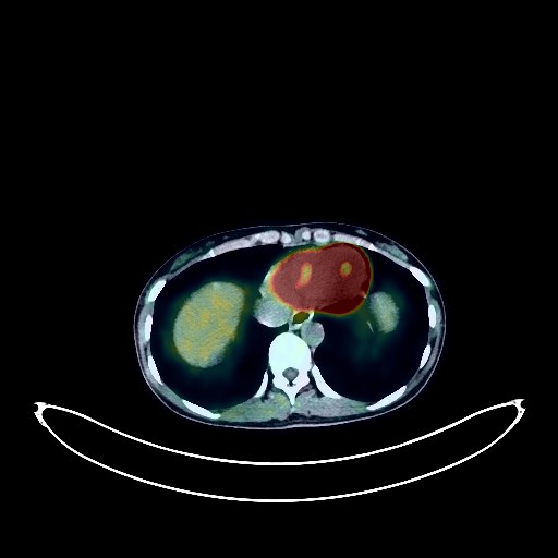
Cervical Cancer PET/CT (case 983824-000065 from PETWB-REP)
1 views10 days agoWhole-body 18F-FDG PET/CT scan in a patient with Cervical Cancer taken from the PETWB-REP dataset. The following English report (translated from original Chinese) is taken verbatim from the public dataset and has not been modified or otherwise checked for accuracy (see the end for citation). Impression a. Cervical mass with increased FDG metabolism, consistent with cervical cancer; reactive hyperplasia of bilateral iliac vessels and bilateral inguinal lymph nodes. b. Possible uterine fibroids, physiological uptake within the uterine cavity; possible left ovarian cyst; ultrasound follow-up recommended. Small amount of pelvic effusion. Chronic inflammatory miliary nodules in the left lung. Calcifications in the lower lobe of the right lung. Anemia. Bilateral renal calcifications. Bilateral renal cysts. No obvious abnormalities seen on cranial scintigraphy. This case is from PETWB-REP, a curated dataset of whole-body 18F-FDG PET/CT scans and corresponding radiology reports from 490 patients with a broad spectrum of malignancies. The data were retrospectively collected from patients who underwent clinically indicated whole-body 18F-FDG PET/CT scans at the Shanghai Universal Medical Imaging Diagnostic Center between 2021 and 2024. License: Creative Commons Attribution 4.0 International (CC BY 4.0) Citation: Xue, L., Feng, G., Wenbo, Z., Zhang, Y., Li, L., Wang, S., Peng, L., Peng, S., & Gao, X. (2026). PETWB-REP: A Multi-Cancer Whole-Body FDG PET/CT Dataset with Corresponding Radiology Reports [Data set]. Zenodo. https://doi.org/10.5281/zenodo.18670487
Whole BodyPET/CT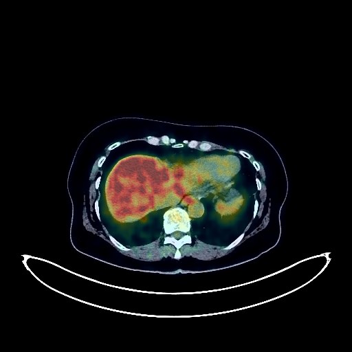
Gastric Cancer PET/CT (case 983824-000138 from PETWB-REP)
2 views10 days agoWhole-body 18F-FDG PET/CT scan in a patient with Gastric Cancer taken from the PETWB-REP dataset. The following English report (translated from original Chinese) is taken verbatim from the public dataset and has not been modified or otherwise checked for accuracy (see the end for citation). Impression a. Post-gastric cancer treatment: Slight thickening of the anastomosis and adjacent gastric wall with increased FDG metabolism, and slight thickening of the lower esophageal wall with increased FDG metabolism, both suggestive of inflammatory changes. Endoscopic follow-up is recommended to rule out other possibilities. b. Partial peritoneal thickening in the abdominopelvic cavity with increased FDG metabolism suggests inflammatory changes, with implantation metastasis to be ruled out. Regular CT follow-up is recommended. c. Mild mesenteric torsion, likely due to reactive hyperplasia of mesenteric lymph nodes; follow-up is advised. Post-colon surgery: No thickening of the anastomosis wall, but increased FDG metabolism in some intestinal segments, suggesting physiological uptake or chronic inflammation; endoscopic follow-up is recommended. Post-bilateral adnexal surgery: No abnormal FDG metabolic foci were observed in the surgical area. Uterine fibroids are possible. Clinical and ultrasound follow-up is recommended. Hydronephrosis and dilation of the left renal pelvis and upper left ureter, suspicious soft tissue shadow with thickened wall in the upper left ureter, and increased FDG metabolism suggest possible inflammatory changes, space-occupying lesion to be ruled out. Please combine with enhanced MRI, and repeat endoscopic examination if necessary. Left kidney fullness with increased parenchymal FDG metabolism, please combine with renal function tests. Ground-glass nodule in the anterior basal segment of the right lower lobe, FDG metabolism normal, suggest chronic inflammatory nodule or atypical adenomatous hyperplasia. Please combine with annual HRCT follow-up. Fibrotic foci in both lungs. Uneven density of the thyroid gland, calcification in the right lobe, low-density nodule in the isthmus with increased FDG metabolism, suggest possible adenomatous nodule. Ultrasound follow-up is recommended, and fine-needle aspiration biopsy if necessary to rule out malignancy. Liver cyst. Osteophyte formation in some cervical, thoracic and lumbar vertebrae. Bilateral pars interarticular rupture at L5. Significantly old changes in the left transverse process of L3. L5/S1 disc herniation. No obvious abnormalities were found on cranial scintigraphy. This case is from PETWB-REP, a curated dataset of whole-body 18F-FDG PET/CT scans and corresponding radiology reports from 490 patients with a broad spectrum of malignancies. The data were retrospectively collected from patients who underwent clinically indicated whole-body 18F-FDG PET/CT scans at the Shanghai Universal Medical Imaging Diagnostic Center between 2021 and 2024. License: Creative Commons Attribution 4.0 International (CC BY 4.0) Citation: Xue, L., Feng, G., Wenbo, Z., Zhang, Y., Li, L., Wang, S., Peng, L., Peng, S., & Gao, X. (2026). PETWB-REP: A Multi-Cancer Whole-Body FDG PET/CT Dataset with Corresponding Radiology Reports [Data set]. Zenodo. https://doi.org/10.5281/zenodo.18670487
Whole BodyPET/CT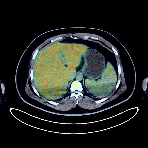
Lymphoma PET/CT (case 983824-000139 from PETWB-REP)
2 views10 days agoWhole-body 18F-FDG PET/CT scan in a patient with Lymphoma taken from the PETWB-REP dataset. The following English report (translated from original Chinese) is taken verbatim from the public dataset and has not been modified or otherwise checked for accuracy (see the end for citation). Impression A mass in the anterior part of the right nasal cavity with increased FDG metabolism, consistent with lymphoma presentation based on the patient's history, involving the right ethmoid sinus, maxillary sinus, orbit, and nasal ala; reactive hyperplasia of the right parotid region, left submandibular region, and bilateral deep cervical spaces is highly probable, with right submandibular lymphoma infiltration to be ruled out. Follow-up is recommended. Chronic inflammatory micronodules in the right lower lobe of the lung. Calcification in the left lower lobe of the lung. A few post-inflammatory remnants in both lungs. Fatty liver. Fatty infiltration of the pancreas. Bilateral renal calculi. Reactive hyperplasia of retroperitoneal and bilateral inguinal lymph nodes. Chronic inflammatory changes or physiological uptake in the gastric antrum and part of the intestine; follow-up with endoscopy is recommended. No obvious abnormalities were found on cranial scintigraphy. Chronic inflammation of the right frontal sinus, ethmoid sinus, and maxillary sinus. This case is from PETWB-REP, a curated dataset of whole-body 18F-FDG PET/CT scans and corresponding radiology reports from 490 patients with a broad spectrum of malignancies. The data were retrospectively collected from patients who underwent clinically indicated whole-body 18F-FDG PET/CT scans at the Shanghai Universal Medical Imaging Diagnostic Center between 2021 and 2024. License: Creative Commons Attribution 4.0 International (CC BY 4.0) Citation: Xue, L., Feng, G., Wenbo, Z., Zhang, Y., Li, L., Wang, S., Peng, L., Peng, S., & Gao, X. (2026). PETWB-REP: A Multi-Cancer Whole-Body FDG PET/CT Dataset with Corresponding Radiology Reports [Data set]. Zenodo. https://doi.org/10.5281/zenodo.18670487
Whole BodyPET/CT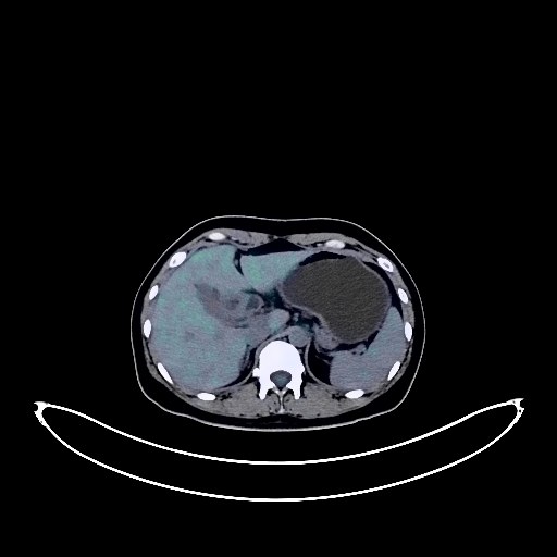
Ovarian Cancer PET/CT (case 983824-000148 from PETWB-REP)
2 views10 days agoWhole-body 18F-FDG PET/CT scan in a patient with Ovarian Cancer taken from the PETWB-REP dataset. The following English report (translated from original Chinese) is taken verbatim from the public dataset and has not been modified or otherwise checked for accuracy (see the end for citation). Impression a. Thickening of the greater omentum and pelvic floor peritoneum with increased FDG metabolism suggests a high probability of metastasis; a mass in the left adnexal region with increased FDG metabolism suggests a possible ovarian tumor; peritoneal tuberculosis caused by the above lesions should be ruled out. Please consider clinicopathology and tumor markers. b. Metastasis to the cardiophrenic angle, hiatal space, and retroperitoneal lymph nodes should be ruled out. c. Pelvic effusion. Mild anemia. Irregular low-density shadow in the myometrium of the posterior wall of the uterus, with no abnormal FDG uptake, suggests uterine fibroid degeneration or adenomyosis. Ground-glass opacity in the right middle lobe of the lung, with no abnormal FDG uptake, suggests chronic inflammation. Calcification in the apical segment of the right upper lobe. Mild osteophyte formation in some vertebrae. No obvious abnormalities were found on cranial FDG imaging. This case is from PETWB-REP, a curated dataset of whole-body 18F-FDG PET/CT scans and corresponding radiology reports from 490 patients with a broad spectrum of malignancies. The data were retrospectively collected from patients who underwent clinically indicated whole-body 18F-FDG PET/CT scans at the Shanghai Universal Medical Imaging Diagnostic Center between 2021 and 2024. License: Creative Commons Attribution 4.0 International (CC BY 4.0) Citation: Xue, L., Feng, G., Wenbo, Z., Zhang, Y., Li, L., Wang, S., Peng, L., Peng, S., & Gao, X. (2026). PETWB-REP: A Multi-Cancer Whole-Body FDG PET/CT Dataset with Corresponding Radiology Reports [Data set]. Zenodo. https://doi.org/10.5281/zenodo.18670487
Whole BodyPET/CT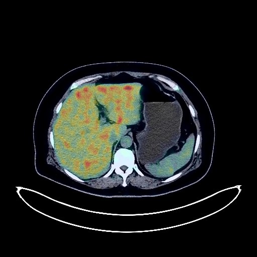
Lung Cancer PET/CT (case 983824-000211 from PETWB-REP)
2 views10 days agoWhole-body 18F-FDG PET/CT scan in a patient with Lung Cancer taken from the PETWB-REP dataset. The following English report (translated from original Chinese) is taken verbatim from the public dataset and has not been modified or otherwise checked for accuracy (see the end for citation). Impression a. A mass in the right middle lobe near the diaphragm pleura with increased FDG metabolism, highly suggestive of peripheral lung cancer; please correlate with clinicopathology. b. Several metastatic tumors in both lungs. Metastatic tumors in the S1-2 vertebral bodies. c. Calcification in the right middle lobe. A few post-inflammatory remnants in both lungs. Reactive hyperplasia of mediastinal lymph nodes. Bilateral breast hyperplasia. Absence after cholecystectomy. Accessory spleen. Possible left renal cyst. Right ovarian cyst. Chronic antral gastritis. Increased FDG metabolism in parts of the colon and rectum, suggestive of physiological uptake or chronic inflammatory changes; please correlate with endoscopic follow-up. Possible hemorrhoids. Degenerative changes in the spine. Schmorl's nodes at the lower margin of T12 vertebral body and the upper margin of L2 vertebral body; L4/5 and L5/S1 intervertebral disc bulges; L5/S1 intervertebral disc pneumatosis and degeneration Deep lacunar infarcts in the brain, MRI follow-up recommended; reactive hyperplasia of bilateral deep cervical intervertebral spaces, submandibular and submental lymph nodes. This case is from PETWB-REP, a curated dataset of whole-body 18F-FDG PET/CT scans and corresponding radiology reports from 490 patients with a broad spectrum of malignancies. The data were retrospectively collected from patients who underwent clinically indicated whole-body 18F-FDG PET/CT scans at the Shanghai Universal Medical Imaging Diagnostic Center between 2021 and 2024. License: Creative Commons Attribution 4.0 International (CC BY 4.0) Citation: Xue, L., Feng, G., Wenbo, Z., Zhang, Y., Li, L., Wang, S., Peng, L., Peng, S., & Gao, X. (2026). PETWB-REP: A Multi-Cancer Whole-Body FDG PET/CT Dataset with Corresponding Radiology Reports [Data set]. Zenodo. https://doi.org/10.5281/zenodo.18670487
Whole BodyPET/CT