Loading...
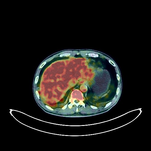
Lung Cancer PET/CT (case 983824-000046 from PETWB-REP)
9 views10 days agoWhole-body 18F-FDG PET/CT scan in a patient with Lung Cancer taken from the PETWB-REP dataset. The following English report (translated from original Chinese) is taken verbatim from the public dataset and has not been modified or otherwise checked for accuracy (see the end for citation). Impression a. A mass in the right upper lobe with increased FDG uptake; multiple enlarged lymph nodes in the right hilum, mediastinum, and bilateral supraclavicular fossa, some fused, with increased FDG metabolism, suggesting a high probability of right lung cancer with multiple lymph node metastases; tuberculosis to be ruled out; airway dissemination in the right upper lobe is also a possibility. b. Small lymph nodes in the hepatogastric interspace with increased FDG metabolism, suggesting reactive hyperplasia. Small amount of pleural effusion and pericardial effusion bilaterally. a. Ground-glass nodule in the left upper lobe, with normal FDG metabolism, suggesting atypical adenomatous hyperplasia or inflammatory nodule; annual HRCT follow-up is recommended. b. Multiple small chronic inflammatory nodules (solid) in the remaining lungs. Paraseptal emphysema in both upper lobes. Minor inflammation in the bilateral ethmoid and maxillary sinuses. Increased FDG uptake in the left maxillary alveolar region, suggesting inflammation. No obvious abnormalities were seen on cranial imaging. Multiple cysts in both kidneys. Benign prostatic hyperplasia with calcifications. Bilateral testicular hydrocele with small amount of fluid in the tunica vaginalis. Spinal osteophyte formation, L4/5 and L5/S1 intervertebral disc bulge. This case is from PETWB-REP, a curated dataset of whole-body 18F-FDG PET/CT scans and corresponding radiology reports from 490 patients with a broad spectrum of malignancies. The data were retrospectively collected from patients who underwent clinically indicated whole-body 18F-FDG PET/CT scans at the Shanghai Universal Medical Imaging Diagnostic Center between 2021 and 2024. License: Creative Commons Attribution 4.0 International (CC BY 4.0) Citation: Xue, L., Feng, G., Wenbo, Z., Zhang, Y., Li, L., Wang, S., Peng, L., Peng, S., & Gao, X. (2026). PETWB-REP: A Multi-Cancer Whole-Body FDG PET/CT Dataset with Corresponding Radiology Reports [Data set]. Zenodo. https://doi.org/10.5281/zenodo.18670487
Whole BodyPET/CT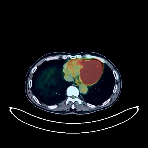
Lung Cancer PET/CT (case 983824-000149 from PETWB-REP)
2 views10 days agoWhole-body 18F-FDG PET/CT scan in a patient with Lung Cancer taken from the PETWB-REP dataset. The following English report (translated from original Chinese) is taken verbatim from the public dataset and has not been modified or otherwise checked for accuracy (see the end for citation). Impression a. A mass in the posterior segment of the right upper lobe, with increased FDG metabolism, suggestive of lung cancer; please correlate with clinicopathology. b. Bone destruction at the anterior margin of the S1 vertebral body and the right femoral head with increased FDG metabolism, suggestive of metastasis; please correlate with MRI. c. Possible reactive hyperplasia of the right hilar and mediastinal lymph nodes; partial metastasis to be ruled out. d. Bilateral emphysema with bullous formation; bullous formation with minor inflammation in the anterior segment of the right upper lobe. Multiple chronic inflammatory nodules in both lungs; CT follow-up recommended. Minor chronic inflammation and sequelae (including calcifications) in both lungs; some adjacent pleural thickening. Partial arteriosclerosis (including coronary arteries). Low-density nodule in the left lobe of the thyroid gland, highly suggestive of adenoma; please correlate with ultrasound. Right renal cyst. Dilatation of the mid-segment of the left ureter. Prostatic calcification. Degenerative changes in the spine. L4/5 and L5/S1 intervertebral disc degeneration and bulging. A few ischemic lesions in the deep bilateral brain regions; MRI is recommended. Bilateral mastoid hypopneumatization. Increased FDG metabolism in the bilateral parotid glands, suggestive of physiological uptake or chronic inflammation. Reactive hyperplasia of small cervical lymph nodes bilaterally. This case is from PETWB-REP, a curated dataset of whole-body 18F-FDG PET/CT scans and corresponding radiology reports from 490 patients with a broad spectrum of malignancies. The data were retrospectively collected from patients who underwent clinically indicated whole-body 18F-FDG PET/CT scans at the Shanghai Universal Medical Imaging Diagnostic Center between 2021 and 2024. License: Creative Commons Attribution 4.0 International (CC BY 4.0) Citation: Xue, L., Feng, G., Wenbo, Z., Zhang, Y., Li, L., Wang, S., Peng, L., Peng, S., & Gao, X. (2026). PETWB-REP: A Multi-Cancer Whole-Body FDG PET/CT Dataset with Corresponding Radiology Reports [Data set]. Zenodo. https://doi.org/10.5281/zenodo.18670487
Whole BodyPET/CT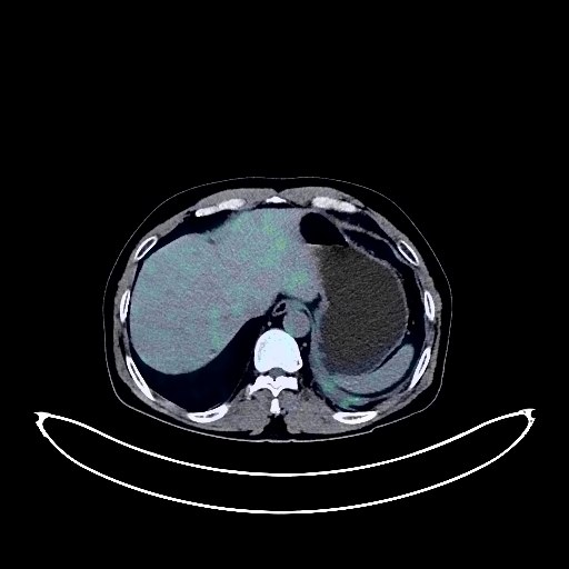
Lung Cancer PET/CT (case 983824-000127 from PETWB-REP)
2 views10 days agoWhole-body 18F-FDG PET/CT scan in a patient with Lung Cancer taken from the PETWB-REP dataset. The following English report (translated from original Chinese) is taken verbatim from the public dataset and has not been modified or otherwise checked for accuracy (see the end for citation). Impression a. Mass in the posterior segment of the left upper lobe, with increased FDG metabolism, suggestive of lung cancer. b. Metastasis to the left hilar, subcarinal, and left internal mammary chain lymph nodes. Reactive hyperplasia of the right hilar lymph nodes. c. Left pleural metastasis. Small amount of pleural effusion on the left side. d. Multiple bone metastases throughout the body. Right adrenal gland metastasis. Several small (solid) chronic inflammatory nodules in both lungs are possible; CT follow-up is recommended to rule out metastasis. A few chronic inflammations and old lesions in both lungs. Some arterial wall calcification. Multiple liver cysts. Gallstones. Left renal cyst. Degenerative changes in the spine. L4/5 and L5/S1 intervertebral disc bulges. A few ischemic lesions in the deep bilateral brain regions. This case is from PETWB-REP, a curated dataset of whole-body 18F-FDG PET/CT scans and corresponding radiology reports from 490 patients with a broad spectrum of malignancies. The data were retrospectively collected from patients who underwent clinically indicated whole-body 18F-FDG PET/CT scans at the Shanghai Universal Medical Imaging Diagnostic Center between 2021 and 2024. License: Creative Commons Attribution 4.0 International (CC BY 4.0) Citation: Xue, L., Feng, G., Wenbo, Z., Zhang, Y., Li, L., Wang, S., Peng, L., Peng, S., & Gao, X. (2026). PETWB-REP: A Multi-Cancer Whole-Body FDG PET/CT Dataset with Corresponding Radiology Reports [Data set]. Zenodo. https://doi.org/10.5281/zenodo.18670487
Whole BodyPET/CT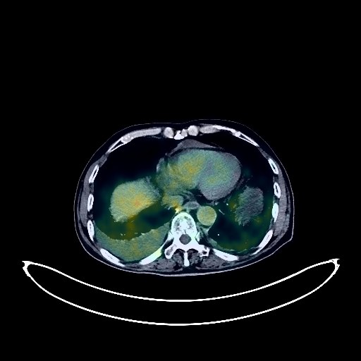
Lung Cancer PET/CT (case 983824-000075 from PETWB-REP)
2 views10 days agoWhole-body 18F-FDG PET/CT scan in a patient with Lung Cancer taken from the PETWB-REP dataset. The following English report (translated from original Chinese) is taken verbatim from the public dataset and has not been modified or otherwise checked for accuracy (see the end for citation). Impression a. A mass in the right upper lobe of the lung with elevated FDG metabolism, suggestive of lung cancer, possibly accompanied by carcinomatous lymphangitis. Please confirm the diagnosis with pathological examination. b. Multiple lymph node metastases in the bilateral hilar, mediastinal, right cardiophrenic angle, bilateral supraclavicular fossa, and left posterior cervical triangle. c. Multiple bone metastases throughout the body. Left adrenal metastasis to be ruled out. Possible subcutaneous metastasis in the right mid-abdomen. d. Multiple solid nodules in both lungs, some with elevated FDG metabolism, suggestive of chronic inflammatory nodules, some metastases to be ruled out. Multiple ground-glass nodules in both lungs with normal FDG metabolism, suggestive of chronic inflammatory nodules or atypical adenomatous hyperplasia. Regular HRCT follow-up is recommended for all of the above. e. Interstitial changes in both lungs with scattered inflammation and remnants. CT follow-up after anti-inflammatory treatment is recommended to rule out other occult lesions. Bilateral pleural effusion. Calcification of some arterial walls (including coronary arteries). Small amount of pericardial effusion. Slightly high-density nodule in the right parietal lobe, with increased FDG metabolism, suggesting a high probability of metastasis; localized decreased density in the left temporal lobe, metastasis to be ruled out. MRI with contrast enhancement is recommended for confirmation. Age-related brain. Bilateral renal cysts. Left renal calculus. Increased FDG metabolism in some intestinal segments, considering inflammatory or physiological uptake; colonoscopy is recommended. Degenerative changes in the spine. L2 vertebral instability. L1 vertebral wedging. L4/5, L5/S1 intervertebral disc bulging. Right abdominal wall muscle swelling with increased FDG metabolism, suggesting possible inflammatory changes; follow-up is needed to rule out other possibilities. Bilateral chronic maxillary sinusitis. This case is from PETWB-REP, a curated dataset of whole-body 18F-FDG PET/CT scans and corresponding radiology reports from 490 patients with a broad spectrum of malignancies. The data were retrospectively collected from patients who underwent clinically indicated whole-body 18F-FDG PET/CT scans at the Shanghai Universal Medical Imaging Diagnostic Center between 2021 and 2024. License: Creative Commons Attribution 4.0 International (CC BY 4.0) Citation: Xue, L., Feng, G., Wenbo, Z., Zhang, Y., Li, L., Wang, S., Peng, L., Peng, S., & Gao, X. (2026). PETWB-REP: A Multi-Cancer Whole-Body FDG PET/CT Dataset with Corresponding Radiology Reports [Data set]. Zenodo. https://doi.org/10.5281/zenodo.18670487
Whole BodyPET/CT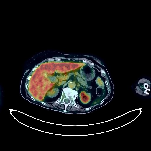
Lymphoma PET/CT (case 983824-000062 from PETWB-REP)
2 views10 days agoWhole-body 18F-FDG PET/CT scan in a patient with Lymphoma taken from the PETWB-REP dataset. The following English report (translated from original Chinese) is taken verbatim from the public dataset and has not been modified or otherwise checked for accuracy (see the end for citation). Impression Post-lymphoma treatment, compared with our center's PET/CT examination on November 4, 2022: a. The gastric antrum wall is thicker, with increased FDG metabolism in some areas, similar to before, suggesting post-treatment changes. Some tumor activity remains to be ruled out; please combine clinical findings with a follow-up gastroscopy. b. Multiple lymph nodes are visible in both cervical, hilar, mediastinal, retroperitoneal, mesenteric, and bilateral inguinal regions. The hilar lymph nodes are larger than before, with increased FDG metabolism; the remaining lymph nodes are similar to before, suggesting possible reactive hyperplasia or inflammation. Follow-up is recommended. c. L2 vertebral compression fracture, pathological cause to be ruled out; possible post-injury changes in the L5 vertebral body, lymphoma infiltration to be ruled out. Follow-up is recommended for all of the above. a. Interstitial lung changes with chronic inflammation and sequelae, chronic inflammatory nodules and plaques in both lungs, similar to previous findings; please follow up with CT scan. Bilateral breast hyperplasia with calcification. b. Post-coronary stent placement, partial arteriosclerosis (including coronary arteries), dilation of the ascending aorta; please consult a specialist. Liver cyst, possible hemangioma in the left lobe of the liver; MRI examination if necessary. Gallstones, chronic cholecystitis. Left renal cyst. Left adrenal hyperplasia, similar to previous findings. Cystic mass in the tail of the pancreas, FDG metabolism normal, similar to previous findings; cystadenoma to be ruled out; enhanced MRI follow-up recommended. Increased FDG metabolism in some intestinal segments, considered physiological uptake or chronic inflammation. 6.a. Osteoporosis, scoliosis with degenerative changes. Old compression fractures of the T11, T12, and L4 vertebrae. L4/5 and L5/S1 intervertebral disc bulges. Old subcutaneous lesions in both buttocks. Bilateral frozen shoulder. b. Post-internal fixation of the right proximal femur fracture, changes following fractures of the right 7th, 8th, and 10th ribs and the left 10th rib. Bilateral deep lacunar infarcts, white matter degeneration, senile encephalopathy. Left maxillary sinusitis. This case is from PETWB-REP, a curated dataset of whole-body 18F-FDG PET/CT scans and corresponding radiology reports from 490 patients with a broad spectrum of malignancies. The data were retrospectively collected from patients who underwent clinically indicated whole-body 18F-FDG PET/CT scans at the Shanghai Universal Medical Imaging Diagnostic Center between 2021 and 2024. License: Creative Commons Attribution 4.0 International (CC BY 4.0) Citation: Xue, L., Feng, G., Wenbo, Z., Zhang, Y., Li, L., Wang, S., Peng, L., Peng, S., & Gao, X. (2026). PETWB-REP: A Multi-Cancer Whole-Body FDG PET/CT Dataset with Corresponding Radiology Reports [Data set]. Zenodo. https://doi.org/10.5281/zenodo.18670487
Whole BodyPET/CT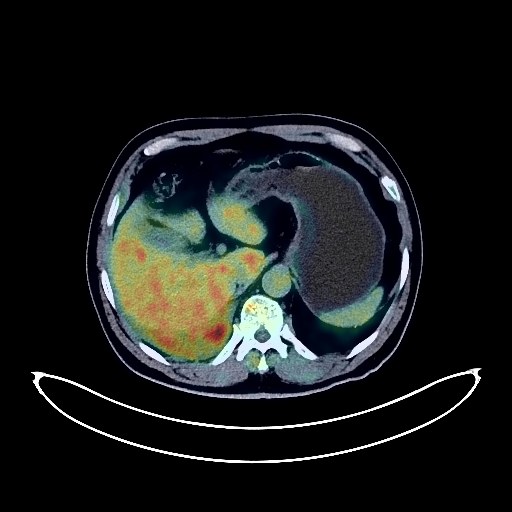
Lung Cancer PET/CT (case 983824-000119 from PETWB-REP)
2 views10 days agoWhole-body 18F-FDG PET/CT scan in a patient with Lung Cancer taken from the PETWB-REP dataset. The following English report (translated from original Chinese) is taken verbatim from the public dataset and has not been modified or otherwise checked for accuracy (see the end for citation). Impression a. A mass near the hilum in the posterior segment of the right lower lobe, with increased FDG metabolism, suggestive of central lung cancer; extensive metastasis to the right pleura (interlobar pleura, mediastinal pleura, costal pleura, diaphragmatic pleura). Multiple lymph node metastases in the right hilum, mediastinum, and right cardiophrenic angle. b. Minor chronic inflammation in the remaining lungs. Small amount of pleural effusion on the right side. Calcification of some arterial walls (including coronary arteries). c. Decreased bone density in the right iliac bone and right scapula, with increased FDG metabolism; bone metastasis to be ruled out, MRI follow-up recommended. Uneven thyroid density, calcification in the right lobe; please combine with ultrasound examination. Benign prostatic hyperplasia with calcification. Degenerative changes in the spine; L3/4, L4/5, L5/S1 intervertebral disc bulge. L5 vertebral body anteriorly slipped, bilateral vertebral arch disintegration. Left iliac bone island. Cranial scintigraphy showed no abnormalities. This case is from PETWB-REP, a curated dataset of whole-body 18F-FDG PET/CT scans and corresponding radiology reports from 490 patients with a broad spectrum of malignancies. The data were retrospectively collected from patients who underwent clinically indicated whole-body 18F-FDG PET/CT scans at the Shanghai Universal Medical Imaging Diagnostic Center between 2021 and 2024. License: Creative Commons Attribution 4.0 International (CC BY 4.0) Citation: Xue, L., Feng, G., Wenbo, Z., Zhang, Y., Li, L., Wang, S., Peng, L., Peng, S., & Gao, X. (2026). PETWB-REP: A Multi-Cancer Whole-Body FDG PET/CT Dataset with Corresponding Radiology Reports [Data set]. Zenodo. https://doi.org/10.5281/zenodo.18670487
Whole BodyPET/CT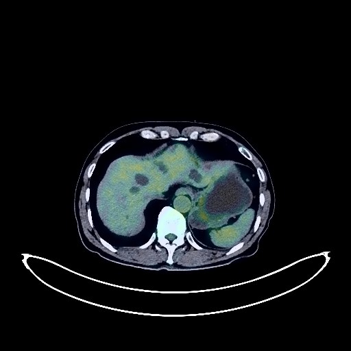
Pancreatic Cancer PET/CT (case 983824-000101 from PETWB-REP)
2 views10 days agoWhole-body 18F-FDG PET/CT scan in a patient with Pancreatic Cancer taken from the PETWB-REP dataset. The following English report (translated from original Chinese) is taken verbatim from the public dataset and has not been modified or otherwise checked for accuracy (see the end for citation). Impression a. A mass in the body of the pancreas, with indistinct borders with adjacent vessels and elevated FDG metabolism, suggestive of pancreatic malignancy. Pathological examination is required for confirmation. b. Blurred fat spaces surrounding the lesion, accompanied by several slightly enlarged lymph nodes with elevated FDG metabolism, suggesting a high probability of metastasis. c. Two nodules metastasized to the left lateral lobe of the liver. Multiple hepatic cysts. Multiple small, solid, chronic inflammatory nodules in both lungs are highly probable; CT scan is recommended. A small amount of chronic inflammation and old lesions in both lungs. Calcification of some arterial walls. Continuous elevated FDG metabolism in the sigmoid colon and rectum, suggestive of inflammatory or physiological uptake; colonoscopy is recommended. Degenerative changes in the spine. L4/5 and L5/S1 intervertebral disc bulges. A few ischemic foci in the deep cerebral regions bilaterally; age-related brain. Minor chronic inflammation of the bilateral maxillary and ethmoid sinuses. Nasal septum deviation. This case is from PETWB-REP, a curated dataset of whole-body 18F-FDG PET/CT scans and corresponding radiology reports from 490 patients with a broad spectrum of malignancies. The data were retrospectively collected from patients who underwent clinically indicated whole-body 18F-FDG PET/CT scans at the Shanghai Universal Medical Imaging Diagnostic Center between 2021 and 2024. License: Creative Commons Attribution 4.0 International (CC BY 4.0) Citation: Xue, L., Feng, G., Wenbo, Z., Zhang, Y., Li, L., Wang, S., Peng, L., Peng, S., & Gao, X. (2026). PETWB-REP: A Multi-Cancer Whole-Body FDG PET/CT Dataset with Corresponding Radiology Reports [Data set]. Zenodo. https://doi.org/10.5281/zenodo.18670487
Whole BodyPET/CT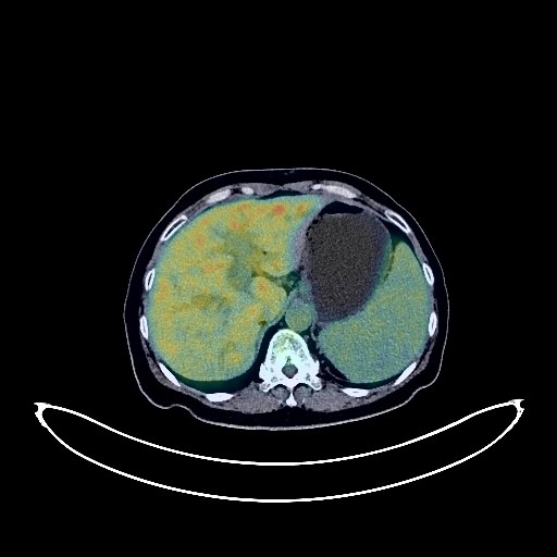
Pancreatic Cancer PET/CT (case 983824-000088 from PETWB-REP)
2 views10 days agoWhole-body 18F-FDG PET/CT scan in a patient with Pancreatic Cancer taken from the PETWB-REP dataset. The following English report (translated from original Chinese) is taken verbatim from the public dataset and has not been modified or otherwise checked for accuracy (see the end for citation). Impression a. Post-chemotherapy for pancreatic cancer, changes following pancreatic duct and bile duct stent placement, with a mass in the pancreatic head accompanied by increased FDG metabolism, suggesting continued tumor activity. It is recommended to compare previous imaging data and follow up. b. Possible metastasis to the hepatogastric space and retroperitoneal lymph nodes; increased peritoneal density with nodules and flocculent shadows, slightly increased FDG metabolism, suggesting possible peritoneal seeding metastasis; abdominopelvic effusion. Irregular thickening of the gallbladder wall with increased FDG metabolism, suggesting possible gallbladder cancer; cholecystitis to be ruled out. Please combine with enhanced MRI for comprehensive analysis. Chronic inflammatory nodules in the lower lobes of both lungs. A few post-inflammatory remnants in both lungs. Slight thickening of the pleura bilaterally. Anemia changes. Right clavicular port placement. Calcification in the left breast; ultrasound follow-up is recommended. Splenomegaly. Uterine fibroids. Chronic inflammatory changes in the gastric antrum, ascending colon, and transverse colon; endoscopic follow-up is recommended. Degenerative changes in the spine, L5/S1 disc herniation with pneumothorax and degeneration. L5/S1 vertebral endplate inflammation. No obvious abnormalities were found on cranial scintigraphy. A small amount of chronic inflammation in both ethmoid sinuses. This case is from PETWB-REP, a curated dataset of whole-body 18F-FDG PET/CT scans and corresponding radiology reports from 490 patients with a broad spectrum of malignancies. The data were retrospectively collected from patients who underwent clinically indicated whole-body 18F-FDG PET/CT scans at the Shanghai Universal Medical Imaging Diagnostic Center between 2021 and 2024. License: Creative Commons Attribution 4.0 International (CC BY 4.0) Citation: Xue, L., Feng, G., Wenbo, Z., Zhang, Y., Li, L., Wang, S., Peng, L., Peng, S., & Gao, X. (2026). PETWB-REP: A Multi-Cancer Whole-Body FDG PET/CT Dataset with Corresponding Radiology Reports [Data set]. Zenodo. https://doi.org/10.5281/zenodo.18670487
Whole BodyPET/CT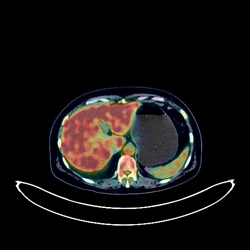
Colon Cancer PET/CT (case 983824-000035 from PETWB-REP)
7 views10 days agoWhole-body 18F-FDG PET/CT scan in a patient with Colon Cancer taken from the PETWB-REP dataset. The following English report (translated from original Chinese) is taken verbatim from the public dataset and has not been modified or otherwise checked for accuracy (see the end for citation). Impression Post-EMR changes in the colon: No abnormal FDG metabolism was observed in the sigmoid colon metal clip area; no obvious space-occupying lesions were seen in the remaining intestinal segments, and FDG metabolism was normal. Follow-up colonoscopy is recommended. Right upper lobe aberrant lobe formation; chronic inflammatory micronodules in the right upper lobe. A few post-inflammatory lesions in both lungs. Partial arteriosclerosis. Possible hemangioma in the liver; please confirm with contrast-enhanced MRI. Gallbladder bile concentration. Chronic inflammatory changes in the gastric antrum; please confirm with endoscopy. Degenerative changes in the spine; L3/4 intervertebral disc bulge. Widespread increased FDG metabolism in the medullary cavity throughout the body, suggesting possible reactive proliferative changes. No obvious abnormalities were seen on cranial scintigraphy. A few chronic inflammations in the left maxillary sinus. Left upper alveolar ulceritis. This case is from PETWB-REP, a curated dataset of whole-body 18F-FDG PET/CT scans and corresponding radiology reports from 490 patients with a broad spectrum of malignancies. The data were retrospectively collected from patients who underwent clinically indicated whole-body 18F-FDG PET/CT scans at the Shanghai Universal Medical Imaging Diagnostic Center between 2021 and 2024. License: Creative Commons Attribution 4.0 International (CC BY 4.0) Citation: Xue, L., Feng, G., Wenbo, Z., Zhang, Y., Li, L., Wang, S., Peng, L., Peng, S., & Gao, X. (2026). PETWB-REP: A Multi-Cancer Whole-Body FDG PET/CT Dataset with Corresponding Radiology Reports [Data set]. Zenodo. https://doi.org/10.5281/zenodo.18670487
Whole BodyPET/CT
Liver Cancer PET/CT (case 983824-000068 from PETWB-REP)
1 views10 days agoWhole-body 18F-FDG PET/CT scan in a patient with Liver Cancer taken from the PETWB-REP dataset. The following English report (translated from original Chinese) is taken verbatim from the public dataset and has not been modified or otherwise checked for accuracy (see the end for citation). Impression a. Liver cirrhosis, partial iodized oil deposition after interventional treatment for liver cancer, multiple space-occupying lesions in the right lobe and left inner lobe of the liver, with increased FDG metabolism near the lesion in the lower segment of the right posterior lobe, suggesting possible residual tumor activity; the tumor activity in the remaining lesions is basically suppressed. Please compare with old films and follow up with enhanced MRI. ? b. Two areas of increased FDG metabolism in the medullary cavity of the upper segment of the left femur, possibly metastatic tumors. Please follow up with enhanced MRI. ? c. Post-L2 vertebral body cementation surgery, changes after radiotherapy to the lower thoracic and lumbar spine. Degenerative changes in the spine. a. Lobulated soft tissue nodules in the anterior segment of the left upper lobe of the lung, no increased FDG metabolism, suggestive of induration. Please compare with old films and follow up with CT. ? b. Chronic inflammatory miliary lesions in the upper lobe of the right lung. Bilateral emphysema with bullae, minor chronic inflammation and sequelae in both lungs, and lower lobe aspiration effect in both lungs. Partial arteriosclerosis (including coronary arteries). Chronic inflammatory changes in the esophagus and stomach, and physiological or inflammatory uptake in some intestinal segments; endoscopic re-examination is necessary if required. Fatty infiltration of the pancreas head and neck. Possible calcification in the left kidney. Calcification in the prostate. Small amount of hydrocele in the right testis. No obvious abnormalities on cranial scintigraphy. Postoperative changes in the left parotid gland; the right submandibular gland is not clearly visualized; clinical correlation is required. This case is from PETWB-REP, a curated dataset of whole-body 18F-FDG PET/CT scans and corresponding radiology reports from 490 patients with a broad spectrum of malignancies. The data were retrospectively collected from patients who underwent clinically indicated whole-body 18F-FDG PET/CT scans at the Shanghai Universal Medical Imaging Diagnostic Center between 2021 and 2024. License: Creative Commons Attribution 4.0 International (CC BY 4.0) Citation: Xue, L., Feng, G., Wenbo, Z., Zhang, Y., Li, L., Wang, S., Peng, L., Peng, S., & Gao, X. (2026). PETWB-REP: A Multi-Cancer Whole-Body FDG PET/CT Dataset with Corresponding Radiology Reports [Data set]. Zenodo. https://doi.org/10.5281/zenodo.18670487
Whole BodyPET/CT