Loading...
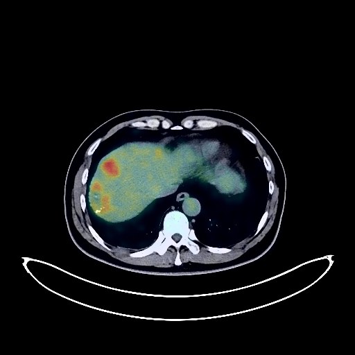
Liver Cancer PET/CT (case 983824-000104 from PETWB-REP)
1 views10 days agoWhole-body 18F-FDG PET/CT scan in a patient with Liver Cancer taken from the PETWB-REP dataset. The following English report (translated from original Chinese) is taken verbatim from the public dataset and has not been modified or otherwise checked for accuracy (see the end for citation). Impression a. Multiple mixed-density lesions in the liver with increased FDG metabolism, highly suggestive of hepatic malignancy with intrahepatic dissemination, with possible metastasis to the hilar region, hilar space, and right cardiophrenic angle lymph nodes. Please combine enhanced MRI and tumor markers for comprehensive judgment. b. Trend towards cirrhosis. Reactive hyperplasia of mediastinal and bilateral hilar lymph nodes. Slight thickening of the gastric wall in the antrum with locally increased FDG metabolism in the posterior wall, highly suggestive of chronic inflammation. A follow-up gastroscopy is recommended to rule out other possibilities. a. Multiple ground-glass opacities in both lungs with no increased FDG metabolism, mostly suggestive of chronic inflammatory nodules, some of which may be atypical adenomatous hyperplasia. Please have a follow-up HRCT 3 months after initial discovery or after regular anti-inflammatory treatment. b. Multiple solid nodules in both lungs with no increased FDG metabolism, suggestive of chronic inflammatory nodules. ? c. Minor chronic inflammation and sequelae in both lungs. Prostatic calcification. Partial vertebral osteophyte formation. L4/5 intervertebral disc bulge. No obvious abnormalities seen on cranial scintigraphy. Minor chronic inflammation in both ethmoid and maxillary sinuses. Reactive hyperplasia of bilateral cervical lymph nodes. This case is from PETWB-REP, a curated dataset of whole-body 18F-FDG PET/CT scans and corresponding radiology reports from 490 patients with a broad spectrum of malignancies. The data were retrospectively collected from patients who underwent clinically indicated whole-body 18F-FDG PET/CT scans at the Shanghai Universal Medical Imaging Diagnostic Center between 2021 and 2024. License: Creative Commons Attribution 4.0 International (CC BY 4.0) Citation: Xue, L., Feng, G., Wenbo, Z., Zhang, Y., Li, L., Wang, S., Peng, L., Peng, S., & Gao, X. (2026). PETWB-REP: A Multi-Cancer Whole-Body FDG PET/CT Dataset with Corresponding Radiology Reports [Data set]. Zenodo. https://doi.org/10.5281/zenodo.18670487
Whole BodyPET/CT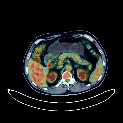
Lung Cancer PET/CT (case 983824-000042 from PETWB-REP)
7 views10 days agoWhole-body 18F-FDG PET/CT scan in a patient with Lung Cancer taken from the PETWB-REP dataset. The following English report (translated from original Chinese) is taken verbatim from the public dataset and has not been modified or otherwise checked for accuracy (see the end for citation). Impression a. A mass in the lower lobe of the left lung with significantly increased FDG metabolism, suggestive of left lung cancer with surrounding obstructive inflammation. Multiple metastases in both lungs. Metastases to the left hilar, mediastinal, and left supraclavicular fossa lymph nodes. b. Multiple bone metastases in the right ribs, sternum, spine, and pelvis. c. Possible left frontal lobe brain metastasis; enhanced MRI is required for confirmation. Chronic inflammatory changes in the gastric antrum; endoscopic re-examination is necessary if needed. Prostatic calcifications. Left inguinal hernia. Reactive hyperplasia of bilateral inguinal lymph nodes. Degenerative changes in the spine. Reactive hyperplasia of bilateral cervical lymph nodes. This case is from PETWB-REP, a curated dataset of whole-body 18F-FDG PET/CT scans and corresponding radiology reports from 490 patients with a broad spectrum of malignancies. The data were retrospectively collected from patients who underwent clinically indicated whole-body 18F-FDG PET/CT scans at the Shanghai Universal Medical Imaging Diagnostic Center between 2021 and 2024. License: Creative Commons Attribution 4.0 International (CC BY 4.0) Citation: Xue, L., Feng, G., Wenbo, Z., Zhang, Y., Li, L., Wang, S., Peng, L., Peng, S., & Gao, X. (2026). PETWB-REP: A Multi-Cancer Whole-Body FDG PET/CT Dataset with Corresponding Radiology Reports [Data set]. Zenodo. https://doi.org/10.5281/zenodo.18670487
Whole BodyPET/CT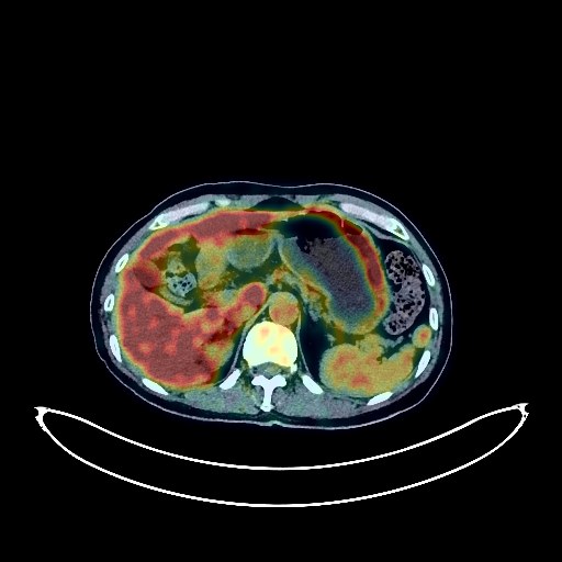
Lung Cancer PET/CT (case 983824-000100 from PETWB-REP)
2 views10 days agoWhole-body 18F-FDG PET/CT scan in a patient with Lung Cancer taken from the PETWB-REP dataset. The following English report (translated from original Chinese) is taken verbatim from the public dataset and has not been modified or otherwise checked for accuracy (see the end for citation). Impression a. A mass in the posterior segment of the right lower lobe, with increased FDG metabolism, strongly suggestive of lung cancer; please correlate with clinicopathology. b. Several small, solid, chronic inflammatory nodules in both lungs are highly probable; follow-up CT scan is recommended. Interstitial inflammation and old lesions in both lower lobes. Alveolar emphysema and bullous formation in both lungs. Reactive hyperplasia of hilar and mediastinal lymph nodes is highly probable; follow-up examination is recommended. Calcification of some arterial walls (including coronary arteries). Multiple cysts in the liver. Slight thickening of the walls of part of the gastric body and antrum, with mildly increased FDG uptake, suggestive of chronic gastritis; increased FDG metabolism in part of the intestines, suggestive of inflammatory or physiological uptake. Follow-up gastroscopy and colonoscopy are recommended for all of the above. Benign prostatic hyperplasia with calcification. Cervical, thoracic, and lumbar spondylosis; L4/5 and L5/S1 intervertebral disc bulge. A few ischemic lesions in the deep bilateral cerebral regions; senile encephalopathy. Chronic inflammation of the bilateral maxillary and ethmoid sinuses. This case is from PETWB-REP, a curated dataset of whole-body 18F-FDG PET/CT scans and corresponding radiology reports from 490 patients with a broad spectrum of malignancies. The data were retrospectively collected from patients who underwent clinically indicated whole-body 18F-FDG PET/CT scans at the Shanghai Universal Medical Imaging Diagnostic Center between 2021 and 2024. License: Creative Commons Attribution 4.0 International (CC BY 4.0) Citation: Xue, L., Feng, G., Wenbo, Z., Zhang, Y., Li, L., Wang, S., Peng, L., Peng, S., & Gao, X. (2026). PETWB-REP: A Multi-Cancer Whole-Body FDG PET/CT Dataset with Corresponding Radiology Reports [Data set]. Zenodo. https://doi.org/10.5281/zenodo.18670487
Whole BodyPET/CT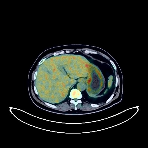
Rectal Cancer PET/CT (case 983824-000113 from PETWB-REP)
2 views10 days agoWhole-body 18F-FDG PET/CT scan in a patient with Rectal Cancer taken from the PETWB-REP dataset. The following English report (translated from original Chinese) is taken verbatim from the public dataset and has not been modified or otherwise checked for accuracy (see the end for citation). Impression a. Post-rectal cancer surgery and chemotherapy, no obvious signs of tumor recurrence were observed at the anastomosis. Multiple lesions with increased FDG uptake in the right middle and lower lobes and left upper lobe, suggestive of malignancy, with a high probability of metastasis, and some lung cancer to be ruled out, along with right middle lobe atelectasis. b. Multiple small nodules in both lungs, FDG metabolism normal, some metastasis to be ruled out, CT follow-up recommended. Bilateral hilar and mediastinal lymph node metastasis to be ruled out. Right paratracheal diverticulum. c. Enlargement of the right adrenal gland with increased FDG metabolism, suggestive of metastasis, CT follow-up recommended. Accessory spleen. Right renal cyst (one complex). Small amount of hydrocele with calcification in the left testicular tunica vaginalis. Spinal osteophyte formation. L4/5 and L5/S1 intervertebral disc bulge. Bilateral femoral head herniation. No obvious abnormalities were found on cranial imaging. Minor inflammation was observed in both ethmoid and maxillary sinuses. This case is from PETWB-REP, a curated dataset of whole-body 18F-FDG PET/CT scans and corresponding radiology reports from 490 patients with a broad spectrum of malignancies. The data were retrospectively collected from patients who underwent clinically indicated whole-body 18F-FDG PET/CT scans at the Shanghai Universal Medical Imaging Diagnostic Center between 2021 and 2024. License: Creative Commons Attribution 4.0 International (CC BY 4.0) Citation: Xue, L., Feng, G., Wenbo, Z., Zhang, Y., Li, L., Wang, S., Peng, L., Peng, S., & Gao, X. (2026). PETWB-REP: A Multi-Cancer Whole-Body FDG PET/CT Dataset with Corresponding Radiology Reports [Data set]. Zenodo. https://doi.org/10.5281/zenodo.18670487
Whole BodyPET/CT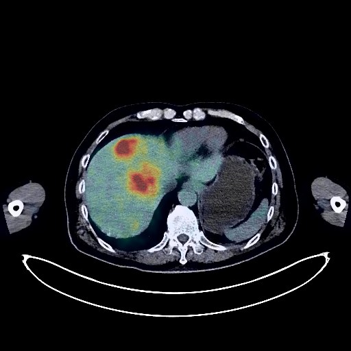
Liver Cancer PET/CT (case 983824-000045 from PETWB-REP)
7 views10 days agoWhole-body 18F-FDG PET/CT scan in a patient with Liver Cancer taken from the PETWB-REP dataset. The following English report (translated from original Chinese) is taken verbatim from the public dataset and has not been modified or otherwise checked for accuracy (see the end for citation). Impression a. Multiple space-occupying lesions in the liver with increased FDG metabolism, suggestive of malignancy, possibly hepatocellular carcinoma with intrahepatic metastasis, metastatic tumors cannot be ruled out; multiple lymph node metastases in the hilar region and retroperitoneum. b. Multiple bone metastases throughout the body. Pathological fracture of the L4 vertebral body. c. Gallstones with chronic cholecystitis. Increased local FDG uptake in the gallbladder, gallbladder cancer to be ruled out, please combine with enhanced MRI for comprehensive analysis. Benign prostatic hyperplasia with calcification, uneven FDG metabolism, please follow up with PSA and MRI. Chronic inflammatory nodules in the upper lobe of the right lung. Scattered post-inflammatory lesions in both lungs. Anemia changes, calcification of some arterial walls (including coronary arteries). Cyst in the right lobe of the liver. Adrenal hyperplasia on the left side. Chronic inflammatory changes in the antrum of the stomach. Osteoporosis, degenerative changes in the spine, multiple intervertebral disc bulges with pneumoconiosis. Postoperative changes in the left femur. The thyroid gland has uneven density and multiple nodules in both lobes. FDG metabolism is normal, suggesting nodular goiter. Please confirm with ultrasound examination. Elderly patient with deep lacunar infarcts. Please confirm with MRI examination. Right maxillary sinus submucosal cyst. This case is from PETWB-REP, a curated dataset of whole-body 18F-FDG PET/CT scans and corresponding radiology reports from 490 patients with a broad spectrum of malignancies. The data were retrospectively collected from patients who underwent clinically indicated whole-body 18F-FDG PET/CT scans at the Shanghai Universal Medical Imaging Diagnostic Center between 2021 and 2024. License: Creative Commons Attribution 4.0 International (CC BY 4.0) Citation: Xue, L., Feng, G., Wenbo, Z., Zhang, Y., Li, L., Wang, S., Peng, L., Peng, S., & Gao, X. (2026). PETWB-REP: A Multi-Cancer Whole-Body FDG PET/CT Dataset with Corresponding Radiology Reports [Data set]. Zenodo. https://doi.org/10.5281/zenodo.18670487
Whole BodyPET/CT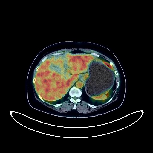
Esophageal Cancer PET/CT (case 983824-000206 from PETWB-REP)
2 views10 days agoWhole-body 18F-FDG PET/CT scan in a patient with Esophageal Cancer taken from the PETWB-REP dataset. The following English report (translated from original Chinese) is taken verbatim from the public dataset and has not been modified or otherwise checked for accuracy (see the end for citation). Impression a. A mass in the cervical segment of the esophagus, with increased FDG metabolism, consistent with esophageal cancer. Possible metastasis to the surrounding lymph nodes and left supraclavicular fossa; please correlate with clinicopathology. The left lobe of the thyroid gland is compressed. b. Slight thickening of the cardia wall, with increased FDG metabolism, consistent with cardia cancer based on the gastroscopy report. c. Slight thickening of the gastric wall on the greater curvature of the gastric body, with increased FDG metabolism, suggesting possible inflammation; follow-up gastroscopy is recommended. d. Reactive hyperplasia of the lesser omental sac and para-aortic lymph nodes; follow-up is recommended. a. Partial active inflammation in the lower lobe of the right lung; follow-up with CT scan after anti-inflammatory treatment is recommended. Reactive hyperplasia of the right hilar and mediastinal lymph nodes; please follow up to rule out partial metastasis. b. Pure ground-glass opacity nodule in the posterior segment of the right lower lobe, suggestive of chronic inflammatory nodule or atypical adenomatous hyperplasia; CT follow-up is recommended. c. Scattered small chronic inflammatory nodules (solid) in both lungs; CT follow-up is recommended. A few post-inflammatory lesions in both lungs. Partial arteriosclerosis (including coronary arteries). Bilateral breast hyperplasia, some showing nodular changes. Slight thickening of the nasopharyngeal wall and possible inflammation of bilateral palatine tonsils in the oropharynx; specialist follow-up is recommended. Reactive hyperplasia of bilateral deep cervical lymph nodes. Small cyst in the right lobe of the liver; calcification in the bile duct of the right lobe of the liver. Calcification in the left kidney. Uterine fibroids are highly probable; Nabothian cysts of the cervix; ultrasound follow-up is recommended. Degenerative changes in the spine. L4/5 and L5/S1 intervertebral disc bulges. Small bony island in the right femoral head. No obvious abnormalities were found on cranial scintigraphy. Minor inflammation of both ethmoid sinuses. This case is from PETWB-REP, a curated dataset of whole-body 18F-FDG PET/CT scans and corresponding radiology reports from 490 patients with a broad spectrum of malignancies. The data were retrospectively collected from patients who underwent clinically indicated whole-body 18F-FDG PET/CT scans at the Shanghai Universal Medical Imaging Diagnostic Center between 2021 and 2024. License: Creative Commons Attribution 4.0 International (CC BY 4.0) Citation: Xue, L., Feng, G., Wenbo, Z., Zhang, Y., Li, L., Wang, S., Peng, L., Peng, S., & Gao, X. (2026). PETWB-REP: A Multi-Cancer Whole-Body FDG PET/CT Dataset with Corresponding Radiology Reports [Data set]. Zenodo. https://doi.org/10.5281/zenodo.18670487
Whole BodyPET/CT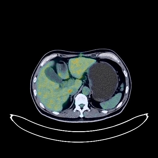
Pancreatic Cancer PET/CT (case 983824-000051 from PETWB-REP)
8 views10 days agoWhole-body 18F-FDG PET/CT scan in a patient with Pancreatic Cancer taken from the PETWB-REP dataset. The following English report (translated from original Chinese) is taken verbatim from the public dataset and has not been modified or otherwise checked for accuracy (see the end for citation). Impression a. After chemotherapy for pancreatic cancer, the pancreatic head and uncinate process show a full and irregular shape with increased FDG metabolism, suggesting that the tumor activity remains after chemotherapy, affecting adjacent blood vessels. The pancreatic body and tail are atrophied, and the main pancreatic duct is dilated. b. Soft tissue nodules near the pancreatic head with increased FDG metabolism are considered to be lymph node metastasis. Reactive hyperplasia of lymph nodes in the portacaval space, intraperitoneal cavity, and retroperitoneum is possible, and partial metastasis cannot be ruled out. Follow-up is recommended. c. Postoperative changes in the anterior abdominal wall. Multiple liver metastases. Right kidney mass, possibly a metastasis, but renal cell carcinoma cannot be ruled out. It is recommended to compare with old films and combine with enhanced MRI for comprehensive analysis. a. Multiple nodules in both lungs, some with slightly increased FDG uptake, suggesting some metastases. It is recommended to compare with old films and repeat the examination. b. A few post-inflammatory lesions in both lungs. Some arterial wall calcifications. Bilateral gynecomastia. Small subcapsular cyst in the right posterior lobe of the liver. Gallstones, chronic cholecystitis. Increased FDG metabolism in parts of the stomach wall and intestines, considered to be physiological uptake or chronic inflammatory changes. Spinal degenerative changes. No obvious abnormalities were seen on cranial scintigraphy. Chronic inflammation of the right maxillary sinus and both palatine tonsils. This case is from PETWB-REP, a curated dataset of whole-body 18F-FDG PET/CT scans and corresponding radiology reports from 490 patients with a broad spectrum of malignancies. The data were retrospectively collected from patients who underwent clinically indicated whole-body 18F-FDG PET/CT scans at the Shanghai Universal Medical Imaging Diagnostic Center between 2021 and 2024. License: Creative Commons Attribution 4.0 International (CC BY 4.0) Citation: Xue, L., Feng, G., Wenbo, Z., Zhang, Y., Li, L., Wang, S., Peng, L., Peng, S., & Gao, X. (2026). PETWB-REP: A Multi-Cancer Whole-Body FDG PET/CT Dataset with Corresponding Radiology Reports [Data set]. Zenodo. https://doi.org/10.5281/zenodo.18670487
Whole BodyPET/CT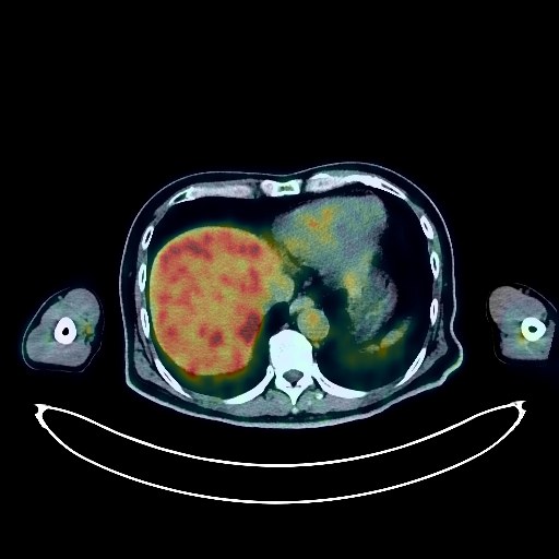
Lung Cancer PET/CT (case 983824-000184 from PETWB-REP)
1 views10 days agoWhole-body 18F-FDG PET/CT scan in a patient with Lung Cancer taken from the PETWB-REP dataset. The following English report (translated from original Chinese) is taken verbatim from the public dataset and has not been modified or otherwise checked for accuracy (see the end for citation). Impression a. Soft tissue mass at the bronchial opening in the right upper lobe of the lung, with increased FDG metabolism, suggestive of lung cancer, accompanied by distal peripheral obstructive changes; please correlate with clinicopathology; multiple lymph node metastases in the right hilum and mediastinum. b. Multiple intracranial metastases, sellar region mass, pituitary adenoma to be ruled out; enhanced MRI follow-up recommended. Elderly brain, large occipital region and cisterna magnum. c. Multiple metastases in the right lobe of the liver; bilateral retrorenal space metastases or lymph node metastases; left adrenal metastasis; multiple bone metastases throughout the body. Localized emphysema in the upper and lower lobes of the right lung. A few fibrotic lesions in both lungs. Gallstones and chronic cholecystitis. Multiple kidney stones in both kidneys. Benign prostatic hyperplasia with calcification. Localized hypermetabolic FDG lesions on the greater curvature of the stomach wall, suggestive of possible inflammatory changes; follow-up gastroscopy is recommended to rule out malignancy. Spinal osteophyte formation. Schmorl's nodes in some thoracic and lumbar vertebrae. Sacral canal cyst. L5/S1 disc herniation. Uneven thyroid density, low-density nodule in the right lobe with increased FDG metabolism, suggestive of adenoma; follow-up ultrasound is recommended. Bilateral ethmoid and maxillary sinusitis. This case is from PETWB-REP, a curated dataset of whole-body 18F-FDG PET/CT scans and corresponding radiology reports from 490 patients with a broad spectrum of malignancies. The data were retrospectively collected from patients who underwent clinically indicated whole-body 18F-FDG PET/CT scans at the Shanghai Universal Medical Imaging Diagnostic Center between 2021 and 2024. License: Creative Commons Attribution 4.0 International (CC BY 4.0) Citation: Xue, L., Feng, G., Wenbo, Z., Zhang, Y., Li, L., Wang, S., Peng, L., Peng, S., & Gao, X. (2026). PETWB-REP: A Multi-Cancer Whole-Body FDG PET/CT Dataset with Corresponding Radiology Reports [Data set]. Zenodo. https://doi.org/10.5281/zenodo.18670487
Whole BodyPET/CT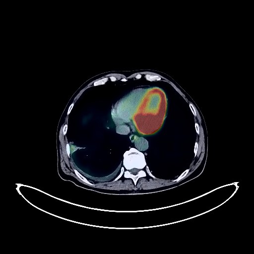
Lung Cancer PET/CT (case 983824-000197 from PETWB-REP)
2 views10 days agoWhole-body 18F-FDG PET/CT scan in a patient with Lung Cancer taken from the PETWB-REP dataset. The following English report (translated from original Chinese) is taken verbatim from the public dataset and has not been modified or otherwise checked for accuracy (see the end for citation). Impression a. A mass in the anterior basal segment of the right lower lobe, with increased FDG metabolism, consistent with lung cancer involving the adjacent oblique fissure pleura. Right-sided pneumothorax. Small amount of right-sided pleural effusion. b. Multiple solid metastases in both lungs. Metastasis to the hilar, mediastinal, and bilateral clavicular fossa lymph nodes. c. Scattered minor inflammatory changes in both lungs, more pronounced in the right lung; minor post-inflammatory remnants (including calcification) in both lungs. Partial arteriosclerosis. d. Multiple bone density changes in the right 2nd rib, localized sternum, right 8th rib, left 3rd rib, left acetabulum, and right proximal femur, with increased FDG metabolism; partial bone metastasis cannot be ruled out. Close follow-up based on clinical findings is recommended. e. Age-related brain changes, deep lacunar infarcts in the brain; contrast-enhanced MRI is recommended. Bilateral adrenal hyperplasia is highly probable; close follow-up with CT is recommended. Hemangioma at the junction of the left and right lobes of the liver is highly probable; enhanced MRI is recommended. Left lateral lobe hepatic cyst. Post-cholecystectomy absence. Left renal parenchymal calcification. Uneven FDG metabolism in the prostate; follow-up with MRI and PSA is recommended to rule out tumors. Mild prostatic hyperplasia with calcification. Degenerative changes in the spine. L4/5 and L5/S1 intervertebral disc bulge. Benign bone disease of the right iliac bone; specialist follow-up is recommended. Minor inflammation of the right ethmoid sinus and bilateral maxillary sinuses. Right deviation of the nasal septum. Reactive hyperplasia of bilateral deep cervical lymph nodes. Uneven thyroid density; follow-up with ultrasound is recommended. This case is from PETWB-REP, a curated dataset of whole-body 18F-FDG PET/CT scans and corresponding radiology reports from 490 patients with a broad spectrum of malignancies. The data were retrospectively collected from patients who underwent clinically indicated whole-body 18F-FDG PET/CT scans at the Shanghai Universal Medical Imaging Diagnostic Center between 2021 and 2024. License: Creative Commons Attribution 4.0 International (CC BY 4.0) Citation: Xue, L., Feng, G., Wenbo, Z., Zhang, Y., Li, L., Wang, S., Peng, L., Peng, S., & Gao, X. (2026). PETWB-REP: A Multi-Cancer Whole-Body FDG PET/CT Dataset with Corresponding Radiology Reports [Data set]. Zenodo. https://doi.org/10.5281/zenodo.18670487
Whole BodyPET/CT
Glioma PET/CT (case 983824-000180 from PETWB-REP)
2 views10 days agoWhole-body 18F-FDG PET/CT scan in a patient with Glioma taken from the PETWB-REP dataset. The following English report (translated from original Chinese) is taken verbatim from the public dataset and has not been modified or otherwise checked for accuracy (see the end for citation). Impression Space-occupying lesions in the right basal ganglia and corona radiata, with decreased or absent FDG uptake, suggestive of malignancy, with glioblastoma being the primary consideration. Softening lesion in the left parietal lobe. Chronic inflammation of the bilateral maxillary sinuses. a. Multiple bronchiectasis with infection in the right middle lobe and both lower lobes; follow-up CT scan after treatment is recommended. b. Scattered post-inflammatory lesions in both lungs. Reactive hyperplasia of mediastinal lymph nodes. Benign lesions in the anterior mediastinum; cysts are the primary consideration. Calcification of some arterial walls (including coronary arteries). Multiple slightly low-density lesions in the liver, with background FDG uptake; hemangioma or cysts are the primary consideration; further enhanced MRI is recommended to rule out other possibilities. Accessory spleen. Right renal cyst (partially complex). Chronic inflammatory changes in the gastric antrum; please follow up with endoscopy. Degenerative changes in the spine, with L3/4, L4/5, and L5/S1 disc bulges. This case is from PETWB-REP, a curated dataset of whole-body 18F-FDG PET/CT scans and corresponding radiology reports from 490 patients with a broad spectrum of malignancies. The data were retrospectively collected from patients who underwent clinically indicated whole-body 18F-FDG PET/CT scans at the Shanghai Universal Medical Imaging Diagnostic Center between 2021 and 2024. License: Creative Commons Attribution 4.0 International (CC BY 4.0) Citation: Xue, L., Feng, G., Wenbo, Z., Zhang, Y., Li, L., Wang, S., Peng, L., Peng, S., & Gao, X. (2026). PETWB-REP: A Multi-Cancer Whole-Body FDG PET/CT Dataset with Corresponding Radiology Reports [Data set]. Zenodo. https://doi.org/10.5281/zenodo.18670487
Whole BodyPET/CT