Loading...
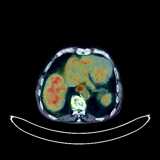
Colon Cancer PET/CT (case 983824-000009 from PETWB-REP)
9 views10 days agoWhole-body 18F-FDG PET/CT scan in a patient with Colon Cancer taken from the PETWB-REP dataset. The following English report (translated from original Chinese) is taken verbatim from the public dataset and has not been modified or otherwise checked for accuracy (see the end for citation). Impression a. Multiple peritoneal metastases in the abdominopelvic cavity (including perihepatic, hilar, and perisplenic areas); right lower abdominal wall metastasis; left inguinal region metastasis. b. A mass in the sigmoid colon with increased FDG uptake, suggestive of malignancy, most likely colon cancer; please follow up with colonoscopy. Liver cirrhosis. Calcification in the right lobe of the liver. A few calcifications in the spleen. Scattered chronic inflammatory nodules in both lungs; please follow up with CT scan. Interstitial lung changes with emphysema in both lungs. Chronic inflammation and sequelae in both lungs. Reactive hyperplasia of bilateral supraclavicular and mediastinal lymph nodes. Cardiac enlargement. Calcification of some arterial walls (including coronary arteries); please follow up with coronary CTA. Highly probable inflammatory uptake in the middle and lower esophagus; please follow up with gastroscopy. Mild renal atrophy in both kidneys. Left renal cyst. Right renal calculus. Calcification and hydrocele in the right testicular tunica vaginalis. Osteoporosis. Compression changes in the T10 vertebral body. Spinal degeneration. Right hip periarthritis. A few ischemic foci deep in the brain, age-related brain changes. This case is from PETWB-REP, a curated dataset of whole-body 18F-FDG PET/CT scans and corresponding radiology reports from 490 patients with a broad spectrum of malignancies. The data were retrospectively collected from patients who underwent clinically indicated whole-body 18F-FDG PET/CT scans at the Shanghai Universal Medical Imaging Diagnostic Center between 2021 and 2024. License: Creative Commons Attribution 4.0 International (CC BY 4.0) Citation: Xue, L., Feng, G., Wenbo, Z., Zhang, Y., Li, L., Wang, S., Peng, L., Peng, S., & Gao, X. (2026). PETWB-REP: A Multi-Cancer Whole-Body FDG PET/CT Dataset with Corresponding Radiology Reports [Data set]. Zenodo. https://doi.org/10.5281/zenodo.18670487
Whole BodyPET/CT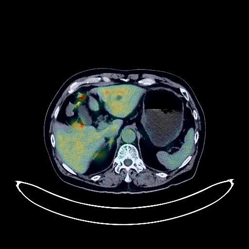
Lung Cancer PET/CT (case 983824-000025 from PETWB-REP)
6 views10 days agoWhole-body 18F-FDG PET/CT scan in a patient with Lung Cancer taken from the PETWB-REP dataset. The following English report (translated from original Chinese) is taken verbatim from the public dataset and has not been modified or otherwise checked for accuracy (see the end for citation). Impression a. A mass in the right middle lobe of the lung, involving the right upper lobe, with increased FDG metabolism, suggestive of lung cancer. Possible right hilar lymph node metastasis. b. Several small, solid, chronic inflammatory nodules in both lungs. A few chronic inflammations and old lesions in both lungs. c. Post-ablation changes after atrial fibrillation; enlarged cardiac silhouette. Calcification of some arterial walls (including coronary arteries). Multiple liver cysts. A small diverticulum in the descending duodenum. Continuous increased FDG metabolism in the descending colon, sigmoid colon, and rectum, suggestive of inflammatory or physiological uptake. Large amount of hydrocele in the right testis. Degenerative changes in the spine. L4/5 and L5/S1 intervertebral disc bulges. A few ischemic lesions in the deep bilateral brain regions, suggestive of age-related brain changes. Chronic inflammation of both ethmoid sinuses. Several slightly high-density nodules in both parotid glands, with increased FDG metabolism, suggestive of adenolymphoma; follow-up MRI is recommended. This case is from PETWB-REP, a curated dataset of whole-body 18F-FDG PET/CT scans and corresponding radiology reports from 490 patients with a broad spectrum of malignancies. The data were retrospectively collected from patients who underwent clinically indicated whole-body 18F-FDG PET/CT scans at the Shanghai Universal Medical Imaging Diagnostic Center between 2021 and 2024. License: Creative Commons Attribution 4.0 International (CC BY 4.0) Citation: Xue, L., Feng, G., Wenbo, Z., Zhang, Y., Li, L., Wang, S., Peng, L., Peng, S., & Gao, X. (2026). PETWB-REP: A Multi-Cancer Whole-Body FDG PET/CT Dataset with Corresponding Radiology Reports [Data set]. Zenodo. https://doi.org/10.5281/zenodo.18670487
Whole BodyPET/CT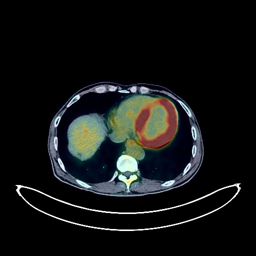
Lung Cancer PET/CT (case 983824-000203 from PETWB-REP)
0 views10 days agoWhole-body 18F-FDG PET/CT scan in a patient with Lung Cancer taken from the PETWB-REP dataset. The following English report (translated from original Chinese) is taken verbatim from the public dataset and has not been modified or otherwise checked for accuracy (see the end for citation). Impression a. A mass near the hilum in the lower lobe of the left lung, with increased FDG metabolism, suggestive of central lung cancer with surrounding obstructive inflammation, possibly involving the left pulmonary artery. Please correlate with clinicopathology. b. Metastasis to the left hilar and mediastinal lymph nodes. Multiple bone metastases throughout the body. c. Chronic bronchitis-like changes in both lungs. Scattered chronic inflammatory nodules (solid) in both lungs; please correlate with CT follow-up. Several calcifications in both lungs. A few post-inflammatory remnants in both lungs. Paraseptal emphysema in both lungs. Slight pleural thickening bilaterally. Partial arteriosclerosis. d. Lacunar infarcts deep in the brain; MRI follow-up is recommended. Multiple liver cysts. Possible gallstones. Possible small splenic angioma. Bilateral renal cysts, small kidney stone in the left kidney. Benign prostatic hyperplasia with calcification. Bilateral hydrocele. Slightly thickened gastric wall, mildly elevated FDG metabolism; please correlate with gastroscopy. Multiple high-density shadows in the colon; please correlate with clinical findings. Degenerative changes in the spine. L4/5 intervertebral disc bulge. L5/S1 vertebral endplate inflammation. Small bony islands within the S1 vertebral body. This case is from PETWB-REP, a curated dataset of whole-body 18F-FDG PET/CT scans and corresponding radiology reports from 490 patients with a broad spectrum of malignancies. The data were retrospectively collected from patients who underwent clinically indicated whole-body 18F-FDG PET/CT scans at the Shanghai Universal Medical Imaging Diagnostic Center between 2021 and 2024. License: Creative Commons Attribution 4.0 International (CC BY 4.0) Citation: Xue, L., Feng, G., Wenbo, Z., Zhang, Y., Li, L., Wang, S., Peng, L., Peng, S., & Gao, X. (2026). PETWB-REP: A Multi-Cancer Whole-Body FDG PET/CT Dataset with Corresponding Radiology Reports [Data set]. Zenodo. https://doi.org/10.5281/zenodo.18670487
Whole BodyPET/CT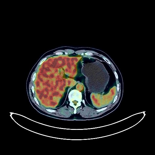
Lung Cancer PET/CT (case 983824-000167 from PETWB-REP)
2 views10 days agoWhole-body 18F-FDG PET/CT scan in a patient with Lung Cancer taken from the PETWB-REP dataset. The following English report (translated from original Chinese) is taken verbatim from the public dataset and has not been modified or otherwise checked for accuracy (see the end for citation). Impression a. A mass in the posterior segment of the right upper lobe, with increased FDG metabolism, suggestive of lung cancer. b. Several solid micronodules of chronic inflammatory origin in both lungs. Minor chronic inflammation and old lesions in both lungs. Partial arteriosclerosis. A low-density nodule in the left lobe of the thyroid gland, with normal FDG metabolism, suggestive of a benign nodule, most likely a lipoma; ultrasound examination recommended. Heterogeneous fatty liver. Degenerative changes in the spine. L4/5 and L5/S1 intervertebral disc bulges. Bilateral L5 vertebral arch disintegration. A few ischemic lesions in the deep bilateral brain regions; follow-up MRI recommended. This case is from PETWB-REP, a curated dataset of whole-body 18F-FDG PET/CT scans and corresponding radiology reports from 490 patients with a broad spectrum of malignancies. The data were retrospectively collected from patients who underwent clinically indicated whole-body 18F-FDG PET/CT scans at the Shanghai Universal Medical Imaging Diagnostic Center between 2021 and 2024. License: Creative Commons Attribution 4.0 International (CC BY 4.0) Citation: Xue, L., Feng, G., Wenbo, Z., Zhang, Y., Li, L., Wang, S., Peng, L., Peng, S., & Gao, X. (2026). PETWB-REP: A Multi-Cancer Whole-Body FDG PET/CT Dataset with Corresponding Radiology Reports [Data set]. Zenodo. https://doi.org/10.5281/zenodo.18670487
Whole BodyPET/CT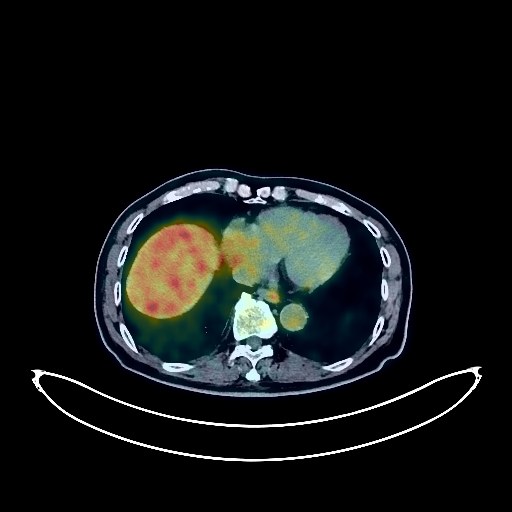
Lung Cancer PET/CT (case 983824-000102 from PETWB-REP)
2 views10 days agoWhole-body 18F-FDG PET/CT scan in a patient with Lung Cancer taken from the PETWB-REP dataset. The following English report (translated from original Chinese) is taken verbatim from the public dataset and has not been modified or otherwise checked for accuracy (see the end for citation). Impression a. Mass in the anterior segment of the left upper lobe, with increased FDG metabolism, suggestive of lung cancer, accompanied by surrounding inflammation. Please correlate with clinicopathology. b. Multiple scattered chronic inflammations in both lungs. Several small chronic inflammatory nodules (solid) in both lungs. Follow-up CT scan recommended. c. Calcification of some arterial walls. Please correlate with coronary CTA. d. Possible left adrenal hyperplasia. Follow-up CT scan recommended. Multiple liver cysts. Small kidney stones in both kidneys. Chronic gastritis. Continuous increased FDG metabolism in the sigmoid colon and rectum, likely due to inflammatory uptake. Colonoscopy recommended. Benign prostatic hyperplasia with calcification; bilateral testicular capsule calcification. Degenerative changes in the spine. Multiple intervertebral disc calcifications in the thoracic and lumbar spine. L4/5 and L5/S1 disc bulges. Mild compression and flattening of L1. A few ischemic foci in the deep bilateral cerebral regions; age-related brain. Chronic inflammation of the bilateral maxillary and ethmoid sinuses. This case is from PETWB-REP, a curated dataset of whole-body 18F-FDG PET/CT scans and corresponding radiology reports from 490 patients with a broad spectrum of malignancies. The data were retrospectively collected from patients who underwent clinically indicated whole-body 18F-FDG PET/CT scans at the Shanghai Universal Medical Imaging Diagnostic Center between 2021 and 2024. License: Creative Commons Attribution 4.0 International (CC BY 4.0) Citation: Xue, L., Feng, G., Wenbo, Z., Zhang, Y., Li, L., Wang, S., Peng, L., Peng, S., & Gao, X. (2026). PETWB-REP: A Multi-Cancer Whole-Body FDG PET/CT Dataset with Corresponding Radiology Reports [Data set]. Zenodo. https://doi.org/10.5281/zenodo.18670487
Whole BodyPET/CT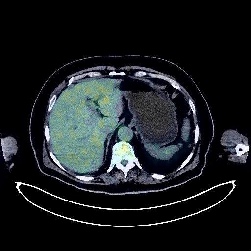
Esophageal Cancer PET/CT (case 983824-000097 from PETWB-REP)
2 views10 days agoWhole-body 18F-FDG PET/CT scan in a patient with Esophageal Cancer taken from the PETWB-REP dataset. The following English report (translated from original Chinese) is taken verbatim from the public dataset and has not been modified or otherwise checked for accuracy (see the end for citation). Impression A mass in the lower thoracic segment of the esophagus with increased FDG metabolism, consistent with esophageal cancer. Reactive hyperplasia of the mediastinal lymph nodes is highly probable; follow-up is recommended. Multiple scattered chronic inflammations in the lower lobe of the right lung; anti-inflammatory treatment followed by a CT scan is recommended to rule out hidden lesions. Chronic inflammatory nodules (solid) in the middle lobe of the right lung. Bilateral pulmonary fibrosis, calcification in the lower lobe of the left lung. Calcification of some arterial walls (including coronary arteries). Benign prostatic hyperplasia with calcification, localized high FDG metabolism in the right peripheral zone, suggestive of inflammatory changes; PSA and enhanced MRI are needed to rule out malignancy. Left renal cyst. Bladder stones or wall calcifications. Small amount of hydrocele in the left testis. Degenerative changes in the spine. L4/5 and L5/S1 intervertebral disc bulges. A few ischemic foci in the deep bilateral cerebral regions; age-related brain. This case is from PETWB-REP, a curated dataset of whole-body 18F-FDG PET/CT scans and corresponding radiology reports from 490 patients with a broad spectrum of malignancies. The data were retrospectively collected from patients who underwent clinically indicated whole-body 18F-FDG PET/CT scans at the Shanghai Universal Medical Imaging Diagnostic Center between 2021 and 2024. License: Creative Commons Attribution 4.0 International (CC BY 4.0) Citation: Xue, L., Feng, G., Wenbo, Z., Zhang, Y., Li, L., Wang, S., Peng, L., Peng, S., & Gao, X. (2026). PETWB-REP: A Multi-Cancer Whole-Body FDG PET/CT Dataset with Corresponding Radiology Reports [Data set]. Zenodo. https://doi.org/10.5281/zenodo.18670487
Whole BodyPET/CT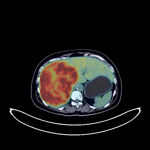
Liver Cancer PET/CT (case 983824-000114 from PETWB-REP)
2 views10 days agoWhole-body 18F-FDG PET/CT scan in a patient with Liver Cancer taken from the PETWB-REP dataset. The following English report (translated from original Chinese) is taken verbatim from the public dataset and has not been modified or otherwise checked for accuracy (see the end for citation). Impression a. Multiple lesions in the liver with increased FDG metabolism, suggestive of malignancy. The larger lesion in the right lobe of the liver is likely primary. Please combine tumor markers and enhanced MRI for comprehensive analysis. b. Multiple metastatic tumors in both lungs. c. Postoperative right palatal tumor, no signs of tumor recurrence observed in the surgical area. Continuous increased FDG metabolism in the colon and rectum, likely due to inflammatory uptake. Colonoscopy is recommended to rule out tumors. A few chronic inflammations and old lesions in both lungs. Calcification of some arterial walls (including coronary arteries). Inflammatory uptake in the middle and lower esophagus. Liver cyst. Left renal cyst. Degenerative changes in the spine. L4/5 and L5/S1 intervertebral disc bulges. Age-related brain changes. This case is from PETWB-REP, a curated dataset of whole-body 18F-FDG PET/CT scans and corresponding radiology reports from 490 patients with a broad spectrum of malignancies. The data were retrospectively collected from patients who underwent clinically indicated whole-body 18F-FDG PET/CT scans at the Shanghai Universal Medical Imaging Diagnostic Center between 2021 and 2024. License: Creative Commons Attribution 4.0 International (CC BY 4.0) Citation: Xue, L., Feng, G., Wenbo, Z., Zhang, Y., Li, L., Wang, S., Peng, L., Peng, S., & Gao, X. (2026). PETWB-REP: A Multi-Cancer Whole-Body FDG PET/CT Dataset with Corresponding Radiology Reports [Data set]. Zenodo. https://doi.org/10.5281/zenodo.18670487
Whole BodyPET/CT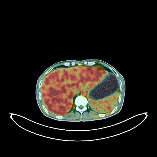
Liver Cancer PET/CT (case 983824-000126 from PETWB-REP)
2 views10 days agoWhole-body 18F-FDG PET/CT scan in a patient with Liver Cancer taken from the PETWB-REP dataset. The following English report (translated from original Chinese) is taken verbatim from the public dataset and has not been modified or otherwise checked for accuracy (see the end for citation). Impression a. A mass in the lower segment of the right anterior lobe of the liver, with unevenly increased FDG metabolism, suggesting possible hepatocellular carcinoma. Please combine with enhanced MRI for comprehensive analysis. b. Cirrhosis. a. An irregular nodule at the opening of the posterior basal segment of the right lower lobe, with increased FDG metabolism, suggesting possible lung cancer. Inflammatory lesions need to be ruled out. Please compare with old films and have a follow-up HRCT in 3 months. b. Several small chronic inflammatory nodules (solid) in both lungs are highly likely. Follow-up CT is recommended. The upper lobe of the right lung contains a pneumocystic cavity. A few chronic inflammations and old lesions in both lungs. Calcification of some arterial walls (including coronary arteries). Gallstones. Stone in the upper segment of the right ureter, with mild hydronephrosis of the proximal ureter and renal pelvis. Osteoporosis. Osteophytes in the cervical, thoracic, and lumbar vertebrae. L4/5 and L5/S1 intervertebral disc bulges. Cranial scintigraphy showed no abnormalities. This case is from PETWB-REP, a curated dataset of whole-body 18F-FDG PET/CT scans and corresponding radiology reports from 490 patients with a broad spectrum of malignancies. The data were retrospectively collected from patients who underwent clinically indicated whole-body 18F-FDG PET/CT scans at the Shanghai Universal Medical Imaging Diagnostic Center between 2021 and 2024. License: Creative Commons Attribution 4.0 International (CC BY 4.0) Citation: Xue, L., Feng, G., Wenbo, Z., Zhang, Y., Li, L., Wang, S., Peng, L., Peng, S., & Gao, X. (2026). PETWB-REP: A Multi-Cancer Whole-Body FDG PET/CT Dataset with Corresponding Radiology Reports [Data set]. Zenodo. https://doi.org/10.5281/zenodo.18670487
Whole BodyPET/CT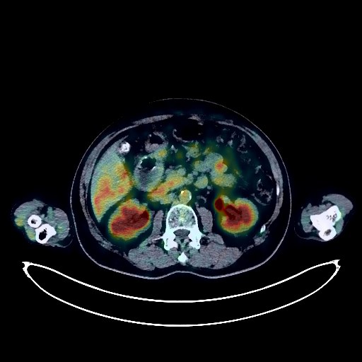
Lymphoma PET/CT (case 983824-000017 from PETWB-REP)
7 views10 days agoWhole-body 18F-FDG PET/CT scan in a patient with Lymphoma taken from the PETWB-REP dataset. The following English report (translated from original Chinese) is taken verbatim from the public dataset and has not been modified or otherwise checked for accuracy (see the end for citation). Impression a. After chemotherapy for lymphoma, no significantly enlarged lymph nodes were observed throughout the body, and FDG metabolism was normal, suggesting that tumor activity was basically suppressed. Specialist follow-up is recommended. b. Small lymph nodes were observed in the bilateral deep cervical spaces, bilateral axillae, and para-aortic region. A mass in the intermuscular space of the left groin with increased FDG uptake, combined with the medical history, suggests a recurrence of leiomyoma. Pathological examination is recommended for confirmation. Several ground-glass nodules in the upper lobe of the right lung, with normal FDG metabolism, suggestive of inflammatory nodules or atypical adenomatous hyperplasia. Annual HRCT follow-up is recommended. Several chronic inflammatory micronodules (solid) in both lungs. A few chronic inflammations and old lesions in both lungs. Calcification of some arterial walls (including coronary arteries). Slight thickening of the cardia and gastric fundus walls, with mildly increased FDG uptake, suggesting chronic gastritis. Gastroscopy follow-up is recommended. Gallstones. Left renal cyst. Prostatic calcification. Spinal degenerative changes. L4/5 disc bulge. Few ischemic lesions in the deep bilateral brain regions, indicative of age-related brain changes. Chronic sphenoid sinusitis. Nodular goiter is highly probable; ultrasound follow-up is recommended. This case is from PETWB-REP, a curated dataset of whole-body 18F-FDG PET/CT scans and corresponding radiology reports from 490 patients with a broad spectrum of malignancies. The data were retrospectively collected from patients who underwent clinically indicated whole-body 18F-FDG PET/CT scans at the Shanghai Universal Medical Imaging Diagnostic Center between 2021 and 2024. License: Creative Commons Attribution 4.0 International (CC BY 4.0) Citation: Xue, L., Feng, G., Wenbo, Z., Zhang, Y., Li, L., Wang, S., Peng, L., Peng, S., & Gao, X. (2026). PETWB-REP: A Multi-Cancer Whole-Body FDG PET/CT Dataset with Corresponding Radiology Reports [Data set]. Zenodo. https://doi.org/10.5281/zenodo.18670487
Whole BodyPET/CT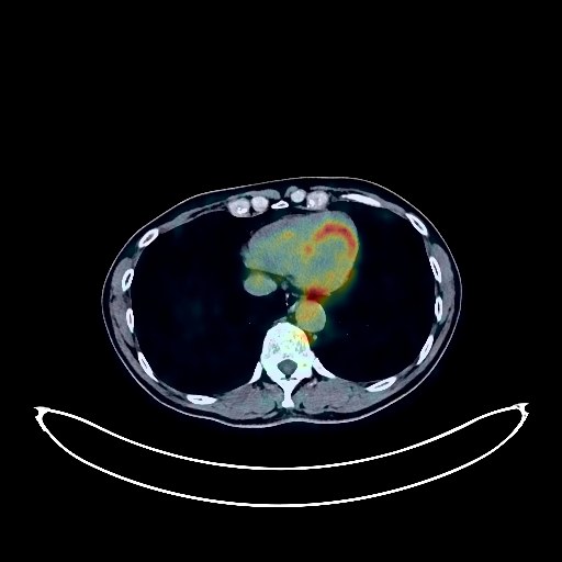
Rectal Cancer PET/CT (case 983824-000106 from PETWB-REP)
2 views10 days agoWhole-body 18F-FDG PET/CT scan in a patient with Rectal Cancer taken from the PETWB-REP dataset. The following English report (translated from original Chinese) is taken verbatim from the public dataset and has not been modified or otherwise checked for accuracy (see the end for citation). Impression Mass in the upper rectum, elevated FDG metabolism, suggestive of rectal cancer; please confirm with pathology. a. Ground-glass nodule in the lateral segment of the right middle lobe, normal FDG metabolism, suggestive of atypical adenomatous hyperplasia or inflammatory nodule; annual HRCT follow-up recommended. b. Multiple chronic inflammatory micronodules in both lungs, calcification in the lateral basal segment of the left lower lobe, and a few fibrotic lesions in both lungs. Partial calcification of the coronary artery wall. Slightly low-density nodules in the left lateral lobe and right anterior lobe of the liver, normal FDG metabolism, suggestive of hemangioma; multiple slightly low-density nodules in the spleen, normal FDG metabolism, suggestive of vascular malformation. Further examination with contrast-enhanced MRI is recommended for all of the above. Complex small cyst in the right kidney. Calcification in the prostate. Small amount of hydrocele in both testes. Spinal osteophyte formation, L4/5 disc bulge. Multiple vertebral bodies and appendages, right 6th posterior rib, and left iliac bone are highly suggestive of benign bone disease; please follow up. Cranial imaging showed no obvious abnormalities. Bilateral ethmoid and maxillary sinusitis. This case is from PETWB-REP, a curated dataset of whole-body 18F-FDG PET/CT scans and corresponding radiology reports from 490 patients with a broad spectrum of malignancies. The data were retrospectively collected from patients who underwent clinically indicated whole-body 18F-FDG PET/CT scans at the Shanghai Universal Medical Imaging Diagnostic Center between 2021 and 2024. License: Creative Commons Attribution 4.0 International (CC BY 4.0) Citation: Xue, L., Feng, G., Wenbo, Z., Zhang, Y., Li, L., Wang, S., Peng, L., Peng, S., & Gao, X. (2026). PETWB-REP: A Multi-Cancer Whole-Body FDG PET/CT Dataset with Corresponding Radiology Reports [Data set]. Zenodo. https://doi.org/10.5281/zenodo.18670487
Whole BodyPET/CT