Loading...
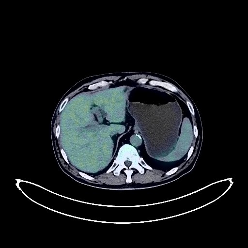
Gastric Cancer PET/CT (case 983824-000182 from PETWB-REP)
2 views10 days agoWhole-body 18F-FDG PET/CT scan in a patient with Gastric Cancer taken from the PETWB-REP dataset. The following English report (translated from original Chinese) is taken verbatim from the public dataset and has not been modified or otherwise checked for accuracy (see the end for citation). Impression a. Space-occupying lesion in the antrum of the stomach, with increased FDG metabolism, consistent with gastric cancer based on clinical findings. b. Localized thickening of the ascending colon wall with increased FDG uptake, consistent with colorectal cancer based on clinical findings. Reactive hyperplasia of surrounding mesenteric lymph nodes. c. Focal increased FDG uptake in the transverse colon and ileocecal region, suggestive of polyps or intestinal contents; follow-up colonoscopy is recommended. Emphysema and bullae in both lungs, multiple chronic inflammatory micronodules in both lungs, and a few post-inflammatory remnants in the lower lobes of both lungs. Slight thickening of the pleura bilaterally. Partial calcification of the aorta and coronary artery walls. Cyst in the left lateral lobe of the liver. Chronic cholecystitis. Multiple cysts in both kidneys. Degenerative changes in the spine. L3/4 and L4/5 disc bulges, mild L5/S1 disc protrusion. Post-left hip replacement surgery changes; right femoral head avascular necrosis. Bilateral deep lacunar infarcts, age-related brain changes. Minor inflammation of the right maxillary sinus. This case is from PETWB-REP, a curated dataset of whole-body 18F-FDG PET/CT scans and corresponding radiology reports from 490 patients with a broad spectrum of malignancies. The data were retrospectively collected from patients who underwent clinically indicated whole-body 18F-FDG PET/CT scans at the Shanghai Universal Medical Imaging Diagnostic Center between 2021 and 2024. License: Creative Commons Attribution 4.0 International (CC BY 4.0) Citation: Xue, L., Feng, G., Wenbo, Z., Zhang, Y., Li, L., Wang, S., Peng, L., Peng, S., & Gao, X. (2026). PETWB-REP: A Multi-Cancer Whole-Body FDG PET/CT Dataset with Corresponding Radiology Reports [Data set]. Zenodo. https://doi.org/10.5281/zenodo.18670487
Whole BodyPET/CT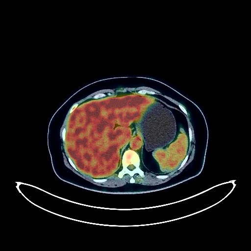
Breast Cancer PET/CT (case 983824-000187 from PETWB-REP)
2 views10 days agoWhole-body 18F-FDG PET/CT scan in a patient with Breast Cancer taken from the PETWB-REP dataset. The following English report (translated from original Chinese) is taken verbatim from the public dataset and has not been modified or otherwise checked for accuracy (see the end for citation). Impression Left breast cancer chemotherapy followed by a mass in the left breast with increased FDG metabolism, suggesting continued tumor activity; follow-up is recommended. Bilateral axillary lymph node reactive hyperplasia is highly probable. Chronic inflammatory micronodules in both lungs; CT follow-up is recommended. A few post-inflammatory lesions in both lungs. Partial arteriosclerosis. Fatty liver. Post-cholecystectomy changes. Small kidney stone in the right kidney. Uterine fibroids. Chronic inflammatory changes in the gastric antrum; endoscopic follow-up is recommended. Mild vertebral osteophyte formation. Possible coccyx fracture. No obvious abnormalities were found on cranial scintigraphy. This case is from PETWB-REP, a curated dataset of whole-body 18F-FDG PET/CT scans and corresponding radiology reports from 490 patients with a broad spectrum of malignancies. The data were retrospectively collected from patients who underwent clinically indicated whole-body 18F-FDG PET/CT scans at the Shanghai Universal Medical Imaging Diagnostic Center between 2021 and 2024. License: Creative Commons Attribution 4.0 International (CC BY 4.0) Citation: Xue, L., Feng, G., Wenbo, Z., Zhang, Y., Li, L., Wang, S., Peng, L., Peng, S., & Gao, X. (2026). PETWB-REP: A Multi-Cancer Whole-Body FDG PET/CT Dataset with Corresponding Radiology Reports [Data set]. Zenodo. https://doi.org/10.5281/zenodo.18670487
Whole BodyPET/CT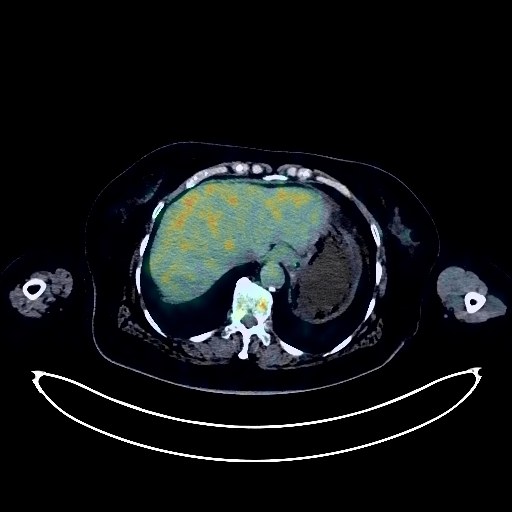
Breast Cancer PET/CT (case 983824-000190 from PETWB-REP)
4 views10 days agoWhole-body 18F-FDG PET/CT scan in a patient with Breast Cancer taken from the PETWB-REP dataset. The following English report (translated from original Chinese) is taken verbatim from the public dataset and has not been modified or otherwise checked for accuracy (see the end for citation). Impression Left breast mass with elevated FDG metabolism, consistent with breast cancer; reactive hyperplasia of small axillary lymph nodes bilaterally. Post-cranial surgery changes, softening lesions in the left basal ganglia and periventricular region, age-related brain changes; MRI follow-up recommended. Thickening of the left nasopharyngeal wall with elevated FDG metabolism; nasopharyngitis is the primary consideration; further specialist examination recommended to rule out tumors. Chronic inflammation of the right ethmoid sinus and right maxillary sinus. Nodular goiter. Reactive hyperplasia of bilateral cervical lymph nodes. Chronic inflammatory nodules in both lungs; CT follow-up recommended to rule out partial metastasis. Scattered post-inflammatory lesions in both lungs. Calcification of some arterial walls (including coronary arteries). Calcifications in the liver. Gallstones. Accessory spleen. Chronic inflammatory changes in part of the gastric wall; please follow up with endoscopy. Osteoporosis, degenerative changes in the spine. L4/5 and L5/S1 intervertebral disc bulge. Multiple subcutaneous calcifications in both buttocks. This case is from PETWB-REP, a curated dataset of whole-body 18F-FDG PET/CT scans and corresponding radiology reports from 490 patients with a broad spectrum of malignancies. The data were retrospectively collected from patients who underwent clinically indicated whole-body 18F-FDG PET/CT scans at the Shanghai Universal Medical Imaging Diagnostic Center between 2021 and 2024. License: Creative Commons Attribution 4.0 International (CC BY 4.0) Citation: Xue, L., Feng, G., Wenbo, Z., Zhang, Y., Li, L., Wang, S., Peng, L., Peng, S., & Gao, X. (2026). PETWB-REP: A Multi-Cancer Whole-Body FDG PET/CT Dataset with Corresponding Radiology Reports [Data set]. Zenodo. https://doi.org/10.5281/zenodo.18670487
Whole BodyPET/CT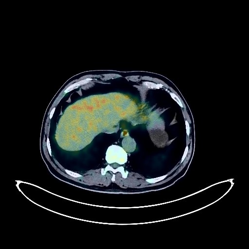
Lung Cancer PET/CT (case 983824-000118 from PETWB-REP)
2 views10 days agoWhole-body 18F-FDG PET/CT scan in a patient with Lung Cancer taken from the PETWB-REP dataset. The following English report (translated from original Chinese) is taken verbatim from the public dataset and has not been modified or otherwise checked for accuracy (see the end for citation). Impression a. A mass near the hilum in the right upper lobe of the lung, with increased FDG metabolism, suggestive of central lung cancer with surrounding obstructive inflammation, and partial carcinomatous lymphangitis in the right upper lobe. b. Right hilar lymph node metastasis; possible reactive hyperplasia of mediastinal lymph nodes, follow-up recommended to rule out metastasis. c. A few ischemic lesions in the deep bilateral brain; age-related encephalopathy. Slightly decreased density in the pancreatic head, mildly increased FDG metabolism, and slightly widened main pancreatic duct, suggesting possible inflammatory uptake; enhanced MRI recommended to rule out tumor. Liver calcifications. Soft tissue nodule in the left palatine tonsil, with increased FDG metabolism, suggesting inflammation; tumor to be ruled out, clinical specialist examination recommended. Bilateral renal cysts. Left renal angiomyolipoma. Slight thickening of the walls of part of the gastric body and antrum, with mildly increased FDG uptake, suggestive of chronic gastritis; continuous increased FDG metabolism in part of the colon and rectum, suggestive of inflammatory or physiological uptake; duodenal diverticulum. Benign prostatic hyperplasia with calcification, increased FDG metabolism in the gland, suggestive of inflammatory or physiological uptake; follow-up PSA and ultrasound examination recommended. Degenerative changes in the spine. L4/5 and L5/S1 intervertebral disc bulge. Low-density nodule in the right lobe of the thyroid gland, no abnormalities in FDG metabolism, suggestive of adenoma; ultrasound examination recommended. This case is from PETWB-REP, a curated dataset of whole-body 18F-FDG PET/CT scans and corresponding radiology reports from 490 patients with a broad spectrum of malignancies. The data were retrospectively collected from patients who underwent clinically indicated whole-body 18F-FDG PET/CT scans at the Shanghai Universal Medical Imaging Diagnostic Center between 2021 and 2024. License: Creative Commons Attribution 4.0 International (CC BY 4.0) Citation: Xue, L., Feng, G., Wenbo, Z., Zhang, Y., Li, L., Wang, S., Peng, L., Peng, S., & Gao, X. (2026). PETWB-REP: A Multi-Cancer Whole-Body FDG PET/CT Dataset with Corresponding Radiology Reports [Data set]. Zenodo. https://doi.org/10.5281/zenodo.18670487
Whole BodyPET/CT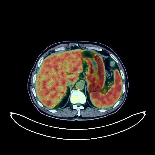
Lung Cancer PET/CT (case 983824-000115 from PETWB-REP)
2 views10 days agoWhole-body 18F-FDG PET/CT scan in a patient with Lung Cancer taken from the PETWB-REP dataset. The following English report (translated from original Chinese) is taken verbatim from the public dataset and has not been modified or otherwise checked for accuracy (see the end for citation). Impression a. A mass near the hilum in the left upper lobe of the lung, with increased FDG metabolism, suggestive of lung cancer with surrounding obstructive inflammation; please correlate with clinicopathology. Multiple lymph node metastases in the left hilum and part of the mediastinum are possible. b. Multiple chronic inflammatory nodules in both lungs; follow-up CT is recommended. Multiple calcifications in the right lung, interstitial inflammation in both lungs. Slight thickening of the pleura bilaterally. Partial calcification of the aorta and coronary artery walls. Multiple soft tissue nodules in the abdomen (paragastric antrum, intermesenteric, retroperitoneal, and presacral), with increased FDG metabolism in some areas, suggestive of lymphoproliferative disorders; regular follow-up CT is recommended, and clinicopathology should be considered if necessary. Small cyst in the left medial lobe of the liver. Accessory spleen. Small cysts in both kidneys (complex cyst on the right). Thickening of the gastric fundus and body mucosa with increased FDG uptake, suggestive of inflammation. Strip-shaped FDG uptake in the terminal ileum suggests inflammation or physiological uptake. Endoscopic follow-up is recommended. Spinal degenerative changes. L3/4, L4/5 intervertebral disc bulge. Possible T9 vertebral hemangioma. Right femoral head hernia fossa. Focal increased FDG uptake at the left acetabular rim, suggestive of inflammation. Bilateral deep lacunar infarcts. Age-related brain changes. Reactive hyperplasia of multiple lymph nodes in the bilateral deep cervical spaces and submandibular region. This case is from PETWB-REP, a curated dataset of whole-body 18F-FDG PET/CT scans and corresponding radiology reports from 490 patients with a broad spectrum of malignancies. The data were retrospectively collected from patients who underwent clinically indicated whole-body 18F-FDG PET/CT scans at the Shanghai Universal Medical Imaging Diagnostic Center between 2021 and 2024. License: Creative Commons Attribution 4.0 International (CC BY 4.0) Citation: Xue, L., Feng, G., Wenbo, Z., Zhang, Y., Li, L., Wang, S., Peng, L., Peng, S., & Gao, X. (2026). PETWB-REP: A Multi-Cancer Whole-Body FDG PET/CT Dataset with Corresponding Radiology Reports [Data set]. Zenodo. https://doi.org/10.5281/zenodo.18670487
Whole BodyPET/CT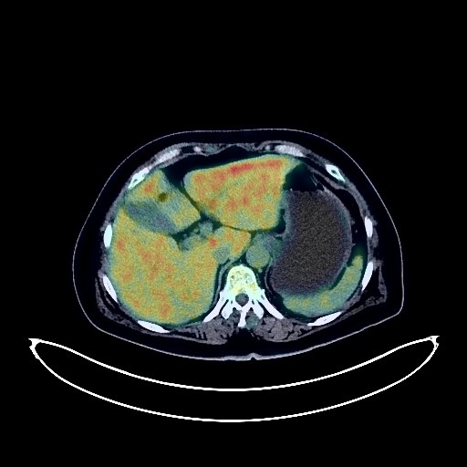
Lung Cancer PET/CT (case 983824-000210 from PETWB-REP)
2 views10 days agoWhole-body 18F-FDG PET/CT scan in a patient with Lung Cancer taken from the PETWB-REP dataset. The following English report (translated from original Chinese) is taken verbatim from the public dataset and has not been modified or otherwise checked for accuracy (see the end for citation). Impression a. A mass near the bronchial opening in the right upper lobe, with unevenly increased FDG metabolism, suggestive of central lung cancer with right upper lobe atelectasis, likely involving the right pulmonary artery. Further clinical and pathological examination is recommended. Mediastinal and right hilar lymph node metastases, some with necrosis. b. Scattered ground-glass opacities in the right lung, some with increased FDG metabolism, suggestive of inflammation. Follow-up CT scan recommended. c. A small, solid, chronic inflammatory nodule in the subpleural region of the apical-posterior segment of the right upper lobe. Further follow-up CT scan recommended. A few post-inflammatory lesions in both lungs. Mild pleural thickening bilaterally. Slight pericardial thickening. Postoperative changes in thyroid nodules: irregular thyroid morphology, uneven density with diffusely increased FDG metabolism, suggestive of thyroiditis or hyperthyroidism. Ultrasound and thyroid function tests recommended. Possible gallbladder polyp; ultrasound follow-up recommended. Uterine fibroids with calcification. Duodenal diverticulum. Degenerative changes in the spine. L4/5 and L5/S1 intervertebral disc bulges. Mild age-related brain changes, deep lacunar infarcts in the brain; MRI is recommended. Minor inflammation of the left maxillary sinus. Reactive hyperplasia of bilateral deep cervical interspace, submandibular, and submental lymph nodes. This case is from PETWB-REP, a curated dataset of whole-body 18F-FDG PET/CT scans and corresponding radiology reports from 490 patients with a broad spectrum of malignancies. The data were retrospectively collected from patients who underwent clinically indicated whole-body 18F-FDG PET/CT scans at the Shanghai Universal Medical Imaging Diagnostic Center between 2021 and 2024. License: Creative Commons Attribution 4.0 International (CC BY 4.0) Citation: Xue, L., Feng, G., Wenbo, Z., Zhang, Y., Li, L., Wang, S., Peng, L., Peng, S., & Gao, X. (2026). PETWB-REP: A Multi-Cancer Whole-Body FDG PET/CT Dataset with Corresponding Radiology Reports [Data set]. Zenodo. https://doi.org/10.5281/zenodo.18670487
Whole BodyPET/CT
Pancreatic Cancer PET/CT (case 983824-000012 from PETWB-REP)
7 views10 days agoWhole-body 18F-FDG PET/CT scan in a patient with Pancreatic Cancer taken from the PETWB-REP dataset. The following English report (translated from original Chinese) is taken verbatim from the public dataset and has not been modified or otherwise checked for accuracy (see the end for citation). Impression a. A slightly low-density mass in the tail of the pancreas with increased FDG metabolism, poorly demarcated from the splenic hilum, highly suggestive of pancreatic cancer; please correlate with clinicopathology. b. Multiple liver metastases. Metastases in the hilar and retroperitoneal lymph nodes. Metastases in the right 3rd and 6th ribs, left T8 vertebral body, left iliac bone, and left pubic bone. Gallbladder wall thickening, soft tissue lesions within the gallbladder with increased FDG metabolism, suggestive of gallbladder adenomyosis; local malignancy to be ruled out; please correlate with pathology. Multiple liver cysts. Small renal cysts. Scattered chronic inflammatory nodules in both lungs. Emphysema in both lungs. Calcification in the right lung. Calcification of some arterial walls. Physiological or inflammatory uptake in some intestinal segments; please correlate with colonoscopy follow-up. Benign prostatic hyperplasia with calcification. Some vertebral osteophytes. Some thoracic intervertebral disc effusion. Age-related brain changes. This case is from PETWB-REP, a curated dataset of whole-body 18F-FDG PET/CT scans and corresponding radiology reports from 490 patients with a broad spectrum of malignancies. The data were retrospectively collected from patients who underwent clinically indicated whole-body 18F-FDG PET/CT scans at the Shanghai Universal Medical Imaging Diagnostic Center between 2021 and 2024. License: Creative Commons Attribution 4.0 International (CC BY 4.0) Citation: Xue, L., Feng, G., Wenbo, Z., Zhang, Y., Li, L., Wang, S., Peng, L., Peng, S., & Gao, X. (2026). PETWB-REP: A Multi-Cancer Whole-Body FDG PET/CT Dataset with Corresponding Radiology Reports [Data set]. Zenodo. https://doi.org/10.5281/zenodo.18670487
Whole BodyPET/CT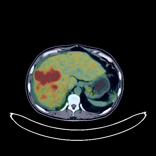
Liver Cancer PET/CT (case 983824-000146 from PETWB-REP)
2 views10 days agoWhole-body 18F-FDG PET/CT scan in a patient with Liver Cancer taken from the PETWB-REP dataset. The following English report (translated from original Chinese) is taken verbatim from the public dataset and has not been modified or otherwise checked for accuracy (see the end for citation). Impression a. A mass in the right lobe of the liver with elevated FDG metabolism, suggestive of malignancy, most likely primary liver cancer. Please combine clinical findings with enhanced MRI for comprehensive analysis. b. Liver cirrhosis. Reactive hyperplasia of the hilar and retroperitoneal lymph nodes. Abdominal and pelvic effusion. c. Possible right portal vein tumor thrombus formation. a. Chronic inflammatory micronodules in both lungs. Chronic inflammation in both lungs (more pronounced in the lower right lobe) and post-inflammatory remnants. b. Right pleural thickening, small amount of right pleural effusion. Reactive hyperplasia of the right hilar and mediastinal lymph nodes. Anemia changes, calcification of some arterial walls (including coronary arteries). Chronic cholecystitis. Benign prostatic hyperplasia with calcification. Bilateral vas deferens calcification. Chronic inflammatory changes in part of the stomach wall and intestines; please follow up with endoscopy. Degenerative changes in the spine. L4/5 and L5/S1 intervertebral disc bulge. Subcutaneous calcification in the right buttock. Soft tissue nodule in the right parotid gland with increased FDG metabolism, suggestive of lymphoma; please follow up with MRI. Deep lacunar infarcts in the brain, mild age-related brain changes. Minor chronic inflammation in both ethmoid sinuses and the left maxillary sinus. This case is from PETWB-REP, a curated dataset of whole-body 18F-FDG PET/CT scans and corresponding radiology reports from 490 patients with a broad spectrum of malignancies. The data were retrospectively collected from patients who underwent clinically indicated whole-body 18F-FDG PET/CT scans at the Shanghai Universal Medical Imaging Diagnostic Center between 2021 and 2024. License: Creative Commons Attribution 4.0 International (CC BY 4.0) Citation: Xue, L., Feng, G., Wenbo, Z., Zhang, Y., Li, L., Wang, S., Peng, L., Peng, S., & Gao, X. (2026). PETWB-REP: A Multi-Cancer Whole-Body FDG PET/CT Dataset with Corresponding Radiology Reports [Data set]. Zenodo. https://doi.org/10.5281/zenodo.18670487
Whole BodyPET/CT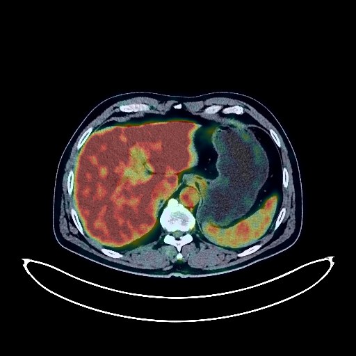
Nasopharyngeal Cancer PET/CT (case 983824-000135 from PETWB-REP)
2 views10 days agoWhole-body 18F-FDG PET/CT scan in a patient with Nasopharyngeal Cancer taken from the PETWB-REP dataset. The following English report (translated from original Chinese) is taken verbatim from the public dataset and has not been modified or otherwise checked for accuracy (see the end for citation). Impression a. After comprehensive treatment of "nasopharyngeal lesion": No significant thickening of the nasopharyngeal wall was observed, and FDG metabolism was not increased, suggesting that tumor activity was basically suppressed after treatment. b. Bilateral deep cervical spaces and submandibular lymph nodes showed no abnormal FDG uptake, suggesting reactive hyperplasia is highly likely; follow-up is recommended. c. Changes after cervical and thoracic spine radiotherapy. Thyroid gland density and FDG uptake are uneven; please follow up with ultrasound. Chronic inflammatory micronodules (solid) in the right upper lobe and posterior segment of the left lower lobe; a few chronic inflammations and remnants in both lower lobes; slight pleural thickening in some areas. Similar to the previous findings. Liver calcifications. Bilateral testicular tunica vaginalis calcifications. Chronic inflammatory or physiological uptake of the stomach and duodenum; please follow up with gastroscopy. Sigmoid colon diverticulum. Degenerative changes in the spine, L4/5 disc bulge. Chronic inflammation with calcifications in the soft tissues surrounding the greater trochanter of the left femur. Age-related brain, deep lacunar infarcts. Chronic inflammation with submucosal cysts in the left maxillary sinus. This case is from PETWB-REP, a curated dataset of whole-body 18F-FDG PET/CT scans and corresponding radiology reports from 490 patients with a broad spectrum of malignancies. The data were retrospectively collected from patients who underwent clinically indicated whole-body 18F-FDG PET/CT scans at the Shanghai Universal Medical Imaging Diagnostic Center between 2021 and 2024. License: Creative Commons Attribution 4.0 International (CC BY 4.0) Citation: Xue, L., Feng, G., Wenbo, Z., Zhang, Y., Li, L., Wang, S., Peng, L., Peng, S., & Gao, X. (2026). PETWB-REP: A Multi-Cancer Whole-Body FDG PET/CT Dataset with Corresponding Radiology Reports [Data set]. Zenodo. https://doi.org/10.5281/zenodo.18670487
Whole BodyPET/CT
Lung Cancer PET/CT (case 983824-000157 from PETWB-REP)
2 views10 days agoWhole-body 18F-FDG PET/CT scan in a patient with Lung Cancer taken from the PETWB-REP dataset. The following English report (translated from original Chinese) is taken verbatim from the public dataset and has not been modified or otherwise checked for accuracy (see the end for citation). Impression a. Space-occupying lesions in the anterior segment of the right upper lobe and the posterior segment of the right lower lobe, accompanied by increased FDG metabolism, consistent with lung cancer. Right hilar lymph node metastasis. Mediastinal lymph node metastasis to be ruled out, follow-up is required. b. Postoperative changes after left lung cancer surgery. Multiple chronic inflammations and sequelae in both lungs. Calcifications in the right hilum, right lower lobe, and right pleura. Calcification of some arterial walls (including coronary arteries). Mild fatty liver. Prostatic calcification. Schistosomiasis intestinal disease. Continuous increased FDG metabolism in the sigmoid colon and rectum, suggesting possible inflammatory uptake; colonoscopy is recommended to rule out tumors. Partial vertebral osteophyte formation. Partial lumbar disc pneumothorax, L3/4 and L4/5 disc bulges. A few ischemic lesions in the deep brain. Age-related brain changes. Left maxillary sinusitis. This case is from PETWB-REP, a curated dataset of whole-body 18F-FDG PET/CT scans and corresponding radiology reports from 490 patients with a broad spectrum of malignancies. The data were retrospectively collected from patients who underwent clinically indicated whole-body 18F-FDG PET/CT scans at the Shanghai Universal Medical Imaging Diagnostic Center between 2021 and 2024. License: Creative Commons Attribution 4.0 International (CC BY 4.0) Citation: Xue, L., Feng, G., Wenbo, Z., Zhang, Y., Li, L., Wang, S., Peng, L., Peng, S., & Gao, X. (2026). PETWB-REP: A Multi-Cancer Whole-Body FDG PET/CT Dataset with Corresponding Radiology Reports [Data set]. Zenodo. https://doi.org/10.5281/zenodo.18670487
Whole BodyPET/CT