Loading...
Lung Cancer PET/CT (case 983824-000031 from PETWB-REP)
7 views10 days agoWhole-body 18F-FDG PET/CT scan in a patient with Lung Cancer taken from the PETWB-REP dataset. The following English report (translated from original Chinese) is taken verbatim from the public dataset and has not been modified or otherwise checked for accuracy (see the end for citation). Impression a. A mass in the posterior segment of the left upper lobe, with increased FDG metabolism, suggestive of peripheral lung cancer. b. Reactive hyperplasia of right hilar and mediastinal lymph nodes, with metastasis to the larger lymph nodes at the aortic window to be ruled out. c. A cystic nodule in the right middle lobe, with thick walls and ground-glass opacity, normal FDG metabolism, suggestive of cystic lung cancer. Multiple inflammatory lymph nodes in the remaining right lung and left lower lobe, follow-up recommended to rule out metastasis. d. A few fibrotic foci in the medial segment of the right middle lobe and the inferior lingular segment of the left upper lobe. Focal increased FDG metabolism in the right lobe of the thyroid gland, suggestive of inflammatory uptake, ultrasound examination recommended. Small cyst in the right anterior lobe of the liver. Gallbladder bile concentration. Post-uterine surgery changes. Spinal osteophyte formation, L4/5 intervertebral disc bulge. Normal FDG metabolism in the brain. Reactive hyperplasia of bilateral deep cervical and submandibular lymph nodes. This case is from PETWB-REP, a curated dataset of whole-body 18F-FDG PET/CT scans and corresponding radiology reports from 490 patients with a broad spectrum of malignancies. The data were retrospectively collected from patients who underwent clinically indicated whole-body 18F-FDG PET/CT scans at the Shanghai Universal Medical Imaging Diagnostic Center between 2021 and 2024. License: Creative Commons Attribution 4.0 International (CC BY 4.0) Citation: Xue, L., Feng, G., Wenbo, Z., Zhang, Y., Li, L., Wang, S., Peng, L., Peng, S., & Gao, X. (2026). PETWB-REP: A Multi-Cancer Whole-Body FDG PET/CT Dataset with Corresponding Radiology Reports [Data set]. Zenodo. https://doi.org/10.5281/zenodo.18670487
Whole BodyPET/CT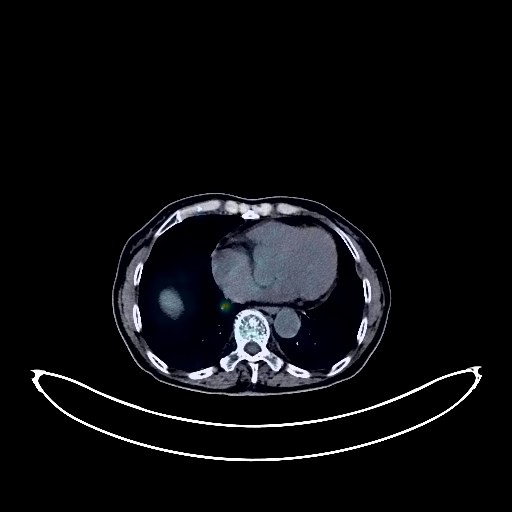
Colon Cancer PET/CT (case 983824-000205 from PETWB-REP)
1 views10 days agoWhole-body 18F-FDG PET/CT scan in a patient with Colon Cancer taken from the PETWB-REP dataset. The following English report (translated from original Chinese) is taken verbatim from the public dataset and has not been modified or otherwise checked for accuracy (see the end for citation). Impression Postoperative changes after colon cancer surgery, anastomotic inflammation is highly likely; specialist follow-up is recommended. Left lobe liver metastasis. Left adrenal gland metastasis. Multiple bone, muscle, and subcutaneous metastases throughout the body (see description for details). Bilateral iliac fossa nodule metastases. High probability of presacral soft tissue metastases; left lower abdominal wall stoma. a. A mass near the right hilum at the bronchial opening in the right upper lobe, with increased FDG metabolism, suggestive of malignancy; primary tumor is more likely than metastatic. Please consider clinicopathology. Atelectasis in part of the anterior segment of the right upper lobe. High probability of metastasis to the right hilum, part of the mediastinum, and right supraclavicular fossa lymph nodes. b. Nodule metastases in the lateral and medial basal segments of the right lower lobe; scattered small chronic inflammatory nodules (solid) in the remaining two lungs are possible; CT re-examination is recommended to rule out metastases. Calcifications in the posterior segment of the left lower lobe and the anterior basal segment of the right lower lobe. A few post-inflammatory lesions in both lungs. Partial arteriosclerosis. Bilateral breast hyperplasia, with calcification in the left breast. Cyst and calcification in the right lobe of the liver. Chronic cholecystitis, gallstones. Slight dilation and hydronephrosis of the renal pelvis, calyces, and upper ureter in both kidneys. Degenerative changes in the spine. L4/5 and L5/S1 intervertebral disc bulge. L4/5 intervertebral disc pneumoconiosis. Age-related brain changes, deep lacunar infarcts, bilateral periventricular white matter lesions; MRI follow-up is recommended. A few inflammations in the right maxillary sinus. Uneven thyroid density; ultrasound follow-up is recommended. This case is from PETWB-REP, a curated dataset of whole-body 18F-FDG PET/CT scans and corresponding radiology reports from 490 patients with a broad spectrum of malignancies. The data were retrospectively collected from patients who underwent clinically indicated whole-body 18F-FDG PET/CT scans at the Shanghai Universal Medical Imaging Diagnostic Center between 2021 and 2024. License: Creative Commons Attribution 4.0 International (CC BY 4.0) Citation: Xue, L., Feng, G., Wenbo, Z., Zhang, Y., Li, L., Wang, S., Peng, L., Peng, S., & Gao, X. (2026). PETWB-REP: A Multi-Cancer Whole-Body FDG PET/CT Dataset with Corresponding Radiology Reports [Data set]. Zenodo. https://doi.org/10.5281/zenodo.18670487
Whole BodyPET/CT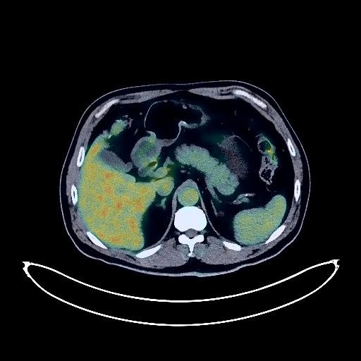
Lung Cancer PET/CT (case 983824-000177 from PETWB-REP)
2 views10 days agoWhole-body 18F-FDG PET/CT scan in a patient with Lung Cancer taken from the PETWB-REP dataset. The following English report (translated from original Chinese) is taken verbatim from the public dataset and has not been modified or otherwise checked for accuracy (see the end for citation). Impression a. A mass near the hilum in the anterior segment of the right upper lobe with increased FDG metabolism, suggestive of lung cancer. Right hilar lymph node metastasis. b. Ground-glass nodule in the apical-posterior segment of the left upper lobe, FDG metabolism normal, suggestive of inflammation or atypical adenomatous hyperplasia; CT follow-up recommended. A few post-inflammatory lesions in both lungs. Calcification of some arterial walls (including coronary arteries). Small cyst in the right lobe of the liver. Accessory spleen. Bilateral renal cysts. Chronic inflammatory changes in the gastric wall; please follow up with endoscopy. Degenerative changes in the spine, L4/5 and L5/S1 intervertebral disc bulges. Benign lesion in the left 6th rib is the primary consideration; follow-up to rule out metastasis is recommended. Deep lacunar infarcts in the brain. Inflammation of the left maxillary sinus. This case is from PETWB-REP, a curated dataset of whole-body 18F-FDG PET/CT scans and corresponding radiology reports from 490 patients with a broad spectrum of malignancies. The data were retrospectively collected from patients who underwent clinically indicated whole-body 18F-FDG PET/CT scans at the Shanghai Universal Medical Imaging Diagnostic Center between 2021 and 2024. License: Creative Commons Attribution 4.0 International (CC BY 4.0) Citation: Xue, L., Feng, G., Wenbo, Z., Zhang, Y., Li, L., Wang, S., Peng, L., Peng, S., & Gao, X. (2026). PETWB-REP: A Multi-Cancer Whole-Body FDG PET/CT Dataset with Corresponding Radiology Reports [Data set]. Zenodo. https://doi.org/10.5281/zenodo.18670487
Whole BodyPET/CT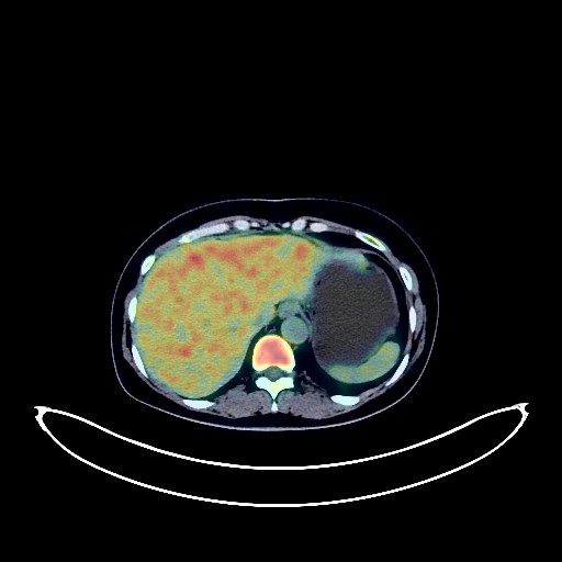
Renal Cancer PET/CT (case 983824-000196 from PETWB-REP)
1 views10 days agoWhole-body 18F-FDG PET/CT scan in a patient with Renal Cancer taken from the PETWB-REP dataset. The following English report (translated from original Chinese) is taken verbatim from the public dataset and has not been modified or otherwise checked for accuracy (see the end for citation). Impression Low-density mass near the left renal pelvis with slightly increased FDG metabolism at the periphery, suggestive of a neoplastic lesion, with cancer as the primary consideration. Further enhanced MRI analysis is recommended. Reactive hyperplasia of retroperitoneal lymph nodes. Chronic inflammatory micronodule in the right upper lobe, calcification in the left lower lobe, and fibrosis in both lungs. Follow-up with CT scan is recommended. Reactive hyperplasia of hilar and mediastinal lymph nodes. Liver cyst. Accessory spleen. Right renal cyst. Right adrenal adenoma. Increased FDG metabolism in part of the gastric wall and intestinal tract, suggestive of physiological uptake or chronic inflammation. Follow-up with endoscopy is recommended. Cervical, thoracic, and lumbar spondylosis. L3/4 and L4/5 intervertebral disc bulge. Absence of the left thyroid lobe; please consider clinical history. Reactive hyperplasia of bilateral deep cervical interspace and supraclavicular lymph nodes. No obvious abnormalities were found on cranial scintigraphy. This case is from PETWB-REP, a curated dataset of whole-body 18F-FDG PET/CT scans and corresponding radiology reports from 490 patients with a broad spectrum of malignancies. The data were retrospectively collected from patients who underwent clinically indicated whole-body 18F-FDG PET/CT scans at the Shanghai Universal Medical Imaging Diagnostic Center between 2021 and 2024. License: Creative Commons Attribution 4.0 International (CC BY 4.0) Citation: Xue, L., Feng, G., Wenbo, Z., Zhang, Y., Li, L., Wang, S., Peng, L., Peng, S., & Gao, X. (2026). PETWB-REP: A Multi-Cancer Whole-Body FDG PET/CT Dataset with Corresponding Radiology Reports [Data set]. Zenodo. https://doi.org/10.5281/zenodo.18670487
Whole BodyPET/CT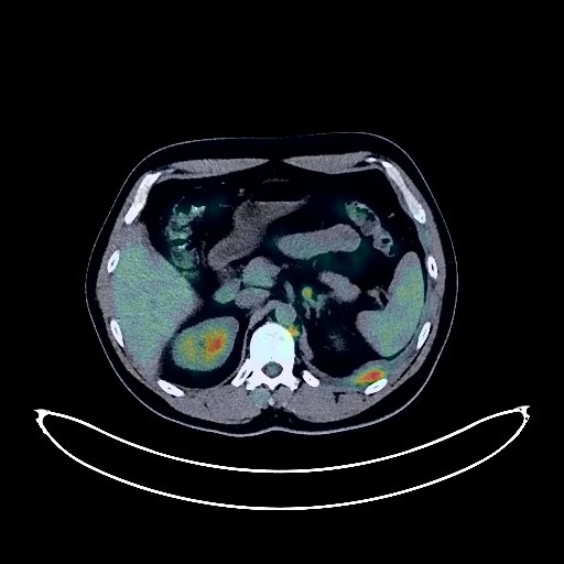
Lung Cancer PET/CT (case 983824-000137 from PETWB-REP)
2 views10 days agoWhole-body 18F-FDG PET/CT scan in a patient with Lung Cancer taken from the PETWB-REP dataset. The following English report (translated from original Chinese) is taken verbatim from the public dataset and has not been modified or otherwise checked for accuracy (see the end for citation). Impression a. A mass near the hilum in the lower lobe of the left lung, with increased FDG metabolism, suggestive of lung cancer. b. Extensive metastasis to the left pleura. Multiple lymph node metastases in the left hilum, mediastinum, bilateral infracervical spaces, bilateral supraclavicular fossa, bilateral internal mammary chains, bilateral posterior diaphragmatic crura, and para-aortic region. Possible reactive hyperplasia of the left axillary lymph nodes, metastasis to be ruled out. c. Emphysema in the upper lobes of both lungs. Scattered inflammation in the left lung. d. Left pleural effusion, partly encapsulated. Slight pericardial thickening. Calcification of some arterial walls (including coronary arteries). Increased local bone density in the left scapula and L2 vertebral body, with increased FDG metabolism, metastasis to be ruled out, close observation recommended. Uneven density in the right temporal lobe and left frontal lobe; no abnormalities observed in FDG metabolism; metastasis needs further investigation. Please combine with contrast-enhanced MRI images from another hospital for comprehensive analysis. A few ischemic lesions in the deep bilateral brain regions. Cervical, thoracic, and lumbar spondylosis. L4/5 and L5/S1 intervertebral disc bulges. This case is from PETWB-REP, a curated dataset of whole-body 18F-FDG PET/CT scans and corresponding radiology reports from 490 patients with a broad spectrum of malignancies. The data were retrospectively collected from patients who underwent clinically indicated whole-body 18F-FDG PET/CT scans at the Shanghai Universal Medical Imaging Diagnostic Center between 2021 and 2024. License: Creative Commons Attribution 4.0 International (CC BY 4.0) Citation: Xue, L., Feng, G., Wenbo, Z., Zhang, Y., Li, L., Wang, S., Peng, L., Peng, S., & Gao, X. (2026). PETWB-REP: A Multi-Cancer Whole-Body FDG PET/CT Dataset with Corresponding Radiology Reports [Data set]. Zenodo. https://doi.org/10.5281/zenodo.18670487
Whole BodyPET/CT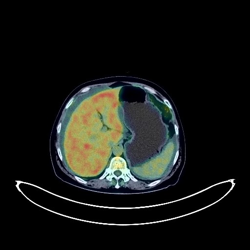
Cervical Cancer PET/CT (case 983824-000162 from PETWB-REP)
1 views10 days agoWhole-body 18F-FDG PET/CT scan in a patient with Cervical Cancer taken from the PETWB-REP dataset. The following English report (translated from original Chinese) is taken verbatim from the public dataset and has not been modified or otherwise checked for accuracy (see the end for citation). Impression a. Cervical mass with unevenly increased FDG metabolism, suggestive of malignancy, with a high probability of vaginal involvement. b. Metastasis to the peritoneum and pelvic mesentery and greater omentum, bilateral periiliac lymph node metastasis. Left cervical root lymph node metastasis cannot be ruled out. c. Bilateral adnexal region poorly visualized, uneven FDG metabolism, metastasis needs to be ruled out; please correlate with clinicopathology. Abdominal and pelvic effusion. d. Uterine fibroid degeneration. Nabothian cyst of the cervix. Multiple chronic inflammatory nodules in both lungs are possible; metastasis of some nodules cannot be ruled out. Close follow-up CT scans are recommended for comparison. Scattered chronic inflammation and sequelae in both lungs. Small cyst in the right lobe of the liver. Chronic cholecystitis. Accessory spleen. Chronic antral gastritis, with increased FDG uptake in parts of the colon and rectum, possibly due to physiological uptake or chronic inflammation. Please follow up with endoscopy. Degenerative changes in the spine. Pneumothorax and degeneration of the T9/10 intervertebral disc. Bulging of the L3/4, L4/5, and L5/S1 intervertebral discs. No obvious abnormalities were found on cranial scintigraphy. This case is from PETWB-REP, a curated dataset of whole-body 18F-FDG PET/CT scans and corresponding radiology reports from 490 patients with a broad spectrum of malignancies. The data were retrospectively collected from patients who underwent clinically indicated whole-body 18F-FDG PET/CT scans at the Shanghai Universal Medical Imaging Diagnostic Center between 2021 and 2024. License: Creative Commons Attribution 4.0 International (CC BY 4.0) Citation: Xue, L., Feng, G., Wenbo, Z., Zhang, Y., Li, L., Wang, S., Peng, L., Peng, S., & Gao, X. (2026). PETWB-REP: A Multi-Cancer Whole-Body FDG PET/CT Dataset with Corresponding Radiology Reports [Data set]. Zenodo. https://doi.org/10.5281/zenodo.18670487
Whole BodyPET/CT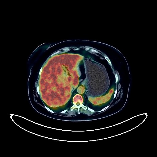
Cervical Cancer PET/CT (case 983824-000185 from PETWB-REP)
2 views10 days agoWhole-body 18F-FDG PET/CT scan in a patient with Cervical Cancer taken from the PETWB-REP dataset. The following English report (translated from original Chinese) is taken verbatim from the public dataset and has not been modified or otherwise checked for accuracy (see the end for citation). Impression a. Cervical mass with elevated FDG metabolism, suggestive of cervical cancer; please correlate with clinicopathology; right iliac lymph node metastasis. b. Benign bone disease of the right frontal bone is the primary consideration; metastatic tumor to be ruled out; close observation is recommended. Chronic inflammatory micronodules in both lungs; CT follow-up is recommended to rule out other possibilities. Right upper lobe contains air sacs; a few post-inflammatory remnants in both lungs. Reactive hyperplasia of hilar and mediastinal lymph nodes. Partial arteriosclerosis. Chronic cholecystitis. Bilateral renal cysts. Bilateral adrenal hyperplasia. Chronic inflammatory changes or physiological uptake in the antrum of the stomach and part of the intestine; please correlate with endoscopic follow-up. Degenerative changes in the spine; L4/5 and L5/S1 intervertebral disc bulge. Left sacral islet. Nodular goiter in the right lobe of the thyroid gland; ultrasound follow-up is recommended. Ischemic lesion in the right basal ganglia region. Chronic inflammation of the base of the tongue and bilateral palatine tonsils. This case is from PETWB-REP, a curated dataset of whole-body 18F-FDG PET/CT scans and corresponding radiology reports from 490 patients with a broad spectrum of malignancies. The data were retrospectively collected from patients who underwent clinically indicated whole-body 18F-FDG PET/CT scans at the Shanghai Universal Medical Imaging Diagnostic Center between 2021 and 2024. License: Creative Commons Attribution 4.0 International (CC BY 4.0) Citation: Xue, L., Feng, G., Wenbo, Z., Zhang, Y., Li, L., Wang, S., Peng, L., Peng, S., & Gao, X. (2026). PETWB-REP: A Multi-Cancer Whole-Body FDG PET/CT Dataset with Corresponding Radiology Reports [Data set]. Zenodo. https://doi.org/10.5281/zenodo.18670487
Whole BodyPET/CT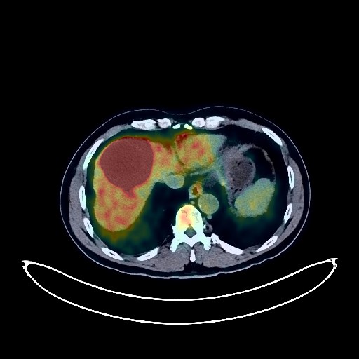
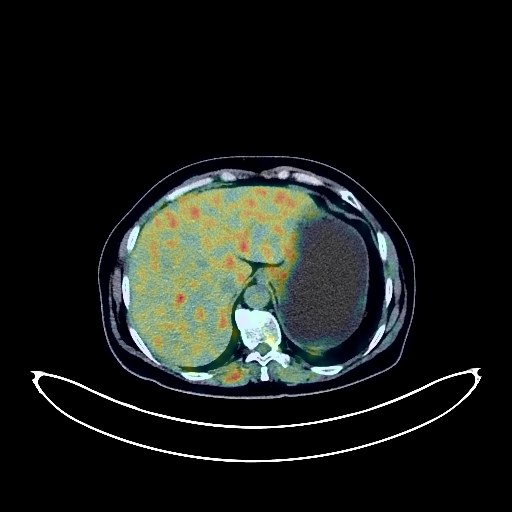
Liver Cancer PET/CT (case 983824-000105 from PETWB-REP)
3 views10 days agoWhole-body 18F-FDG PET/CT scan in a patient with Liver Cancer taken from the PETWB-REP dataset. The following English report (translated from original Chinese) is taken verbatim from the public dataset and has not been modified or otherwise checked for accuracy (see the end for citation). Impression A mass in the right anterior lobe of the liver, a mass in the left adrenal gland, and multiple enlarged lymph nodes in the abdominal cavity, retroperitoneum, left hilum, mediastinum, and right infracervical space, all with increased FDG metabolism. Malignancy is suspected, possibly a primary liver tumor with multiple metastases; lymphoma cannot be ruled out. A biopsy is recommended. A slightly high-density nodule with increased FDG metabolism is seen under the right parietal cranial plate, suggesting a possible meningioma; a slightly high-density nodule with increased FDG uptake is seen in the right occipital lobe, suggesting a possible benign lesion. Further enhanced MRI is recommended. Chronic inflammatory nodules in the posterior segment of the right upper lobe and beside the oblique fissure of the left lower lobe. Calcification in the right upper lobe. A few chronic inflammations and old lesions in both lungs. Calcification of some arterial walls. Increased FDG metabolism in some intestinal segments, suggesting inflammatory or physiological uptake. Left renal cyst. Benign prostatic hyperplasia with calcification. Degenerative changes in the spine. L4/5 and L5/S1 intervertebral disc bulge. Chronic inflammation of the left maxillary sinus. This case is from PETWB-REP, a curated dataset of whole-body 18F-FDG PET/CT scans and corresponding radiology reports from 490 patients with a broad spectrum of malignancies. The data were retrospectively collected from patients who underwent clinically indicated whole-body 18F-FDG PET/CT scans at the Shanghai Universal Medical Imaging Diagnostic Center between 2021 and 2024. License: Creative Commons Attribution 4.0 International (CC BY 4.0) Citation: Xue, L., Feng, G., Wenbo, Z., Zhang, Y., Li, L., Wang, S., Peng, L., Peng, S., & Gao, X. (2026). PETWB-REP: A Multi-Cancer Whole-Body FDG PET/CT Dataset with Corresponding Radiology Reports [Data set]. Zenodo. https://doi.org/10.5281/zenodo.18670487
Whole BodyPET/CT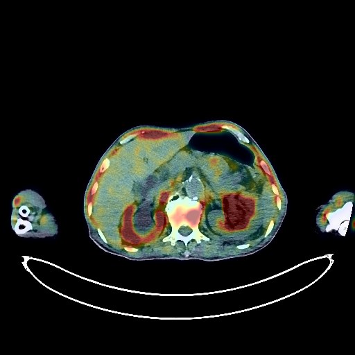
Prostate Cancer PET/CT (case 983824-000186 from PETWB-REP)
2 views10 days agoWhole-body 18F-FDG PET/CT scan in a patient with Prostate Cancer taken from the PETWB-REP dataset. The following English report (translated from original Chinese) is taken verbatim from the public dataset and has not been modified or otherwise checked for accuracy (see the end for citation). Impression Benign prostatic hyperplasia with calcification; a mass on the right side of the prostate with increased FDG metabolism; multiple enlarged lymph nodes in the right iliac vessels, retroperitoneum, and left supraclavicular fossa with increased FDG metabolism. Malignancy is suspected, with prostate cancer with multiple metastases being the primary consideration. Please confirm with pathology. Chronic inflammatory nodules in both lungs; CT follow-up is recommended to rule out other possibilities. Bilateral emphysema. Scattered post-inflammatory lesions in both lungs. Reactive hyperplasia of hilar lymph nodes bilaterally; mediastinal lymph node metastasis to be ruled out; follow-up is recommended. Small amount of pleural effusion bilaterally. Pericardial effusion. Anemia changes; calcification of some arterial walls (including coronary arteries). Bilateral gynecomastia. Small liver cyst. Left renal cyst. Bilateral dilated effusion of the renal pelvis and upper ureter. Degenerative changes in the spine, L4/5 and L5/S1 disc bulges. Right sacral islet. Possible old fracture of the right clavicle; please refer to medical history. Age-related brain abnormalities, deep cerebral ischemia; please follow up with MRI. Chronic inflammation of the left maxillary sinus. This case is from PETWB-REP, a curated dataset of whole-body 18F-FDG PET/CT scans and corresponding radiology reports from 490 patients with a broad spectrum of malignancies. The data were retrospectively collected from patients who underwent clinically indicated whole-body 18F-FDG PET/CT scans at the Shanghai Universal Medical Imaging Diagnostic Center between 2021 and 2024. License: Creative Commons Attribution 4.0 International (CC BY 4.0) Citation: Xue, L., Feng, G., Wenbo, Z., Zhang, Y., Li, L., Wang, S., Peng, L., Peng, S., & Gao, X. (2026). PETWB-REP: A Multi-Cancer Whole-Body FDG PET/CT Dataset with Corresponding Radiology Reports [Data set]. Zenodo. https://doi.org/10.5281/zenodo.18670487
Whole BodyPET/CT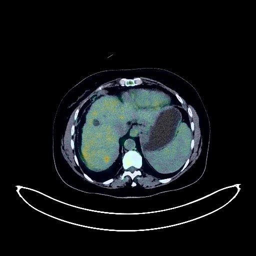
Liver Cancer PET/CT (case 983824-000090 from PETWB-REP)
4 views10 days agoWhole-body 18F-FDG PET/CT scan in a patient with Liver Cancer taken from the PETWB-REP dataset. The following English report (translated from original Chinese) is taken verbatim from the public dataset and has not been modified or otherwise checked for accuracy (see the end for citation). Impression a. Multiple lesions in the liver, increased FDG metabolism, suggestive of malignancy, most likely primary liver cancer with intrahepatic metastasis. b. Multiple metastatic tumors in both lungs. Most likely a bone metastasis in the upper right humerus. c. Cirrhosis; slightly enlarged spleen; portal hypertension. Scattered chronic inflammation and old lesions in both lungs. Calcification of some arterial walls (including coronary arteries). Liver cysts; chronic cholecystitis. Increased FDG metabolism in the lower rectum, suggestive of inflammatory or physiological uptake. Highly likely uterine fibroid degeneration; ultrasound follow-up recommended. Degenerative changes in the spine. L4/5 and L5/S1 intervertebral disc bulge. L5/S1 intervertebral disc pneumoconiosis and degeneration. Old fracture of the left second anterior rib. A few ischemic lesions in the deep bilateral cerebral regions, indicative of age-related brain changes. This case is from PETWB-REP, a curated dataset of whole-body 18F-FDG PET/CT scans and corresponding radiology reports from 490 patients with a broad spectrum of malignancies. The data were retrospectively collected from patients who underwent clinically indicated whole-body 18F-FDG PET/CT scans at the Shanghai Universal Medical Imaging Diagnostic Center between 2021 and 2024. License: Creative Commons Attribution 4.0 International (CC BY 4.0) Citation: Xue, L., Feng, G., Wenbo, Z., Zhang, Y., Li, L., Wang, S., Peng, L., Peng, S., & Gao, X. (2026). PETWB-REP: A Multi-Cancer Whole-Body FDG PET/CT Dataset with Corresponding Radiology Reports [Data set]. Zenodo. https://doi.org/10.5281/zenodo.18670487
Whole BodyPET/CT