Loading...
Bladder Cancer PET/CT (case 983824-000128 from PETWB-REP)
2 views10 days agoWhole-body 18F-FDG PET/CT scan in a patient with Bladder Cancer taken from the PETWB-REP dataset. The following English report (translated from original Chinese) is taken verbatim from the public dataset and has not been modified or otherwise checked for accuracy (see the end for citation). Impression a. Post-bladder cancer treatment: Slight thickening of the right bladder wall with localized increased FDG metabolism, considered a post-treatment change. Residual tumor activity needs further investigation. Please combine clinical findings with enhanced MRI for comprehensive analysis. Mild hydronephrosis and dilatation of the right upper urinary tract. ? b. Reactive hyperplasia of bilateral inguinal lymph nodes. Inflammatory changes in the right lower abdominal wall. Sellar region lesion with increased FDG uptake, suggestive of possible pituitary adenoma. Further enhanced pituitary MRI is recommended. Minor inflammation in the upper lobes of both lungs. CT follow-up is recommended after anti-inflammatory treatment. Chronic inflammatory micronodules in both lungs, fibrotic lesions in both lungs. Chronic inflammatory lymph nodes in both hilar and mediastinal regions. Partial arteriosclerosis (including coronary arteries). Liver cirrhosis, splenomegaly. Gallstones. Right adrenal adenoma. Prostatic calcification. Bilateral hydrocele. Increased FDG metabolism in some intestinal segments, possibly due to physiological uptake or chronic inflammation; please follow up with endoscopy. Spinal degenerative changes. Bilateral deep lacunar infarcts, senile encephalopathy. Chronic inflammation of bilateral ethmoid sinuses and left maxillary sinus. This case is from PETWB-REP, a curated dataset of whole-body 18F-FDG PET/CT scans and corresponding radiology reports from 490 patients with a broad spectrum of malignancies. The data were retrospectively collected from patients who underwent clinically indicated whole-body 18F-FDG PET/CT scans at the Shanghai Universal Medical Imaging Diagnostic Center between 2021 and 2024. License: Creative Commons Attribution 4.0 International (CC BY 4.0) Citation: Xue, L., Feng, G., Wenbo, Z., Zhang, Y., Li, L., Wang, S., Peng, L., Peng, S., & Gao, X. (2026). PETWB-REP: A Multi-Cancer Whole-Body FDG PET/CT Dataset with Corresponding Radiology Reports [Data set]. Zenodo. https://doi.org/10.5281/zenodo.18670487
Whole BodyPET/CT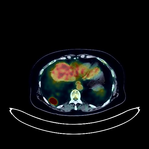
Rectal Cancer PET/CT (case 983824-000037 from PETWB-REP)
7 views10 days agoWhole-body 18F-FDG PET/CT scan in a patient with Rectal Cancer taken from the PETWB-REP dataset. The following English report (translated from original Chinese) is taken verbatim from the public dataset and has not been modified or otherwise checked for accuracy (see the end for citation). Impression a. Post-rectal cancer surgery, increased FDG metabolism in the lower rectum to anal canal suggests possible inflammatory uptake; tumor recurrence needs to be ruled out. Please have a follow-up colonoscopy. b. Retroperitoneal and right posterior diaphragmatic lymph node metastasis, involving the upper and middle segments of the left ureter, dilation and effusion of the left renal pelvis and upper ureter, and left kidney atrophy. The right kidney shows compensatory slight enlargement. c. Metastasis to the left upper cervical deep space and left supraclavicular lymph nodes. d. Multiple metastatic tumors in both lungs. Multiple bronchiectasis in both lungs, predominantly in the lower lobe of the left lung. Possible mass in the lower part of the left kidney; enhanced MRI is recommended. A few ischemic lesions deep in the brain, senile cerebral changes. Cholestasis in the gallbladder. Fatty infiltration of the pancreas. Osteophyte formation in some vertebrae. This case is from PETWB-REP, a curated dataset of whole-body 18F-FDG PET/CT scans and corresponding radiology reports from 490 patients with a broad spectrum of malignancies. The data were retrospectively collected from patients who underwent clinically indicated whole-body 18F-FDG PET/CT scans at the Shanghai Universal Medical Imaging Diagnostic Center between 2021 and 2024. License: Creative Commons Attribution 4.0 International (CC BY 4.0) Citation: Xue, L., Feng, G., Wenbo, Z., Zhang, Y., Li, L., Wang, S., Peng, L., Peng, S., & Gao, X. (2026). PETWB-REP: A Multi-Cancer Whole-Body FDG PET/CT Dataset with Corresponding Radiology Reports [Data set]. Zenodo. https://doi.org/10.5281/zenodo.18670487
Whole BodyPET/CT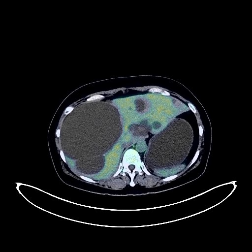
Cervical Cancer PET/CT (case 983824-000044 from PETWB-REP)
8 views10 days agoWhole-body 18F-FDG PET/CT scan in a patient with Cervical Cancer taken from the PETWB-REP dataset. The following English report (translated from original Chinese) is taken verbatim from the public dataset and has not been modified or otherwise checked for accuracy (see the end for citation). Impression Cervical mass with increased FDG metabolism, involving adjacent uterine body and vagina, consistent with cervical cancer. Multiple lymph node metastases in bilateral iliac vessels, bilateral pelvic walls, para-aortic region, posterior diaphragmatic crura, left infradeep cervical space, left posterior cervical triangle, and left supraclavicular fossa. Ground-glass nodule in the anterior segment of the right upper lobe, FDG metabolism normal, suggestive of inflammatory nodule or atypical adenomatous hyperplasia; annual HRCT follow-up recommended. Calcification in the right middle lobe. Bilateral breast proliferative changes; several soft tissue nodules in both breasts, FDG metabolism normal, suggestive of fibroadenoma or proliferative nodules; ultrasound follow-up recommended. Multiple liver cysts. Right kidney stone or calcification. Complex cyst in the left kidney. Increased FDG metabolism in parts of the intestine, suggestive of inflammatory or physiological uptake. Degenerative changes in the spine. L4/5 and L5/S1 intervertebral disc bulges. A subcutaneous soft tissue nodule in the midline of the buttock; FDG metabolism was normal, suggestive of a sebaceous cyst. Cranial scintigraphy showed no abnormalities. A small amount of chronic inflammation was observed in the left maxillary sinus. This case is from PETWB-REP, a curated dataset of whole-body 18F-FDG PET/CT scans and corresponding radiology reports from 490 patients with a broad spectrum of malignancies. The data were retrospectively collected from patients who underwent clinically indicated whole-body 18F-FDG PET/CT scans at the Shanghai Universal Medical Imaging Diagnostic Center between 2021 and 2024. License: Creative Commons Attribution 4.0 International (CC BY 4.0) Citation: Xue, L., Feng, G., Wenbo, Z., Zhang, Y., Li, L., Wang, S., Peng, L., Peng, S., & Gao, X. (2026). PETWB-REP: A Multi-Cancer Whole-Body FDG PET/CT Dataset with Corresponding Radiology Reports [Data set]. Zenodo. https://doi.org/10.5281/zenodo.18670487
Whole BodyPET/CT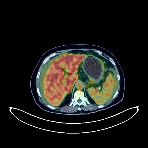
Pancreatic Cancer PET/CT (case 983824-000039 from PETWB-REP)
7 views10 days agoWhole-body 18F-FDG PET/CT scan in a patient with Pancreatic Cancer taken from the PETWB-REP dataset. The following English report (translated from original Chinese) is taken verbatim from the public dataset and has not been modified or otherwise checked for accuracy (see the end for citation). Impression a. A mass in the neck and body of the pancreas, with elevated FDG metabolism, suggestive of pancreatic cancer. Extensive peritoneal metastasis in the abdominopelvic cavity. b. Metastasis to the peripancreatic, hepatic hilum, retroperitoneum, left supraclavicular fossa, and left posterior cervical triangle lymph nodes. c. Diffuse metastases in both lungs. Abdominal and pelvic effusions. Small amounts of pleural effusion bilaterally. Chronic cholecystitis. Degenerative changes in the spine. L4/5 and L5/S1 intervertebral disc bulges. Post-left total hip replacement surgery changes. Left basal ganglia softening lesion, a few ischemic lesions in the deep bilateral brain regions, senile encephalopathy. This case is from PETWB-REP, a curated dataset of whole-body 18F-FDG PET/CT scans and corresponding radiology reports from 490 patients with a broad spectrum of malignancies. The data were retrospectively collected from patients who underwent clinically indicated whole-body 18F-FDG PET/CT scans at the Shanghai Universal Medical Imaging Diagnostic Center between 2021 and 2024. License: Creative Commons Attribution 4.0 International (CC BY 4.0) Citation: Xue, L., Feng, G., Wenbo, Z., Zhang, Y., Li, L., Wang, S., Peng, L., Peng, S., & Gao, X. (2026). PETWB-REP: A Multi-Cancer Whole-Body FDG PET/CT Dataset with Corresponding Radiology Reports [Data set]. Zenodo. https://doi.org/10.5281/zenodo.18670487
Whole BodyPET/CT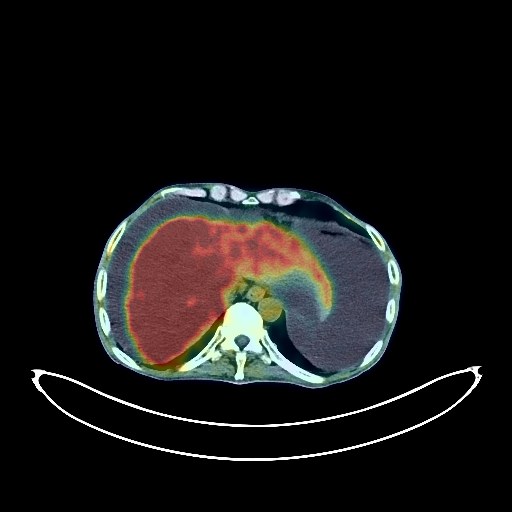
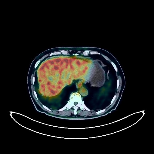
Liver Cancer PET/CT (case 983824-000176 from PETWB-REP)
3 views10 days agoWhole-body 18F-FDG PET/CT scan in a patient with Liver Cancer taken from the PETWB-REP dataset. The following English report (translated from original Chinese) is taken verbatim from the public dataset and has not been modified or otherwise checked for accuracy (see the end for citation). Impression a. After liver cancer treatment, multiple space-occupying lesions in the liver with increased FDG metabolism suggest the tumor may still be active. Portal vein tumor thrombus formation is also possible; please confirm with an MRI. Slightly increased FDG metabolism is observed in the hepatogastric space, retroperitoneum, and left supradiaphragmatic lymph nodes, suggesting reactive hyperplasia. Metastasis is not ruled out; follow-up is recommended. b. Liver cirrhosis, splenomegaly, portal hypertension with collateral circulation formation. Large amount of effusion in the abdominopelvic cavity. Chronic inflammatory micronodules in both lungs. Emphysema in both lungs, a few post-inflammatory remnants in both lungs. Slight thickening of the pleura bilaterally. Anemic changes, partial arteriosclerosis. Small hepatic cysts. Concentrated bile or sludge-like stones in the gallbladder. Splenic infarction. Prostatic calcification. Partial chronic inflammatory changes in the gastric wall; please confirm with endoscopic follow-up. Mild vertebral osteophyte formation. Nuchal ligament calcification. Small nodule in the left parotid gland with increased FDG metabolism, suggestive of lymphoma; please combine with contrast-enhanced MRI for comprehensive analysis. No obvious abnormalities were found on cranial scintigraphy. This case is from PETWB-REP, a curated dataset of whole-body 18F-FDG PET/CT scans and corresponding radiology reports from 490 patients with a broad spectrum of malignancies. The data were retrospectively collected from patients who underwent clinically indicated whole-body 18F-FDG PET/CT scans at the Shanghai Universal Medical Imaging Diagnostic Center between 2021 and 2024. License: Creative Commons Attribution 4.0 International (CC BY 4.0) Citation: Xue, L., Feng, G., Wenbo, Z., Zhang, Y., Li, L., Wang, S., Peng, L., Peng, S., & Gao, X. (2026). PETWB-REP: A Multi-Cancer Whole-Body FDG PET/CT Dataset with Corresponding Radiology Reports [Data set]. Zenodo. https://doi.org/10.5281/zenodo.18670487
Whole BodyPET/CT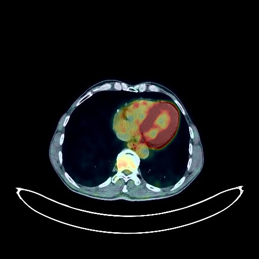
Lung Cancer PET/CT (case 983824-000023 from PETWB-REP)
7 views10 days agoWhole-body 18F-FDG PET/CT scan in a patient with Lung Cancer taken from the PETWB-REP dataset. The following English report (translated from original Chinese) is taken verbatim from the public dataset and has not been modified or otherwise checked for accuracy (see the end for citation). Impression a. Space-occupying lesions near the hilum in the posterior segment of the right upper lobe and in the posterior segment of the left upper lobe, both with increased FDG metabolism, suggestive of central lung cancer; multiple lymph node metastases in the right hilum and mediastinum. b. Multiple small nodules in both lungs, some with slightly increased FDG metabolism, suggesting possible metastasis, while others are chronic inflammatory nodules. c. Small patchy low-density lesions in the left frontal, parietal, and temporal lobes, with normal FDG metabolism, metastasis to be ruled out; contrast-enhanced MRI is recommended for clarification. Bilateral emphysema. Scattered post-inflammatory lesions in both lungs. Minor arteriosclerosis in some arteries. Benign prostatic hyperplasia. Calcification of the tunica vaginalis in the right testis. Reactive hyperplasia of retroperitoneal and mesenteric lymph nodes. Chronic inflammatory changes in the cardia, antrum of the stomach, and part of the intestines; please follow up with endoscopy. Degenerative changes in the spine, L4/5 and L5/S1 intervertebral disc bulges. Right femoral head hernia. Chronic inflammation of the left lateral wall of the nasopharynx. This case is from PETWB-REP, a curated dataset of whole-body 18F-FDG PET/CT scans and corresponding radiology reports from 490 patients with a broad spectrum of malignancies. The data were retrospectively collected from patients who underwent clinically indicated whole-body 18F-FDG PET/CT scans at the Shanghai Universal Medical Imaging Diagnostic Center between 2021 and 2024. License: Creative Commons Attribution 4.0 International (CC BY 4.0) Citation: Xue, L., Feng, G., Wenbo, Z., Zhang, Y., Li, L., Wang, S., Peng, L., Peng, S., & Gao, X. (2026). PETWB-REP: A Multi-Cancer Whole-Body FDG PET/CT Dataset with Corresponding Radiology Reports [Data set]. Zenodo. https://doi.org/10.5281/zenodo.18670487
Whole BodyPET/CT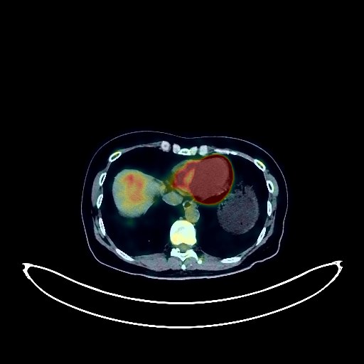
Colon Cancer PET/CT (case 983824-000079 from PETWB-REP)
2 views10 days agoWhole-body 18F-FDG PET/CT scan in a patient with Colon Cancer taken from the PETWB-REP dataset. The following English report (translated from original Chinese) is taken verbatim from the public dataset and has not been modified or otherwise checked for accuracy (see the end for citation). Impression a. Postoperative changes after colon cancer surgery; no signs of tumor recurrence were observed in the surgical area. Colonoscopy follow-up is recommended. b. Postoperative changes after resection of liver metastases; no significant space-occupying lesions were observed in the remaining liver, and FDG metabolism was normal. Chronic inflammatory micronodules in the right lung; CT follow-up is recommended. A few post-inflammatory remnants in the right lung. Partial arteriosclerosis. Bilateral breast hyperplasia; calcification in the left breast; ultrasound follow-up is recommended. Chronic gastritis; please combine with endoscopic follow-up. Mild vertebral osteophyte formation. L4/5 and L5/S1 intervertebral disc bulges. No significant abnormalities were found on cranial scintigraphy. This case is from PETWB-REP, a curated dataset of whole-body 18F-FDG PET/CT scans and corresponding radiology reports from 490 patients with a broad spectrum of malignancies. The data were retrospectively collected from patients who underwent clinically indicated whole-body 18F-FDG PET/CT scans at the Shanghai Universal Medical Imaging Diagnostic Center between 2021 and 2024. License: Creative Commons Attribution 4.0 International (CC BY 4.0) Citation: Xue, L., Feng, G., Wenbo, Z., Zhang, Y., Li, L., Wang, S., Peng, L., Peng, S., & Gao, X. (2026). PETWB-REP: A Multi-Cancer Whole-Body FDG PET/CT Dataset with Corresponding Radiology Reports [Data set]. Zenodo. https://doi.org/10.5281/zenodo.18670487
Whole BodyPET/CT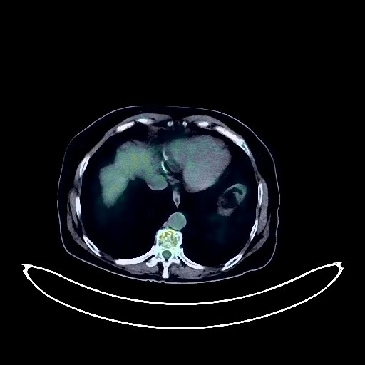
Ovarian Cancer PET/CT (case 983824-000112 from PETWB-REP)
1 views10 days agoWhole-body 18F-FDG PET/CT scan in a patient with Ovarian Cancer taken from the PETWB-REP dataset. The following English report (translated from original Chinese) is taken verbatim from the public dataset and has not been modified or otherwise checked for accuracy (see the end for citation). Impression a. A cystic mass in the right pelvic cavity (upper right side of the uterus) with slightly increased FDG metabolism in the cyst wall, highly suggestive of an adnexal malignancy, such as ovarian cancer. Please combine tumor markers and enhanced MRI for comprehensive analysis. b. High probability of peritoneal seeding metastasis, tuberculosis to be ruled out. Please correlate with clinical findings. Abdominal and pelvic effusion, partially encapsulated. a. High probability of subpleural inflammation in the right lung apex; possible chronic inflammatory ground-glass nodule in the apical segment of the right upper lobe, atypical adenomatous hyperplasia to be ruled out. HRCT follow-up is recommended for the above. b. Chronic inflammatory solid micronodules in the left upper lobe and right middle lobe, please follow up with CT. c. Slight bronchiectasis in the apical segment of the right upper lobe, a few fibrotic lesions in both lungs. Reactive hyperplasia of the left hilar and mediastinal lymph nodes. Partial arteriosclerosis (including coronary arteries). Manifestations of chronic gastritis. Liver cysts. Fatty infiltration of the pancreatic head. Left adrenal hyperplasia. Changes following bilateral fallopian tube ligation. Degenerative changes in the spine. L3/4 disc bulge, L4/5 disc herniation. Bilateral physiological uptake of the masseter muscles. Deep lacunar infarcts, age-related encephalopathy. Physiological uptake in the nasopharynx is highly probable; please correlate with clinical findings. This case is from PETWB-REP, a curated dataset of whole-body 18F-FDG PET/CT scans and corresponding radiology reports from 490 patients with a broad spectrum of malignancies. The data were retrospectively collected from patients who underwent clinically indicated whole-body 18F-FDG PET/CT scans at the Shanghai Universal Medical Imaging Diagnostic Center between 2021 and 2024. License: Creative Commons Attribution 4.0 International (CC BY 4.0) Citation: Xue, L., Feng, G., Wenbo, Z., Zhang, Y., Li, L., Wang, S., Peng, L., Peng, S., & Gao, X. (2026). PETWB-REP: A Multi-Cancer Whole-Body FDG PET/CT Dataset with Corresponding Radiology Reports [Data set]. Zenodo. https://doi.org/10.5281/zenodo.18670487
Whole BodyPET/CT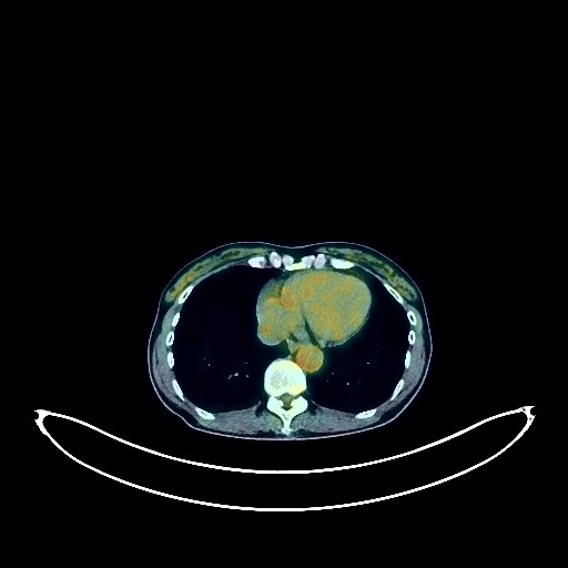
Ovarian Cancer PET/CT (case 983824-000144 from PETWB-REP)
2 views10 days agoWhole-body 18F-FDG PET/CT scan in a patient with Ovarian Cancer taken from the PETWB-REP dataset. The following English report (translated from original Chinese) is taken verbatim from the public dataset and has not been modified or otherwise checked for accuracy (see the end for citation). Impression a. Postoperative changes in the abdominal wall following bilateral adnexal and peritoneal lesion resection, thickening of the greater omentum, mesentery, and pelvic floor fascia with a few flocculent shadows. FDG metabolism was normal. Postoperative changes are considered possible, but metastatic tumors need to be ruled out. Please correlate with clinical findings and conduct regular follow-up examinations. Small amount of pelvic effusion. b. No obvious space-occupying lesions were found in the liver, gallbladder, or pancreas. Enhanced MRI of the upper abdomen should be performed if necessary to rule out occult lesions. Chronic inflammatory micronodules in both lungs. Calcification in the left upper lobe of the lung, a few post-inflammatory remnants in both lungs. Mediastinal calcification. Partial arteriosclerosis. Bilateral breast hyperplasia, calcification in the right breast. Multiple liver cysts. Right kidney cyst. Possible chronic inflammatory changes in the gastric wall; please combine with endoscopic examination to rule out other possibilities. Degenerative changes in the spine, L4/5 intervertebral disc bulge with posterior calcification. Schmorl's node at the superior margin of the T12 vertebral body. No obvious abnormalities were found on cranial scintigraphy. This case is from PETWB-REP, a curated dataset of whole-body 18F-FDG PET/CT scans and corresponding radiology reports from 490 patients with a broad spectrum of malignancies. The data were retrospectively collected from patients who underwent clinically indicated whole-body 18F-FDG PET/CT scans at the Shanghai Universal Medical Imaging Diagnostic Center between 2021 and 2024. License: Creative Commons Attribution 4.0 International (CC BY 4.0) Citation: Xue, L., Feng, G., Wenbo, Z., Zhang, Y., Li, L., Wang, S., Peng, L., Peng, S., & Gao, X. (2026). PETWB-REP: A Multi-Cancer Whole-Body FDG PET/CT Dataset with Corresponding Radiology Reports [Data set]. Zenodo. https://doi.org/10.5281/zenodo.18670487
Whole BodyPET/CT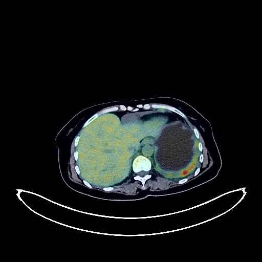
Cervical Cancer PET/CT (case 983824-000198 from PETWB-REP)
2 views10 days agoWhole-body 18F-FDG PET/CT scan in a patient with Cervical Cancer taken from the PETWB-REP dataset. The following English report (translated from original Chinese) is taken verbatim from the public dataset and has not been modified or otherwise checked for accuracy (see the end for citation). Impression a. A mass in the left upper lobe of the lung with increased FDG metabolism, suggestive of lung cancer; please refer to pathology. b. Metastatic lesions in the left pleura and pericardium, multiple lymph node metastases in the bilateral hilar, mediastinal, and bilateral supraclavicular fossae. c. Liver metastases. Bilateral adrenal metastases. d. Multiple bone metastases throughout the body (see description for details), muscle metastases in the left buttock and left shoulder. e. Suspected soft tissue nodule in the left parietal lobe, with background FDG uptake; metastasis to be ruled out; MRI is recommended. a. After cervical cancer treatment, the cervical density is uneven, but FDG metabolism is normal, suggesting suppressed tumor activity after treatment. b. Multiple foci of increased FDG metabolism in the greater omentum and mesenteric region; metastasis to be ruled out; close observation is recommended. a. Mixed ground-glass opacity in the subpleural region of the apical segment of the right upper lobe with increased FDG metabolism, suggestive of lung cancer; please correlate with clinicopathology. b. Possible chronic inflammatory nodules in both lungs; follow-up CT is recommended. Scattered post-inflammatory lesions in both lungs. Left pleural and pericardial effusion. Anemia. Chronic cholecystitis, gallstones, or residual contrast agent. Residual contrast agent in the urinary tract. Left renal cyst. Left adnexal cyst. No obvious pelvic effusion. Chronic inflammatory changes in the antrum of the stomach; please correlate with endoscopic follow-up. Degenerative changes in the spine, multiple intervertebral disc bulges, L3/4 disc herniation. Post-radiotherapy changes in some lumbar and sacral vertebrae. Benign bone disease in the upper right femur. Bilateral chronic maxillary sinusitis. This case is from PETWB-REP, a curated dataset of whole-body 18F-FDG PET/CT scans and corresponding radiology reports from 490 patients with a broad spectrum of malignancies. The data were retrospectively collected from patients who underwent clinically indicated whole-body 18F-FDG PET/CT scans at the Shanghai Universal Medical Imaging Diagnostic Center between 2021 and 2024. License: Creative Commons Attribution 4.0 International (CC BY 4.0) Citation: Xue, L., Feng, G., Wenbo, Z., Zhang, Y., Li, L., Wang, S., Peng, L., Peng, S., & Gao, X. (2026). PETWB-REP: A Multi-Cancer Whole-Body FDG PET/CT Dataset with Corresponding Radiology Reports [Data set]. Zenodo. https://doi.org/10.5281/zenodo.18670487
Whole BodyPET/CT