Loading...
Lung Cancer PET/CT (case 983824-000204 from PETWB-REP)
1 views10 days agoWhole-body 18F-FDG PET/CT scan in a patient with Lung Cancer taken from the PETWB-REP dataset. The following English report (translated from original Chinese) is taken verbatim from the public dataset and has not been modified or otherwise checked for accuracy (see the end for citation). Impression a. Irregular soft tissue nodules with increased FDG metabolism in the posterior segment of the right upper lobe, suggestive of peripheral lung cancer; please correlate with clinicopathology. b. Chronic inflammatory nodules in the remaining two lungs; follow-up CT scan recommended. Emphysema in both lungs; scattered post-inflammatory lesions in both lungs. Right pleural thickening. Mild anemia; partial arterial wall calcification (including coronary arteries). Liver calcifications. Chronic cholecystitis; gallstones. Postoperative changes in the spleen. Cystic mass in the right inguinal region, suggestive of benign; ultrasound follow-up recommended. Benign prostatic hyperplasia with calcification. Partial chronic inflammatory changes in the gastric wall; please correlate with endoscopy. Osteoporosis; degenerative changes in the spine; L4/5 and L5/S1 intervertebral disc bulges. L3 vertebral body wedge deformity, benign osteopathy of the L3 vertebral body. Calcifications in the soft tissue around the right iliac crest. Senile brain, deep lacunar infarcts. Bilateral maxillary sinus submucosal cysts, chronic inflammation of the right maxillary sinus. This case is from PETWB-REP, a curated dataset of whole-body 18F-FDG PET/CT scans and corresponding radiology reports from 490 patients with a broad spectrum of malignancies. The data were retrospectively collected from patients who underwent clinically indicated whole-body 18F-FDG PET/CT scans at the Shanghai Universal Medical Imaging Diagnostic Center between 2021 and 2024. License: Creative Commons Attribution 4.0 International (CC BY 4.0) Citation: Xue, L., Feng, G., Wenbo, Z., Zhang, Y., Li, L., Wang, S., Peng, L., Peng, S., & Gao, X. (2026). PETWB-REP: A Multi-Cancer Whole-Body FDG PET/CT Dataset with Corresponding Radiology Reports [Data set]. Zenodo. https://doi.org/10.5281/zenodo.18670487
Whole BodyPET/CT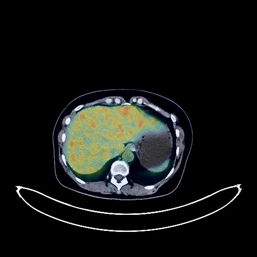
Lung Cancer PET/CT (case 983824-000021 from PETWB-REP)
7 views10 days agoWhole-body 18F-FDG PET/CT scan in a patient with Lung Cancer taken from the PETWB-REP dataset. The following English report (translated from original Chinese) is taken verbatim from the public dataset and has not been modified or otherwise checked for accuracy (see the end for citation). Impression a. A soft tissue mass in the lower lobe of the right lung with increased FDG metabolism, suggestive of lung cancer involving the adjacent pleura, accompanied by multiple metastases in both lungs. Please confirm with pathology. b. Right hilar and mediastinal lymph node metastases. Bilateral supraclavicular lymph node metastases are possible. c. A mixed ground-glass opacity nodule in the apical segment of the right upper lobe with increased FDG metabolism, suggestive of early lung cancer, but inflammatory lesions cannot be ruled out; ground-glass opacities in the anterior segment of the right upper lobe and the apical-posterior segment of the left upper lobe, with normal FDG metabolism, suggestive of atypical adenomatous hyperplasia or chronic inflammatory nodules. Annual HRCT follow-up is recommended. d. Chronic inflammation and sequelae in both lungs. Multiple intracranial metastases. Please combine with enhanced MRI for comprehensive analysis. Postoperative left breast surgery: Bilateral breast hyperplasia. A slightly high-density nodule in the upper inner quadrant of the left breast, with normal FDG metabolism, suggests possible fibroadenoma or lobular hyperplasia nodule. Please confirm with ultrasound or enhanced MRI. Accessory breast in the right axilla. Localized elevated FDG metabolism in the pancreatic tail, with no abnormal density shadows seen on the same CT scan, suggesting a possible physiological change. Please rule out space-occupying lesions based on clinical findings and MRI. Liver calcifications. Increased FDG metabolism in parts of the intestine, suggesting physiological uptake or chronic inflammation. Please follow up with endoscopy. Partial vertebral osteophyte formation. L5/S1 intervertebral disc bulge. This case is from PETWB-REP, a curated dataset of whole-body 18F-FDG PET/CT scans and corresponding radiology reports from 490 patients with a broad spectrum of malignancies. The data were retrospectively collected from patients who underwent clinically indicated whole-body 18F-FDG PET/CT scans at the Shanghai Universal Medical Imaging Diagnostic Center between 2021 and 2024. License: Creative Commons Attribution 4.0 International (CC BY 4.0) Citation: Xue, L., Feng, G., Wenbo, Z., Zhang, Y., Li, L., Wang, S., Peng, L., Peng, S., & Gao, X. (2026). PETWB-REP: A Multi-Cancer Whole-Body FDG PET/CT Dataset with Corresponding Radiology Reports [Data set]. Zenodo. https://doi.org/10.5281/zenodo.18670487
Whole BodyPET/CT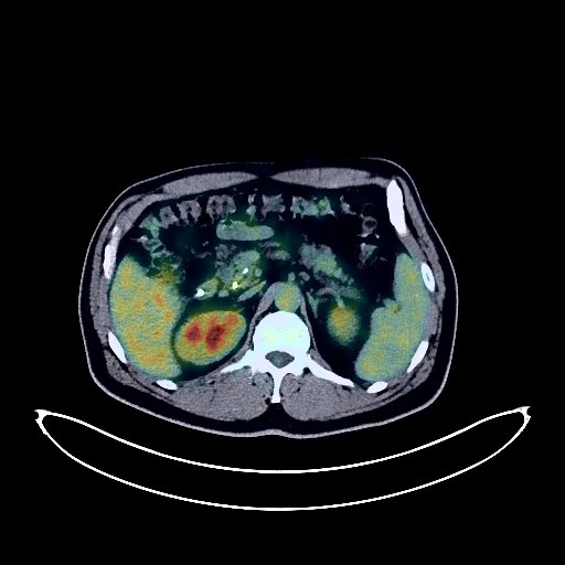
Cholangiocarcinoma PET/CT (case 983824-000053 from PETWB-REP)
5 views10 days agoWhole-body 18F-FDG PET/CT scan in a patient with Cholangiocarcinoma taken from the PETWB-REP dataset. The following English report (translated from original Chinese) is taken verbatim from the public dataset and has not been modified or otherwise checked for accuracy (see the end for citation). Impression a. Post-nasobiliary drainage + pancreatic duct stent placement changes: No dilation of the common bile duct, no obvious space-occupying lesion in the lower segment of the common bile duct, mild FDG metabolism in the course of the lower segment of the common bile duct. Based on pathology and MRI from another hospital, inflammation with local carcinogenesis in the lower segment of the common bile duct is suspected. Please correlate with clinical findings. b. Reactive hyperplasia of the lymph nodes near the gastric antrum, pancreatic head, and retroperitoneum is highly probable. Please follow up. c. Chronic cholecystitis. Pancreatic fat infiltration. Increased local FDG uptake in the course of the nasobiliary drainage tube near the cardia, likely due to inflammation. Please follow up with endoscopic findings. A few aspiration changes in the posterior right lung. Calcification of the coronary artery wall. Bilateral gynecomastia. Spinal degenerative changes. L2/3, L3/4, L4/5, and L5/S1 intervertebral disc bulges. Atrophy with fatty changes in the right buttock, right thigh muscles (shown), and part of the left thigh muscles. No obvious abnormalities were found on cranial scintigraphy. Minor inflammation was observed in both maxillary sinuses. This case is from PETWB-REP, a curated dataset of whole-body 18F-FDG PET/CT scans and corresponding radiology reports from 490 patients with a broad spectrum of malignancies. The data were retrospectively collected from patients who underwent clinically indicated whole-body 18F-FDG PET/CT scans at the Shanghai Universal Medical Imaging Diagnostic Center between 2021 and 2024. License: Creative Commons Attribution 4.0 International (CC BY 4.0) Citation: Xue, L., Feng, G., Wenbo, Z., Zhang, Y., Li, L., Wang, S., Peng, L., Peng, S., & Gao, X. (2026). PETWB-REP: A Multi-Cancer Whole-Body FDG PET/CT Dataset with Corresponding Radiology Reports [Data set]. Zenodo. https://doi.org/10.5281/zenodo.18670487
Whole BodyPET/CT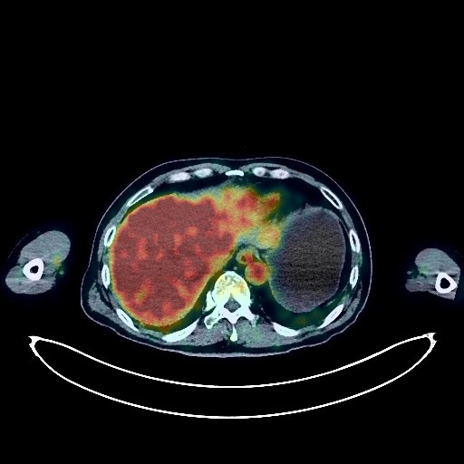
Pancreatic Cancer PET/CT (case 983824-000003 from PETWB-REP)
8 views10 days agoWhole-body 18F-FDG PET/CT scan in a patient with Pancreatic Cancer taken from the PETWB-REP dataset. The following English report (translated from original Chinese) is taken verbatim from the public dataset and has not been modified or otherwise checked for accuracy (see the end for citation). Impression a. After chemotherapy for pancreatic cancer, a soft tissue mass in the pancreatic body with increased FDG metabolism suggests continued tumor activity; peripancreatic lymph nodes show slightly increased FDG metabolism. It is recommended to compare these findings with pre-treatment imaging data and follow up. b. After radiofrequency ablation of liver metastases, multiple low-density nodules and masses were observed in the liver. The lesion in the right lobe showed loss of FDG uptake, but no significant increase in FDG metabolism, suggesting suppressed tumor activity after treatment. c. No space-occupying lesions were observed in either kidney, and FDG metabolism was normal. Enhanced MRI follow-up is recommended. a. Ground-glass nodules in the apical segment of the right upper lobe, the posterior basal segment of the left lower lobe, and the lateral basal segment of the right lower lobe, with normal FDG metabolism, suggest inflammatory nodules or atypical adenomatous hyperplasia. Annual HRCT follow-up is recommended. b. Calcification in the right upper lobe, and a few post-inflammatory remnants in both lungs. Calcification of some arterial walls (including coronary arteries). Multiple liver cysts. Calcification in the right inguinal canal. Degenerative changes in the spine, with L4/5 and L5/S1 intervertebral disc bulges. Post-fracture changes of the left 7th-10th ribs. No obvious abnormalities were found on cranial scintigraphy. This case is from PETWB-REP, a curated dataset of whole-body 18F-FDG PET/CT scans and corresponding radiology reports from 490 patients with a broad spectrum of malignancies. The data were retrospectively collected from patients who underwent clinically indicated whole-body 18F-FDG PET/CT scans at the Shanghai Universal Medical Imaging Diagnostic Center between 2021 and 2024. License: Creative Commons Attribution 4.0 International (CC BY 4.0) Citation: Xue, L., Feng, G., Wenbo, Z., Zhang, Y., Li, L., Wang, S., Peng, L., Peng, S., & Gao, X. (2026). PETWB-REP: A Multi-Cancer Whole-Body FDG PET/CT Dataset with Corresponding Radiology Reports [Data set]. Zenodo. https://doi.org/10.5281/zenodo.18670487
Whole BodyPET/CT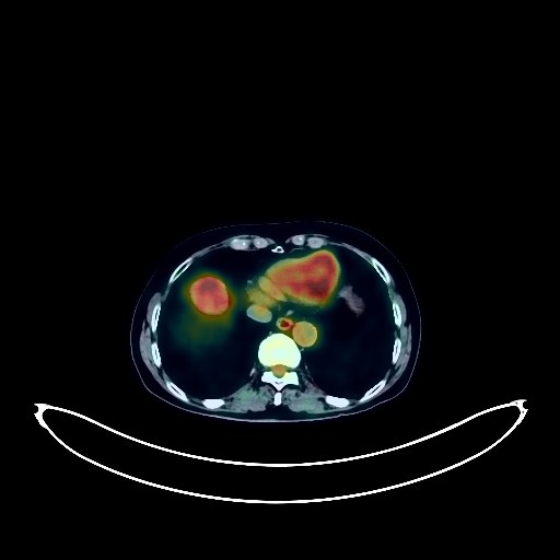
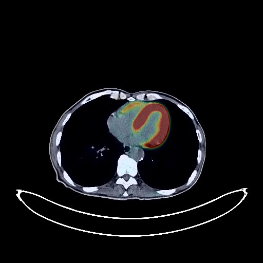
Liver Cancer PET/CT (case 983824-000178 from PETWB-REP)
2 views10 days agoWhole-body 18F-FDG PET/CT scan in a patient with Liver Cancer taken from the PETWB-REP dataset. The following English report (translated from original Chinese) is taken verbatim from the public dataset and has not been modified or otherwise checked for accuracy (see the end for citation). Impression a. A mass at the junction of the left and right lobes of the liver with increased FDG metabolism; malignancy is the primary consideration, hemangioma to be ruled out. Further analysis with enhanced MRI is recommended. Reactive hyperplasia of the hepatogastric space and retroperitoneal lymph nodes. b. Calcification in the right lobe of the liver. Cyst in the right lobe of the liver. Adenomyosis of the gallbladder to be ruled out; ultrasound follow-up is recommended. Chronic inflammatory micronodules (solid) in the left upper lobe of the lung. Fibrous lesions in the right lung. Reactive hyperplasia of bilateral axillary lymph nodes. Partial vertebral osteophyte formation. L3/4, L4/5, and L5/S1 intervertebral disc bulges. Subcutaneous calcification in the left buttock. No significant abnormalities were found on cranial FDG imaging. This case is from PETWB-REP, a curated dataset of whole-body 18F-FDG PET/CT scans and corresponding radiology reports from 490 patients with a broad spectrum of malignancies. The data were retrospectively collected from patients who underwent clinically indicated whole-body 18F-FDG PET/CT scans at the Shanghai Universal Medical Imaging Diagnostic Center between 2021 and 2024. License: Creative Commons Attribution 4.0 International (CC BY 4.0) Citation: Xue, L., Feng, G., Wenbo, Z., Zhang, Y., Li, L., Wang, S., Peng, L., Peng, S., & Gao, X. (2026). PETWB-REP: A Multi-Cancer Whole-Body FDG PET/CT Dataset with Corresponding Radiology Reports [Data set]. Zenodo. https://doi.org/10.5281/zenodo.18670487
Whole BodyPET/CT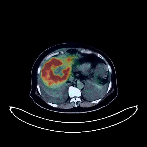
Liver Cancer PET/CT (case 983824-000124 from PETWB-REP)
2 views10 days agoWhole-body 18F-FDG PET/CT scan in a patient with Liver Cancer taken from the PETWB-REP dataset. The following English report (translated from original Chinese) is taken verbatim from the public dataset and has not been modified or otherwise checked for accuracy (see the end for citation). Impression a. Multiple space-occupying lesions in the liver with increased FDG metabolism suggest malignancy, possibly metastatic tumors. Please combine tumor markers and enhanced MRI for comprehensive analysis. b. Multiple lymph node metastases in the hepatic hilum, hepatogastric space, retroperitoneum, and right cardiophrenic angle; increased density in the greater omentum and mesentery, multiple nodules and flocculent shadows, slightly increased FDG metabolism, suggesting possible implantation metastasis; abdominopelvic effusion. Thickening of the gastric wall in the antrum with increased FDG metabolism suggests inflammation. Please combine gastroscopy to rule out tumors. Chronic inflammation or physiological uptake of some intestinal segments is possible; please follow up with colonoscopy. a. Chronic inflammatory nodules in both lungs are highly probable; CT follow-up is recommended. Chronic inflammation and post-inflammatory remnants in both lungs. Reactive hyperplasia of hilar and mediastinal lymph nodes in both lungs. Bilateral pleural effusion (partially loculated on the left). b. Pericardial thickening, mild anemia changes, partial arterial wall calcification (including coronary arteries). Bilateral breast hyperplasia. Post-cholecystectomy changes, splenic capsule calcification, accessory spleen. Left adrenal hyperplasia. Bilateral renal calculi, bilateral renal cysts. Osteoporosis, degenerative changes in the spine, L4/5, L5/S1 intervertebral disc herniation with pneumothorax and degeneration. Subcutaneous edema in the abdomen and buttocks. Senile brain, deep lacunar infarcts. This case is from PETWB-REP, a curated dataset of whole-body 18F-FDG PET/CT scans and corresponding radiology reports from 490 patients with a broad spectrum of malignancies. The data were retrospectively collected from patients who underwent clinically indicated whole-body 18F-FDG PET/CT scans at the Shanghai Universal Medical Imaging Diagnostic Center between 2021 and 2024. License: Creative Commons Attribution 4.0 International (CC BY 4.0) Citation: Xue, L., Feng, G., Wenbo, Z., Zhang, Y., Li, L., Wang, S., Peng, L., Peng, S., & Gao, X. (2026). PETWB-REP: A Multi-Cancer Whole-Body FDG PET/CT Dataset with Corresponding Radiology Reports [Data set]. Zenodo. https://doi.org/10.5281/zenodo.18670487
Whole BodyPET/CT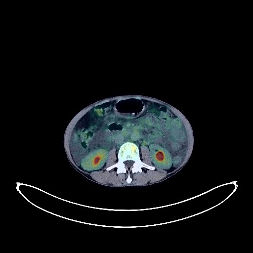
Liver Cancer PET/CT (case 983824-000008 from PETWB-REP)
8 views10 days agoWhole-body 18F-FDG PET/CT scan in a patient with Liver Cancer taken from the PETWB-REP dataset. The following English report (translated from original Chinese) is taken verbatim from the public dataset and has not been modified or otherwise checked for accuracy (see the end for citation). Impression a. Multiple space-occupying lesions in the liver, with increased FDG metabolism, suggestive of malignancy, most likely primary liver cancer. b. Multiple lymph node metastases in the hilum and retroperitoneum. Diffuse metastases in both lungs. Extensive bone metastases throughout the body (pathological fracture of the right 3rd posterior rib). c. Liver cirrhosis; splenomegaly. Large amounts of fluid in the abdomen and pelvis. A few chronic lesions and old lesions in both lungs. Degenerative changes in the spine. L4/5 and L5/S1 intervertebral disc bulges. Mild age-related brain changes. Sclerotic mastoid process on the left side. This case is from PETWB-REP, a curated dataset of whole-body 18F-FDG PET/CT scans and corresponding radiology reports from 490 patients with a broad spectrum of malignancies. The data were retrospectively collected from patients who underwent clinically indicated whole-body 18F-FDG PET/CT scans at the Shanghai Universal Medical Imaging Diagnostic Center between 2021 and 2024. License: Creative Commons Attribution 4.0 International (CC BY 4.0) Citation: Xue, L., Feng, G., Wenbo, Z., Zhang, Y., Li, L., Wang, S., Peng, L., Peng, S., & Gao, X. (2026). PETWB-REP: A Multi-Cancer Whole-Body FDG PET/CT Dataset with Corresponding Radiology Reports [Data set]. Zenodo. https://doi.org/10.5281/zenodo.18670487
Whole BodyPET/CT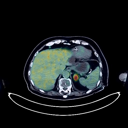
Lung Cancer PET/CT (case 983824-000136 from PETWB-REP)
2 views10 days agoWhole-body 18F-FDG PET/CT scan in a patient with Lung Cancer taken from the PETWB-REP dataset. The following English report (translated from original Chinese) is taken verbatim from the public dataset and has not been modified or otherwise checked for accuracy (see the end for citation). Impression a. A soft tissue mass near the hilum of the left upper lobe with increased FDG metabolism, strongly suggestive of lung cancer; please correlate with clinical findings and pathology. Left pleural effusion. b. Possible reactive hyperplasia of mediastinal lymph nodes; metastasis to the right anterior diaphragmatic lymph nodes needs to be ruled out; follow-up is recommended. c. Multiple solid nodular plaque lesions in both lungs, some with increased FDG metabolism, suggesting possible metastases; some chronic inflammatory lesions cannot be ruled out; regular follow-up with CT scans is recommended. d. Bone metastases in the right 1st and 4th ribs and T7 vertebral appendages. Left adrenal metastasis; right adrenal metastasis needs to be ruled out. e. Multiple metastases are highly likely in the subcutaneous tissue of the left nasal root, right pectoralis major muscle, subcutaneous tissue of the upper medial aspect of the right thigh, behind the right nipple, left perirenal fascia, and left paracolic gutter; please correlate with clinical findings and follow-up. Benign prostatic hyperplasia with calcification, nodular FDG hypermetabolic lesions in the left peripheral zone, prostate cancer to be ruled out; further examination with PSA and enhanced MRI is recommended. Ground-glass nodule in the anterior basal segment of the right lower lobe, FDG metabolism normal, suggestive of chronic inflammatory nodule or atypical adenomatous hyperplasia; please combine with annual follow-up via HRCT. Chronic inflammation and sequelae in both lungs. Full cardiac silhouette, post-pacemaker implantation. Partial arteriosclerosis (including coronary arteries). Liver cysts. Gallstones, chronic cholecystitis. Bilateral renal cysts. Spinal degenerative changes. L4/5 disc herniation, L5/S1 disc bulge. A slightly high-density nodule on the left side of the sellar region with increased FDG metabolism, suggesting a possible space-occupying lesion. Enhanced MRI of the sellar region is recommended. Bilateral deep cerebral ischemic lesions, age-related brain. Chronic inflammation of the left maxillary sinus. Possible physiological or inflammatory changes in the left submandibular gland; please follow up to rule out other possibilities. This case is from PETWB-REP, a curated dataset of whole-body 18F-FDG PET/CT scans and corresponding radiology reports from 490 patients with a broad spectrum of malignancies. The data were retrospectively collected from patients who underwent clinically indicated whole-body 18F-FDG PET/CT scans at the Shanghai Universal Medical Imaging Diagnostic Center between 2021 and 2024. License: Creative Commons Attribution 4.0 International (CC BY 4.0) Citation: Xue, L., Feng, G., Wenbo, Z., Zhang, Y., Li, L., Wang, S., Peng, L., Peng, S., & Gao, X. (2026). PETWB-REP: A Multi-Cancer Whole-Body FDG PET/CT Dataset with Corresponding Radiology Reports [Data set]. Zenodo. https://doi.org/10.5281/zenodo.18670487
Whole BodyPET/CT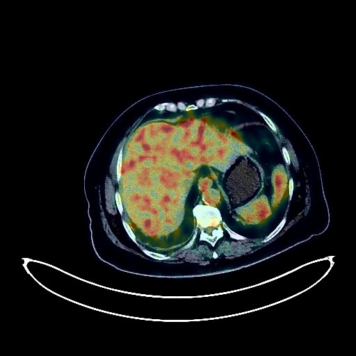
Lung Cancer PET/CT (case 983824-000132 from PETWB-REP)
2 views10 days agoWhole-body 18F-FDG PET/CT scan in a patient with Lung Cancer taken from the PETWB-REP dataset. The following English report (translated from original Chinese) is taken verbatim from the public dataset and has not been modified or otherwise checked for accuracy (see the end for citation). Impression a. Irregular soft tissue nodule near the horizontal fissure in the medial segment of the right middle lobe, with increased FDG metabolism, highly suggestive of lung cancer; please refer to pathology. b. Chronic inflammatory nodules in the right horizontal fissure and left lower lobe. Old lesion in the anterior segment of the right upper lobe, with a few chronic inflammations and remnants in both lungs. c. Small amount of pleural effusion on the right side. Reactive hyperplasia of hilar and mediastinal lymph nodes in both lungs. Low-density lesion in the right frontal lobe, possibly ischemic, space-occupying lesion to be ruled out; enhanced MRI is recommended for further examination. Chronic inflammatory changes in the nasopharynx, possibly left-sided periodontitis; please refer to specialist examination. A few chronic inflammations in the right maxillary sinus. Reactive hyperplasia of bilateral cervical lymph nodes. Manifestations of chronic gastritis; please refer to endoscopic follow-up. Physiological uptake of some intestinal segments is highly likely. Small kidney stones in both kidneys. Uterine fibroids with calcification. Degenerative changes in the spine, multiple thoracic and lumbar intervertebral disc effusions, multiple lumbar intervertebral disc bulges. Peritoneitis of the left hip joint and both ischial tuberosities. Partial arteriosclerosis (including coronary arteries). This case is from PETWB-REP, a curated dataset of whole-body 18F-FDG PET/CT scans and corresponding radiology reports from 490 patients with a broad spectrum of malignancies. The data were retrospectively collected from patients who underwent clinically indicated whole-body 18F-FDG PET/CT scans at the Shanghai Universal Medical Imaging Diagnostic Center between 2021 and 2024. License: Creative Commons Attribution 4.0 International (CC BY 4.0) Citation: Xue, L., Feng, G., Wenbo, Z., Zhang, Y., Li, L., Wang, S., Peng, L., Peng, S., & Gao, X. (2026). PETWB-REP: A Multi-Cancer Whole-Body FDG PET/CT Dataset with Corresponding Radiology Reports [Data set]. Zenodo. https://doi.org/10.5281/zenodo.18670487
Whole BodyPET/CT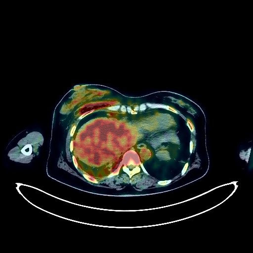
Lung Cancer PET/CT (case 983824-000109 from PETWB-REP)
2 views10 days agoWhole-body 18F-FDG PET/CT scan in a patient with Lung Cancer taken from the PETWB-REP dataset. The following English report (translated from original Chinese) is taken verbatim from the public dataset and has not been modified or otherwise checked for accuracy (see the end for citation). Impression a. A mass in the upper lobe of the right lung, with increased FDG metabolism, suggestive of lung cancer. Multiple metastatic tumors in both lungs and the right interlobar pleura. b. Metastasis to the right hilar and some mediastinal lymph nodes. Small amount of pleural effusion on the right side. c. A few old lesions in both lungs. Right pneumothorax (15% lung compression). Subcutaneous pneumothorax in the right axilla and right chest wall. d. Left adrenal hyperplasia is highly probable; follow-up CT scan is recommended to rule out metastasis. A slightly low-density nodule in the right lobe of the liver, with normal FDG uptake, suggestive of a cyst or hemangioma; ultrasound follow-up is recommended. Cyst in the left lobe of the liver. Postoperative changes after uterine surgery, cyst in the cervical region. Chronic gastritis. Mild osteophyte formation in the cervical, thoracic, and lumbar spine. L4/5 and L5/S1 intervertebral disc bulge. Cranial scintigraphy showed no abnormalities. This case is from PETWB-REP, a curated dataset of whole-body 18F-FDG PET/CT scans and corresponding radiology reports from 490 patients with a broad spectrum of malignancies. The data were retrospectively collected from patients who underwent clinically indicated whole-body 18F-FDG PET/CT scans at the Shanghai Universal Medical Imaging Diagnostic Center between 2021 and 2024. License: Creative Commons Attribution 4.0 International (CC BY 4.0) Citation: Xue, L., Feng, G., Wenbo, Z., Zhang, Y., Li, L., Wang, S., Peng, L., Peng, S., & Gao, X. (2026). PETWB-REP: A Multi-Cancer Whole-Body FDG PET/CT Dataset with Corresponding Radiology Reports [Data set]. Zenodo. https://doi.org/10.5281/zenodo.18670487
Whole BodyPET/CT