Loading...
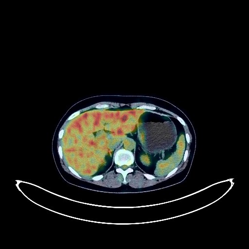
Cervical Cancer PET/CT (case 983827-000084 from PETWB-REP)
0 views10 days agoWhole-body 18F-FDG PET/CT scan in a patient with Cervical Cancer taken from the PETWB-REP dataset. The following English report (translated from original Chinese) is taken verbatim from the public dataset and has not been modified or otherwise checked for accuracy (see the end for citation). Impression a. Cervical mass, indistinctly demarcated from the adjacent uterine body, with increased FDG metabolism, consistent with cervical cancer. b. Focal increased FDG uptake on the anterior wall of the uterine body, suggestive of fibroid with degeneration; ultrasound is recommended. Intrauterine device (IUD) present. Several small, solid, chronic inflammatory nodules in both lungs. A few chronic inflammatory lesions and old lesions in both lungs. Proliferative changes in both breasts; follow-up ultrasound is recommended. Mild osteophyte formation in the cervical, thoracic, and lumbar spine. No abnormalities found on cranial scintigraphy. This case is from PETWB-REP, a curated dataset of whole-body 18F-FDG PET/CT scans and corresponding radiology reports from 490 patients with a broad spectrum of malignancies. The data were retrospectively collected from patients who underwent clinically indicated whole-body 18F-FDG PET/CT scans at the Shanghai Universal Medical Imaging Diagnostic Center between 2021 and 2024. License: Creative Commons Attribution 4.0 International (CC BY 4.0) Citation: Xue, L., Feng, G., Wenbo, Z., Zhang, Y., Li, L., Wang, S., Peng, L., Peng, S., & Gao, X. (2026). PETWB-REP: A Multi-Cancer Whole-Body FDG PET/CT Dataset with Corresponding Radiology Reports [Data set]. Zenodo. https://doi.org/10.5281/zenodo.18670487
Whole BodyPET/CT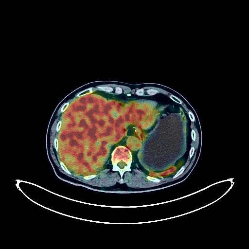
Lung Cancer PET/CT (case 983827-000191 from PETWB-REP)
0 views10 days agoWhole-body 18F-FDG PET/CT scan in a patient with Lung Cancer taken from the PETWB-REP dataset. The following English report (translated from original Chinese) is taken verbatim from the public dataset and has not been modified or otherwise checked for accuracy (see the end for citation). Impression a. A mass near the hilum in the anterior segment of the right upper lobe, with unclear boundaries from the adjacent pericardium and great vessels. Increased FDG metabolism is consistent with lung cancer, accompanied by obstructive pneumonia. A small amount of pleural effusion on the right side with partial atelectasis in the right lower lobe. b. Metastasis to the right hilar and pretracheal vena cava lymph nodes. Possible metastasis to the right upper mediastinal paratracheal, aortopulmonary window, and subcarinal lymph nodes. c. Several solid micronodules of chronic inflammation in both lungs. Chronic inflammation and old lesions in the remaining lungs. Calcification of some arterial walls. A few ischemic lesions in the deep bilateral brain, indicative of senile encephalopathy. Unclear visualization of the left lens. Small renal calculi in the right kidney. Calcification of the prostate. Slight thickening of the gastric antrum wall and mildly increased FDG uptake suggest chronic gastritis; continuous increased FDG metabolism in parts of the colon and rectum suggest inflammatory or physiological uptake. Follow-up gastroscopy and colonoscopy are recommended. Cervical, thoracic, and lumbar spondylosis. L4/5 and L5/S1 intervertebral disc bulges. Sacral canal cyst. A few ischemic lesions in the deep bilateral cerebral regions; age-related encephalopathy. This case is from PETWB-REP, a curated dataset of whole-body 18F-FDG PET/CT scans and corresponding radiology reports from 490 patients with a broad spectrum of malignancies. The data were retrospectively collected from patients who underwent clinically indicated whole-body 18F-FDG PET/CT scans at the Shanghai Universal Medical Imaging Diagnostic Center between 2021 and 2024. License: Creative Commons Attribution 4.0 International (CC BY 4.0) Citation: Xue, L., Feng, G., Wenbo, Z., Zhang, Y., Li, L., Wang, S., Peng, L., Peng, S., & Gao, X. (2026). PETWB-REP: A Multi-Cancer Whole-Body FDG PET/CT Dataset with Corresponding Radiology Reports [Data set]. Zenodo. https://doi.org/10.5281/zenodo.18670487
Whole BodyPET/CT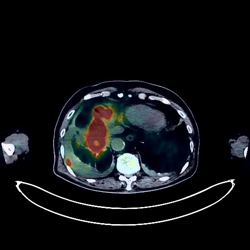
Lung Cancer PET/CT (case 983827-000134 from PETWB-REP)
1 views10 days agoWhole-body 18F-FDG PET/CT scan in a patient with Lung Cancer taken from the PETWB-REP dataset. The following English report (translated from original Chinese) is taken verbatim from the public dataset and has not been modified or otherwise checked for accuracy (see the end for citation). Impression a. A soft tissue mass on the diaphragmatic surface of the right lung with increased FDG metabolism, highly suggestive of lung cancer; please confirm with pathology. Bilateral pleural metastases, small amounts of pleural effusion bilaterally. b. Multiple soft tissue nodules in the upper lobe of the left lung with increased FDG metabolism, highly suggestive of malignancy; both metastatic and primary lesions are possible. Please confirm with clinical findings; biopsy may be necessary. c. Metastasis to the left hilar, mediastinal, hepatogastric space, and retroperitoneal lymph nodes in the upper abdomen. d. Multiple liver metastases; please confirm with contrast-enhanced MRI. T12 vertebral bone metastasis to be ruled out. Chronic inflammation and sequelae in both lungs. Partial arteriosclerosis (including coronary arteries). Small amount of pericardial effusion. Highly suggestive of right adrenal adenoma; please follow up. Highly suggestive of left inguinal hernia. Prostatic calcifications, with a nodular FDG hypermetabolic lesion at the right anterior margin in the central part, suggestive of inflammatory changes. Please confirm with PSA and enhanced MRI to rule out space-occupying lesions. Spinal degeneration. L2-3 vertebral instability. T12-L1 vertebral wedging. L2-S1 intervertebral disc bulge. Bilateral frozen shoulder. Bilateral deep cerebral ischemic lesions, age-related brain; please confirm with MRI. Solid nodule in the deep lobe of the right parotid gland with increased FDG metabolism, suggestive of adenolymphoma or mixed tumor; please follow up to rule out other possibilities. This case is from PETWB-REP, a curated dataset of whole-body 18F-FDG PET/CT scans and corresponding radiology reports from 490 patients with a broad spectrum of malignancies. The data were retrospectively collected from patients who underwent clinically indicated whole-body 18F-FDG PET/CT scans at the Shanghai Universal Medical Imaging Diagnostic Center between 2021 and 2024. License: Creative Commons Attribution 4.0 International (CC BY 4.0) Citation: Xue, L., Feng, G., Wenbo, Z., Zhang, Y., Li, L., Wang, S., Peng, L., Peng, S., & Gao, X. (2026). PETWB-REP: A Multi-Cancer Whole-Body FDG PET/CT Dataset with Corresponding Radiology Reports [Data set]. Zenodo. https://doi.org/10.5281/zenodo.18670487
Whole BodyPET/CT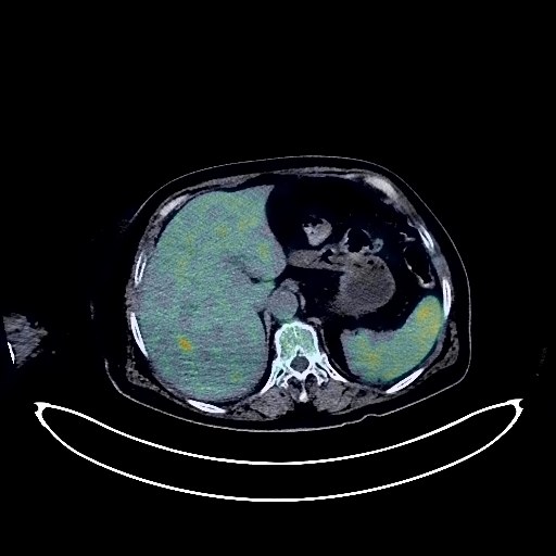
Gallbladder Cancer PET/CT (case 983827-000176 from PETWB-REP)
2 views10 days agoWhole-body 18F-FDG PET/CT scan in a patient with Gallbladder Cancer taken from the PETWB-REP dataset. The following English report (translated from original Chinese) is taken verbatim from the public dataset and has not been modified or otherwise checked for accuracy (see the end for citation). Impression a. Gallbladder lesion with elevated FDG metabolism, suggestive of malignancy, most likely gallbladder cancer; please confirm clinicopathologically. b. Multiple liver metastases. Hilar lymph node metastasis. Possible reactive hyperplasia of retroperitoneal lymph nodes; follow-up recommended. Chronic inflammation and post-inflammatory remnants in both lungs. Bilateral pleural thickening. Reactive hyperplasia of hilar and mediastinal lymph nodes. Calcification of some arterial walls (including coronary arteries). Possible bilateral adrenal hyperplasia. Chronic inflammatory changes in the entire esophagus and antrum of the stomach, hemorrhoidal changes; please confirm endoscopic follow-up. Osteoporosis, degenerative changes in the spine, L4/5 intervertebral disc bulge. T3/L5 vertebral hemangioma. Age-related brain lesions with deep lacunar infarcts; please include an MRI scan. This case is from PETWB-REP, a curated dataset of whole-body 18F-FDG PET/CT scans and corresponding radiology reports from 490 patients with a broad spectrum of malignancies. The data were retrospectively collected from patients who underwent clinically indicated whole-body 18F-FDG PET/CT scans at the Shanghai Universal Medical Imaging Diagnostic Center between 2021 and 2024. License: Creative Commons Attribution 4.0 International (CC BY 4.0) Citation: Xue, L., Feng, G., Wenbo, Z., Zhang, Y., Li, L., Wang, S., Peng, L., Peng, S., & Gao, X. (2026). PETWB-REP: A Multi-Cancer Whole-Body FDG PET/CT Dataset with Corresponding Radiology Reports [Data set]. Zenodo. https://doi.org/10.5281/zenodo.18670487
Whole BodyPET/CT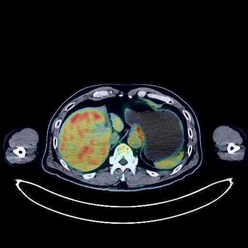
Colon Cancer PET/CT (case 983827-000232 from PETWB-REP)
4 views10 days agoWhole-body 18F-FDG PET/CT scan in a patient with Colon Cancer taken from the PETWB-REP dataset. The following English report (translated from original Chinese) is taken verbatim from the public dataset and has not been modified or otherwise checked for accuracy (see the end for citation). Impression Post-treatment for sigmoid colon cancer: No signs of tumor recurrence were observed in the surgical area; please follow up with colonoscopy. Anterior abdominal wall hernia. Reactive hyperplasia of bilateral inguinal lymph nodes. a. Sellar region mass; enhanced MRI is recommended for further examination. b. Bilateral deep lacunar infarcts, senile encephalopathy. Bilateral ethmoid sinusitis. Chronic inflammatory micronodules in the upper lobes of both lungs, chronic inflammation and sequelae in both lungs, calcification in the lower lobe of the right lung, para-apical emphysema in both lungs. Reactive hyperplasia of hilar and mediastinal lymph nodes in both lungs. Mild pleural thickening on both sides. Liver cyst. Gallstones, chronic cholecystitis. Complex right renal cyst, right renal stones. Prostatic calcification. Spinal degenerative changes. L4/5 vertebral endplate inflammation. L4/5 intervertebral disc pneumatosis and degeneration; L2/3 and L3/4 intervertebral disc bulging. Decreased thyroid density, increased FDG metabolism, suggestive of inflammatory uptake; please follow up with thyroid function tests and ultrasound. Reactive hyperplasia of cervical lymph nodes. This case is from PETWB-REP, a curated dataset of whole-body 18F-FDG PET/CT scans and corresponding radiology reports from 490 patients with a broad spectrum of malignancies. The data were retrospectively collected from patients who underwent clinically indicated whole-body 18F-FDG PET/CT scans at the Shanghai Universal Medical Imaging Diagnostic Center between 2021 and 2024. License: Creative Commons Attribution 4.0 International (CC BY 4.0) Citation: Xue, L., Feng, G., Wenbo, Z., Zhang, Y., Li, L., Wang, S., Peng, L., Peng, S., & Gao, X. (2026). PETWB-REP: A Multi-Cancer Whole-Body FDG PET/CT Dataset with Corresponding Radiology Reports [Data set]. Zenodo. https://doi.org/10.5281/zenodo.18670487
Whole BodyPET/CT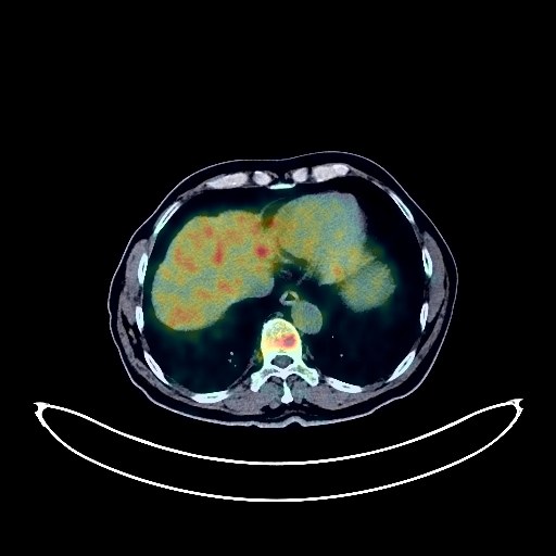
Prostate Cancer PET/CT (case 983827-000252 from PETWB-REP)
3 views10 days agoWhole-body 18F-FDG PET/CT scan in a patient with Prostate Cancer taken from the PETWB-REP dataset. The following English report (translated from original Chinese) is taken verbatim from the public dataset and has not been modified or otherwise checked for accuracy (see the end for citation). Impression a. Benign prostatic hyperplasia, prostatic mass with increased FDG metabolism, suggestive of prostate cancer; please correlate with PSA and pathology. b. Multiple lymph node metastases in the bilateral pelvic walls, bilateral iliac vessels, presacral region, and retroperitoneum. c. Multiple bone metastases throughout the body (see description for details). Pathological fractures of the T6 and L1 vertebrae. Chronic inflammatory micronodules in both lungs; CT follow-up is recommended. Bilateral emphysema, scattered post-inflammatory lesions in both lungs. Calcification of some arterial walls (including coronary arteries). Fatty liver, calcification in the right lobe of the liver, small hepatic cysts. Calcification in the head of the pancreas. Bilateral renal cysts (partially complex cyst in the right kidney). Small amount of hydrocele in both testes. Chronic inflammatory changes in part of the gastric wall; please follow up with endoscopy. Degenerative changes in the spine, L4/5 and L5/S1 intervertebral disc bulges. Schmorl's nodes at the lower margins of the T12 and L1 vertebral bodies. Enlargement of the right thyroid lobe with several low-density nodules, mildly elevated FDG metabolism, suggestive of nodular goiter; please follow up with ultrasound. Age-related brain changes, deep lacunar infarcts in the brain. Right sclerotic mastoid process. This case is from PETWB-REP, a curated dataset of whole-body 18F-FDG PET/CT scans and corresponding radiology reports from 490 patients with a broad spectrum of malignancies. The data were retrospectively collected from patients who underwent clinically indicated whole-body 18F-FDG PET/CT scans at the Shanghai Universal Medical Imaging Diagnostic Center between 2021 and 2024. License: Creative Commons Attribution 4.0 International (CC BY 4.0) Citation: Xue, L., Feng, G., Wenbo, Z., Zhang, Y., Li, L., Wang, S., Peng, L., Peng, S., & Gao, X. (2026). PETWB-REP: A Multi-Cancer Whole-Body FDG PET/CT Dataset with Corresponding Radiology Reports [Data set]. Zenodo. https://doi.org/10.5281/zenodo.18670487
Whole BodyPET/CT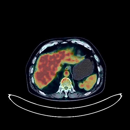
Lung Cancer PET/CT (case 983827-000077 from PETWB-REP)
2 views10 days agoWhole-body 18F-FDG PET/CT scan in a patient with Lung Cancer taken from the PETWB-REP dataset. The following English report (translated from original Chinese) is taken verbatim from the public dataset and has not been modified or otherwise checked for accuracy (see the end for citation). Impression a. Soft tissue nodules in the apical-posterior segment of the left upper lobe, with increased FDG metabolism, suggestive of peripheral lung cancer. Please confirm the diagnosis with pathological examination. b. Left hilar lymph node metastasis to be ruled out; reactive hyperplasia of right hilar and mediastinal lymph nodes. Please follow up on the above. c. Chronic inflammatory nodules and plaque-like foci (solid) in both lungs. Bilateral pulmonary fibrosis, emphysema, bullae. Bilateral pleural nodular thickening with calcification. Partial arteriosclerosis (including coronary arteries). Liver calcifications. Gallstones, chronic cholecystitis. Bilateral renal malrotation. Prostatic calcifications, with localized high FDG metabolism in the central zone, suggesting possible urinary retention. Please rule out space-occupying lesions with PSA and MRI. Bilateral hydrocele. Increased FDG metabolism in some intestinal segments, suggestive of physiological uptake or chronic inflammation. Please follow up with endoscopy. Spinal degenerative changes. L5/S1 intervertebral disc bulge. Bilateral deep lacunar infarcts, age-related brain abnormalities. Chronic inflammation of the right maxillary sinus. This case is from PETWB-REP, a curated dataset of whole-body 18F-FDG PET/CT scans and corresponding radiology reports from 490 patients with a broad spectrum of malignancies. The data were retrospectively collected from patients who underwent clinically indicated whole-body 18F-FDG PET/CT scans at the Shanghai Universal Medical Imaging Diagnostic Center between 2021 and 2024. License: Creative Commons Attribution 4.0 International (CC BY 4.0) Citation: Xue, L., Feng, G., Wenbo, Z., Zhang, Y., Li, L., Wang, S., Peng, L., Peng, S., & Gao, X. (2026). PETWB-REP: A Multi-Cancer Whole-Body FDG PET/CT Dataset with Corresponding Radiology Reports [Data set]. Zenodo. https://doi.org/10.5281/zenodo.18670487
Whole BodyPET/CT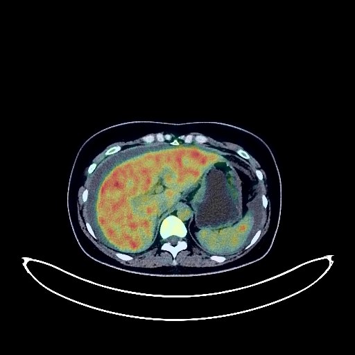
Ovarian Cancer PET/CT (case 983827-000223 from PETWB-REP)
2 views10 days agoWhole-body 18F-FDG PET/CT scan in a patient with Ovarian Cancer taken from the PETWB-REP dataset. The following English report (translated from original Chinese) is taken verbatim from the public dataset and has not been modified or otherwise checked for accuracy (see the end for citation). Impression a. A large, mixed-density mass in the pelvic cavity with increased FDG metabolism in the solid portion, highly suggestive of ovarian cancer; please confirm with pathology. b. Large amounts of fluid in the abdomen and pelvis. a. A small amount of inflammation in the lower lobe of the right lung is highly likely; anti-inflammatory treatment followed by a CT scan is recommended to rule out other possibilities. A few post-inflammatory lesions in both lungs. Localized bulging of the left diaphragm. b. Dense fibroadenomas in some parts of both breasts; a cystic mass in the lower part of the left breast, suggestive of a cyst. Please follow up with specialist examination and ultrasound. Gallbladder cholestasis or sludge-like stones. Mild hyperplasia of the left adrenal gland. Slight thickening of the gastric wall in the gastric body with mildly increased FDG metabolism, suggestive of chronic inflammatory changes; please confirm with gastroscopy. Osteophyte formation in some vertebral bodies of the cervical, thoracic, and lumbar vertebrae; slight calcification of the nuchal ligament. L4/5 and L5/S1 intervertebral disc herniation. No obvious abnormalities were found on cranial scintigraphy. This case is from PETWB-REP, a curated dataset of whole-body 18F-FDG PET/CT scans and corresponding radiology reports from 490 patients with a broad spectrum of malignancies. The data were retrospectively collected from patients who underwent clinically indicated whole-body 18F-FDG PET/CT scans at the Shanghai Universal Medical Imaging Diagnostic Center between 2021 and 2024. License: Creative Commons Attribution 4.0 International (CC BY 4.0) Citation: Xue, L., Feng, G., Wenbo, Z., Zhang, Y., Li, L., Wang, S., Peng, L., Peng, S., & Gao, X. (2026). PETWB-REP: A Multi-Cancer Whole-Body FDG PET/CT Dataset with Corresponding Radiology Reports [Data set]. Zenodo. https://doi.org/10.5281/zenodo.18670487
Whole BodyPET/CT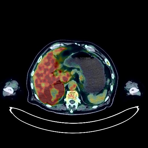
Lung Cancer PET/CT (case 983824-000064 from PETWB-REP)
2 views10 days agoWhole-body 18F-FDG PET/CT scan in a patient with Lung Cancer taken from the PETWB-REP dataset. The following English report (translated from original Chinese) is taken verbatim from the public dataset and has not been modified or otherwise checked for accuracy (see the end for citation). Impression a. After treatment for right lung cancer, an irregular soft tissue mass with increased FDG metabolism in the upper lobe of the right lung suggests that the tumor is still active. b. Multiple metastases in both lungs. Possible metastases to the right hilar and some mediastinal lymph nodes. c. Emphysema in both lungs. Chronic inflammation and post-inflammatory remnants in both lungs. Bilateral pleural thickening, with a small amount of pleural effusion on the right side. d. Pericardial thickening with effusion. Anemic changes, calcification of some arterial walls (including coronary arteries). a. A mass in the right kidney with increased FDG metabolism, suggestive of malignancy, with the metastasis larger than the primary tumor. b. Metastasis in the right lobe of the liver, with a metastatic lesion below the right kidney. Possible metastasis to the left hip. Cyst in the right lobe of the liver. Calcification in the pancreas. Left kidney stone, left adrenal hyperplasia. Benign prostatic hyperplasia with calcification. Bilateral inguinal hernia. Small amount of hydrocele in both testes. Chronic inflammatory changes in the gastric antrum and part of the intestine. Osteoporosis, degenerative changes in the spine, multiple intervertebral disc bulges. Left femoral head-neck cystic lesion. Low-density thyroid nodules and calcifications with increased FDG metabolism, suggestive of nodular goiter; please combine with ultrasound examination. Age-related brain, deep lacunar infarcts, left cerebellar softening. Right lens poorly visualized; please combine with specialist examination. This case is from PETWB-REP, a curated dataset of whole-body 18F-FDG PET/CT scans and corresponding radiology reports from 490 patients with a broad spectrum of malignancies. The data were retrospectively collected from patients who underwent clinically indicated whole-body 18F-FDG PET/CT scans at the Shanghai Universal Medical Imaging Diagnostic Center between 2021 and 2024. License: Creative Commons Attribution 4.0 International (CC BY 4.0) Citation: Xue, L., Feng, G., Wenbo, Z., Zhang, Y., Li, L., Wang, S., Peng, L., Peng, S., & Gao, X. (2026). PETWB-REP: A Multi-Cancer Whole-Body FDG PET/CT Dataset with Corresponding Radiology Reports [Data set]. Zenodo. https://doi.org/10.5281/zenodo.18670487
Whole BodyPET/CT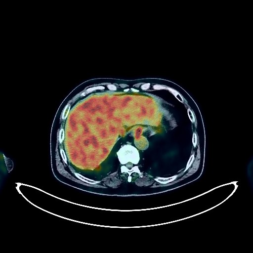
Renal Cancer PET/CT (case 983824-000073 from PETWB-REP)
2 views10 days agoWhole-body 18F-FDG PET/CT scan in a patient with Renal Cancer taken from the PETWB-REP dataset. The following English report (translated from original Chinese) is taken verbatim from the public dataset and has not been modified or otherwise checked for accuracy (see the end for citation). Impression a. Postoperative left renal cell carcinoma showed no signs of tumor recurrence in the surgical area. b. Postoperative thoracic spine lesion, after internal fixation of T5/6/8/9 vertebrae, a soft tissue mass with increased FDG metabolism at T7, which was larger than before, showed slightly decreased FDG metabolism, suggesting the tumor is still active. c. FDG metabolism of the left iliac wing metastasis was decreased compared to before, suggesting suppressed tumor activity. d. Bilateral inguinal lymph node metastases, liver metastases, and right acetabular bone metastases were all newly developed compared to before. Thyroid gland density was uneven, but FDG uptake was normal; ultrasound follow-up is recommended. a. Multiple chronic inflammatory nodules in both lungs, similar to previous findings. Scattered chronic inflammation and sequelae in both lungs. Slight thickening of the pleura bilaterally. Reactive hyperplasia of the hilar and mediastinal lymph nodes bilaterally. ? b. Bilateral gynecomastia. Reactive hyperplasia of the axillary lymph nodes bilaterally. Chronic gastritis, increased FDG metabolism in parts of the colon and rectum, considered physiological uptake or chronic inflammatory changes; please follow up with endoscopy. Prostatic calcification. Degenerative changes in the spine. L4/5 and L5/S1 intervertebral disc bulge. L5/S1 intervertebral disc pneumoconiosis. Bilateral frozen shoulder. Bilateral hip synovitis. No obvious abnormalities seen on cranial scintigraphy. Minor inflammation of the right sphenoid sinus and bilateral ethmoid sinuses. This case is from PETWB-REP, a curated dataset of whole-body 18F-FDG PET/CT scans and corresponding radiology reports from 490 patients with a broad spectrum of malignancies. The data were retrospectively collected from patients who underwent clinically indicated whole-body 18F-FDG PET/CT scans at the Shanghai Universal Medical Imaging Diagnostic Center between 2021 and 2024. License: Creative Commons Attribution 4.0 International (CC BY 4.0) Citation: Xue, L., Feng, G., Wenbo, Z., Zhang, Y., Li, L., Wang, S., Peng, L., Peng, S., & Gao, X. (2026). PETWB-REP: A Multi-Cancer Whole-Body FDG PET/CT Dataset with Corresponding Radiology Reports [Data set]. Zenodo. https://doi.org/10.5281/zenodo.18670487
Whole BodyPET/CT