Loading...
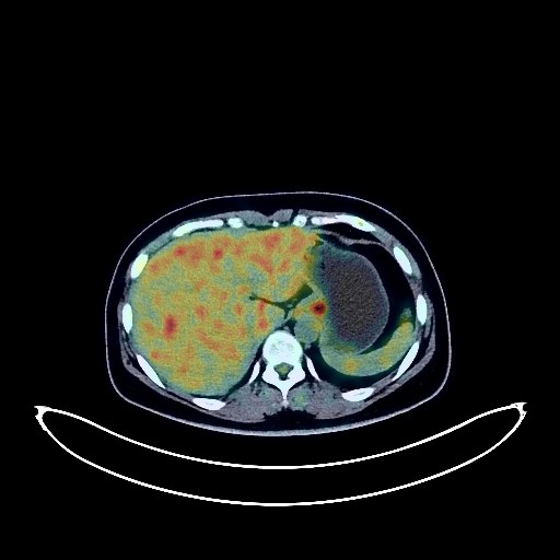
Cervical Cancer PET/CT (case 983827-000153 from PETWB-REP)
0 views10 days agoWhole-body 18F-FDG PET/CT scan in a patient with Cervical Cancer taken from the PETWB-REP dataset. The following English report (translated from original Chinese) is taken verbatim from the public dataset and has not been modified or otherwise checked for accuracy (see the end for citation). Impression a. Cervical mass with increased FDG metabolism, consistent with cervical cancer, involving the lower segment of the cervix; reactive hyperplasia of bilateral iliac vessels and bilateral inguinal lymph nodes. b. Physiological uptake in the uterine cavity and left ovary. Chronic inflammatory micronodules in both lungs. Calcification in the lower lobe of the right lung, and a few post-inflammatory remnants in the upper lobe of the left lung. Anemia. Calcification in the right lobe of the liver, and a cyst in the left lobe of the liver. Chronic inflammatory changes in the cardia and antrum of the stomach, and hemorrhoidal changes; please follow up with endoscopy. Degenerative changes in the spine, with L4/5 and L5/S1 intervertebral disc bulges. Inflammation of the right hip joint space. A low-density nodule with elevated FDG metabolism in the right lobe of the thyroid gland is suggestive of an adenoma; please confirm with ultrasound examination. Cranial scintigraphy showed no obvious abnormalities. Minor chronic inflammation of both ethmoid sinuses. Bilateral palatine tonsillitis. This case is from PETWB-REP, a curated dataset of whole-body 18F-FDG PET/CT scans and corresponding radiology reports from 490 patients with a broad spectrum of malignancies. The data were retrospectively collected from patients who underwent clinically indicated whole-body 18F-FDG PET/CT scans at the Shanghai Universal Medical Imaging Diagnostic Center between 2021 and 2024. License: Creative Commons Attribution 4.0 International (CC BY 4.0) Citation: Xue, L., Feng, G., Wenbo, Z., Zhang, Y., Li, L., Wang, S., Peng, L., Peng, S., & Gao, X. (2026). PETWB-REP: A Multi-Cancer Whole-Body FDG PET/CT Dataset with Corresponding Radiology Reports [Data set]. Zenodo. https://doi.org/10.5281/zenodo.18670487
Whole BodyPET/CT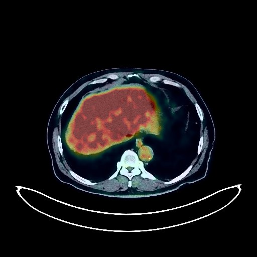
Colon Cancer PET/CT (case 983827-000262 from PETWB-REP)
0 views10 days agoWhole-body 18F-FDG PET/CT scan in a patient with Colon Cancer taken from the PETWB-REP dataset. The following English report (translated from original Chinese) is taken verbatim from the public dataset and has not been modified or otherwise checked for accuracy (see the end for citation). Impression After treatment for colorectal cancer, no obvious space-occupying lesions were found in the intestinal tract, and FDG metabolism was normal, suggesting suppressed tumor activity. Colonoscopy follow-up is recommended. Post-hemorrhoidectomy changes. Chronic inflammatory micronodules in both lungs are highly probable; CT follow-up is recommended to rule out other possibilities. A few post-inflammatory lesions in both lungs. Reactive hyperplasia of the right hilar and mediastinal lymph nodes. Calcification of some arterial walls (including coronary arteries). Calcification in the right lobe of the liver. Benign prostatic hyperplasia with calcification. Small amount of hydrocele in the left testis. Possible reactive hyperplasia of retroperitoneal lymph nodes. Possible chronic inflammatory changes in the gastric wall; please follow up with endoscopy. Osteoporosis, degenerative changes in the spine, L4/5 disc herniation. Inflammation of the left shoulder and left hip. A low-density nodule in the right lobe of the thyroid gland with elevated FDG metabolism suggests a possible adenoma; please have a follow-up ultrasound examination. A lacunar infarct in the deep brain region of an elderly patient; please have an MRI examination. Chronic inflammation of both maxillary sinuses. This case is from PETWB-REP, a curated dataset of whole-body 18F-FDG PET/CT scans and corresponding radiology reports from 490 patients with a broad spectrum of malignancies. The data were retrospectively collected from patients who underwent clinically indicated whole-body 18F-FDG PET/CT scans at the Shanghai Universal Medical Imaging Diagnostic Center between 2021 and 2024. License: Creative Commons Attribution 4.0 International (CC BY 4.0) Citation: Xue, L., Feng, G., Wenbo, Z., Zhang, Y., Li, L., Wang, S., Peng, L., Peng, S., & Gao, X. (2026). PETWB-REP: A Multi-Cancer Whole-Body FDG PET/CT Dataset with Corresponding Radiology Reports [Data set]. Zenodo. https://doi.org/10.5281/zenodo.18670487
Whole BodyPET/CT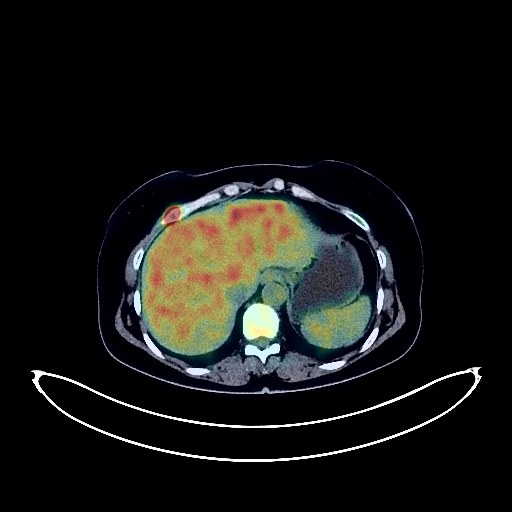
Cervical Cancer PET/CT (case 983827-000146 from PETWB-REP)
0 views10 days agoWhole-body 18F-FDG PET/CT scan in a patient with Cervical Cancer taken from the PETWB-REP dataset. The following English report (translated from original Chinese) is taken verbatim from the public dataset and has not been modified or otherwise checked for accuracy (see the end for citation). Impression a. Cervical mass with elevated FDG metabolism, consistent with cervical cancer; bilateral iliac lymph node metastasis. Uterine cavity effusion, uterine fibroids. b. Hypermetabolic lesions in the right 6th anterior rib and L3 vertebral body, metastasis to be ruled out; please confirm with MRI. a. Ground-glass opacity in the medial segment of the right middle lobe, FDG metabolism normal, suggest inflammation or atypical adenomatous hyperplasia; CT follow-up recommended. b. Chronic inflammatory micronodule (solid) in the right upper lobe; CT follow-up recommended. A few post-inflammatory lesions in both lungs. Reactive hyperplasia of the left hilar lymph nodes. Anemia changes, slight arteriosclerosis in some arteries. Small cyst in the left kidney. Chronic inflammatory changes in the gastric antrum; please confirm with endoscopy. Degenerative changes in the spine, L5/S1 disc bulge. T3 vertebral hemangioma. No obvious abnormalities were found on cranial scintigraphy. Chronic inflammation of the left maxillary sinus. Reactive hyperplasia of bilateral cervical lymph nodes. This case is from PETWB-REP, a curated dataset of whole-body 18F-FDG PET/CT scans and corresponding radiology reports from 490 patients with a broad spectrum of malignancies. The data were retrospectively collected from patients who underwent clinically indicated whole-body 18F-FDG PET/CT scans at the Shanghai Universal Medical Imaging Diagnostic Center between 2021 and 2024. License: Creative Commons Attribution 4.0 International (CC BY 4.0) Citation: Xue, L., Feng, G., Wenbo, Z., Zhang, Y., Li, L., Wang, S., Peng, L., Peng, S., & Gao, X. (2026). PETWB-REP: A Multi-Cancer Whole-Body FDG PET/CT Dataset with Corresponding Radiology Reports [Data set]. Zenodo. https://doi.org/10.5281/zenodo.18670487
Whole BodyPET/CT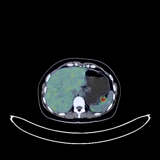
Breast Cancer PET/CT (case 983827-000230 from PETWB-REP)
0 views10 days agoWhole-body 18F-FDG PET/CT scan in a patient with Breast Cancer taken from the PETWB-REP dataset. The following English report (translated from original Chinese) is taken verbatim from the public dataset and has not been modified or otherwise checked for accuracy (see the end for citation). Impression a. Multiple lesions in the left breast with increased FDG metabolism, consistent with breast cancer; reactive hyperplasia of bilateral axillary lymph nodes is possible, follow-up is recommended. b. Bilateral breast hyperplasia, calcification in the left breast, ultrasound follow-up is recommended. Cystic-solid mass in the right adnexal region, with increased FDG metabolism in the solid portion; solid nodule in the left adnexal region with increased FDG metabolism, both considered malignant tumors, most likely ovarian origin, metastasis to be ruled out, please combine clinical and enhanced MRI analysis; extensive peritoneal seeding metastasis, small amount of effusion in the abdominopelvic cavity. Calcification in the right middle lobe of the lung. Mild anemia changes. Possible right lobe hepatic cyst. Gallbladder bile concentration. Intrauterine device insertion. Possible chronic inflammatory changes in the cardia and antrum of the stomach, please combine endoscopic follow-up. No obvious abnormalities were found on cranial scintigraphy. This case is from PETWB-REP, a curated dataset of whole-body 18F-FDG PET/CT scans and corresponding radiology reports from 490 patients with a broad spectrum of malignancies. The data were retrospectively collected from patients who underwent clinically indicated whole-body 18F-FDG PET/CT scans at the Shanghai Universal Medical Imaging Diagnostic Center between 2021 and 2024. License: Creative Commons Attribution 4.0 International (CC BY 4.0) Citation: Xue, L., Feng, G., Wenbo, Z., Zhang, Y., Li, L., Wang, S., Peng, L., Peng, S., & Gao, X. (2026). PETWB-REP: A Multi-Cancer Whole-Body FDG PET/CT Dataset with Corresponding Radiology Reports [Data set]. Zenodo. https://doi.org/10.5281/zenodo.18670487
Whole BodyPET/CT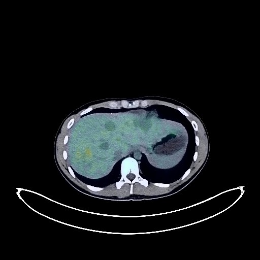
Gastric Cancer PET/CT (case 983827-000127 from PETWB-REP)
0 views10 days agoWhole-body 18F-FDG PET/CT scan in a patient with Gastric Cancer taken from the PETWB-REP dataset. The following English report (translated from original Chinese) is taken verbatim from the public dataset and has not been modified or otherwise checked for accuracy (see the end for citation). Impression After treatment for gastric cancer with multiple metastases, the activity of most tumors was suppressed, while some tumors retained activity, specifically as follows: a. Extensive and uneven thickening of the gastric body and wall, with decreased FDG metabolism and a smaller affected area compared to before. b. Significantly smaller lymph nodes in the left supraclavicular fossa and left upper thoracic inlet, with significantly decreased FDG metabolism (still above background levels). Some of the previously enlarged thoracic and abdominal lymph nodes disappeared, and some significantly decreased in size, with FDG metabolism reduced to background levels. c. Slight peritoneal thickening with omental soft tissue nodules in multiple areas, significantly smaller in area than before, with significantly reduced FDG metabolism to background levels. d. The volume and density of multiple liver lesions have decreased compared to before, and FDG metabolism has decreased (most to background levels, a few still above the liver background level). e. The bilateral adrenal glands have slightly thickened after treatment, and FDG metabolism has significantly decreased compared to before. f. The previously focal FDG metabolism increase in multiple bones was not clearly visible in this examination. The previously right-sided FDG metabolism increase in the spinal canal at the T5 vertebral level was not clearly visible in this examination. The gastric wall at the cardia is thickened with increased FDG metabolism; combined with the previous gastroscopy and PET/CT scan, this is considered to be physiological or inflammatory uptake. Physiological uptake in part of the intestine. Small amount of pelvic effusion. The previously solid miliary nodule in the right lung was not clearly visible in this examination. Anemia is present. Physiological or inflammatory uptake in the nasopharynx, base of the tongue, and bilateral palatine tonsils. The previously observed low-density lesion in the head of the left caudate nucleus was not clearly visualized this time; please follow up with MRI. This case is from PETWB-REP, a curated dataset of whole-body 18F-FDG PET/CT scans and corresponding radiology reports from 490 patients with a broad spectrum of malignancies. The data were retrospectively collected from patients who underwent clinically indicated whole-body 18F-FDG PET/CT scans at the Shanghai Universal Medical Imaging Diagnostic Center between 2021 and 2024. License: Creative Commons Attribution 4.0 International (CC BY 4.0) Citation: Xue, L., Feng, G., Wenbo, Z., Zhang, Y., Li, L., Wang, S., Peng, L., Peng, S., & Gao, X. (2026). PETWB-REP: A Multi-Cancer Whole-Body FDG PET/CT Dataset with Corresponding Radiology Reports [Data set]. Zenodo. https://doi.org/10.5281/zenodo.18670487
Whole BodyPET/CT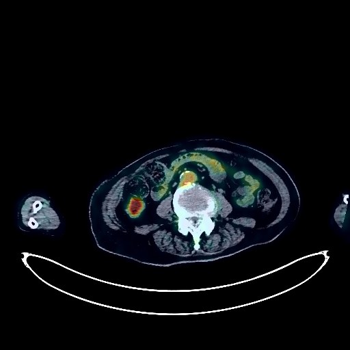
Lymphoma PET/CT (case 983827-000101 from PETWB-REP)
0 views10 days agoWhole-body 18F-FDG PET/CT scan in a patient with Lymphoma taken from the PETWB-REP dataset. The following English report (translated from original Chinese) is taken verbatim from the public dataset and has not been modified or otherwise checked for accuracy (see the end for citation). Impression Post-lymphoma treatment: a. Thickened gastric antrum wall, with increased FDG metabolism in some gastric walls, suggesting post-treatment changes. Some tumor activity may still exist; please correlate with clinical findings and conduct follow-up examinations. b. Multiple lymph nodes are visible in the bilateral neck, hilum, mediastinum, retroperitoneum, mesentery, and bilateral groins. Increased FDG metabolism in the hilar lymph nodes suggests reactive lymph node hyperplasia or post-treatment changes; please compare with previous data and conduct follow-up examinations. a. Interstitial lung changes with chronic inflammation and sequelae, chronic inflammatory nodules and plaque nodules in both lungs; please follow up with CT scans. Bilateral breast hyperplasia with calcification. b. Post-coronary artery stenting, partial arteriosclerosis (including coronary arteries), and ascending aortic dilation; please conduct specialist examinations. Liver cysts, possible left lobe hemangioma; MRI examination if necessary. Gallstones, chronic cholecystitis. Left renal cyst. Left adrenal hyperplasia. Cystic mass in the tail of the pancreas, FDG metabolism normal, cystadenoma to be ruled out, enhanced MRI recommended. Increased FDG metabolism in some intestinal segments, considered physiological uptake or chronic inflammation, please follow up with endoscopy. a. Scoliosis with degenerative changes. Old compression fractures of the T11, T12, and L4 vertebrae. L4/5 and L5/S1 intervertebral disc bulges. b. Post-internal fixation of the right upper femoral fracture, changes following fractures of the right 7th, 8th, and 10th ribs and the left 10th rib. Bilateral deep lacunar infarcts, white matter degeneration, senile encephalopathy. Left maxillary sinusitis. This case is from PETWB-REP, a curated dataset of whole-body 18F-FDG PET/CT scans and corresponding radiology reports from 490 patients with a broad spectrum of malignancies. The data were retrospectively collected from patients who underwent clinically indicated whole-body 18F-FDG PET/CT scans at the Shanghai Universal Medical Imaging Diagnostic Center between 2021 and 2024. License: Creative Commons Attribution 4.0 International (CC BY 4.0) Citation: Xue, L., Feng, G., Wenbo, Z., Zhang, Y., Li, L., Wang, S., Peng, L., Peng, S., & Gao, X. (2026). PETWB-REP: A Multi-Cancer Whole-Body FDG PET/CT Dataset with Corresponding Radiology Reports [Data set]. Zenodo. https://doi.org/10.5281/zenodo.18670487
Whole BodyPET/CT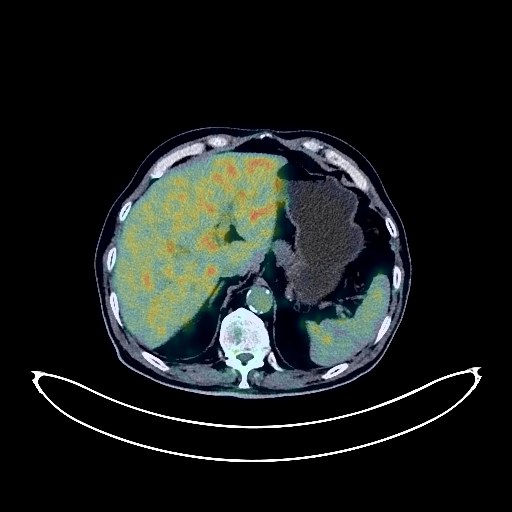
Prostate Cancer PET/CT (case 983827-000010 from PETWB-REP)
0 views10 days agoWhole-body 18F-FDG PET/CT scan in a patient with Prostate Cancer taken from the PETWB-REP dataset. The following English report (translated from original Chinese) is taken verbatim from the public dataset and has not been modified or otherwise checked for accuracy (see the end for citation). Impression a. Benign prostatic hyperplasia with calcification, prostatic mass with increased FDG metabolism, consistent with prostate cancer presentation, invading the left seminal vesicle and bladder; multiple metastatic lesions in the surrounding fat space, pelvic floor, mesentery, and bilateral iliac vessels. b. Left scapular metastasis is highly probable; left iliac bone metastasis is pending, right iliac bone island is highly probable, please confirm with contrast-enhanced MRI. Bilateral chronic inflammatory nodules are highly probable, CT follow-up is recommended. Left upper lobe contains air sac cavity. A few post-inflammatory remnants in both lungs. Reactive hyperplasia of hilar and mediastinal lymph nodes in both lungs. Calcification of some arterial walls (including coronary arteries). Gallstones. Accessory spleen. Calcification lesion in the pancreatic tail. Bilateral kidney stones. Calcification lesion in the right testicular tunica vaginalis. Left inguinal hernia. Chronic inflammatory changes in the lower esophagus and gastric antrum, please confirm with endoscopy. Osteoporosis, degenerative changes in the spine, multiple intervertebral disc bulges, and T11 vertebral body wedging. Age-related brain abnormalities, deep lacunar infarcts, and white matter degeneration; please correlate with MRI. Chronic inflammation of the left maxillary sinus. High-density nodule below the left eyeball; please correlate with clinical findings. This case is from PETWB-REP, a curated dataset of whole-body 18F-FDG PET/CT scans and corresponding radiology reports from 490 patients with a broad spectrum of malignancies. The data were retrospectively collected from patients who underwent clinically indicated whole-body 18F-FDG PET/CT scans at the Shanghai Universal Medical Imaging Diagnostic Center between 2021 and 2024. License: Creative Commons Attribution 4.0 International (CC BY 4.0) Citation: Xue, L., Feng, G., Wenbo, Z., Zhang, Y., Li, L., Wang, S., Peng, L., Peng, S., & Gao, X. (2026). PETWB-REP: A Multi-Cancer Whole-Body FDG PET/CT Dataset with Corresponding Radiology Reports [Data set]. Zenodo. https://doi.org/10.5281/zenodo.18670487
Whole BodyPET/CT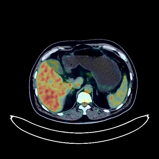
Renal Cancer PET/CT (case 983827-000112 from PETWB-REP)
0 views10 days agoWhole-body 18F-FDG PET/CT scan in a patient with Renal Cancer taken from the PETWB-REP dataset. The following English report (translated from original Chinese) is taken verbatim from the public dataset and has not been modified or otherwise checked for accuracy (see the end for citation). Impression a. A mass in the lower pole of the left kidney involving the renal calyces and pelvis, with FDG showing background metabolism. Combined with contrast-enhanced CT images from another hospital, this is considered a malignant tumor, most likely renal cell carcinoma. Please confirm with pathology. b. Reactive hyperplasia of small retroperitoneal lymph nodes. Slight thickening of the perirenal fascia bilaterally. c. Bilateral renal cysts, right renal calculus. A few post-inflammatory lesions in both lungs. Liver cysts. Accessory splenic nodules. Benign prostatic hyperplasia with calcification. Increased FDG metabolism in parts of the stomach wall and intestines, likely due to chronic inflammation. Please follow up with endoscopy. Hemorrhoidal changes, please correlate with clinical findings. Spinal degenerative changes. L4/5 disc bulge, L5/S1 disc herniation. Cranial scintigraphy showed no obvious abnormalities. Bilateral maxillary sinusitis and ethmoid sinusitis. Bilateral chronic palatine tonsillitis. This case is from PETWB-REP, a curated dataset of whole-body 18F-FDG PET/CT scans and corresponding radiology reports from 490 patients with a broad spectrum of malignancies. The data were retrospectively collected from patients who underwent clinically indicated whole-body 18F-FDG PET/CT scans at the Shanghai Universal Medical Imaging Diagnostic Center between 2021 and 2024. License: Creative Commons Attribution 4.0 International (CC BY 4.0) Citation: Xue, L., Feng, G., Wenbo, Z., Zhang, Y., Li, L., Wang, S., Peng, L., Peng, S., & Gao, X. (2026). PETWB-REP: A Multi-Cancer Whole-Body FDG PET/CT Dataset with Corresponding Radiology Reports [Data set]. Zenodo. https://doi.org/10.5281/zenodo.18670487
Whole BodyPET/CT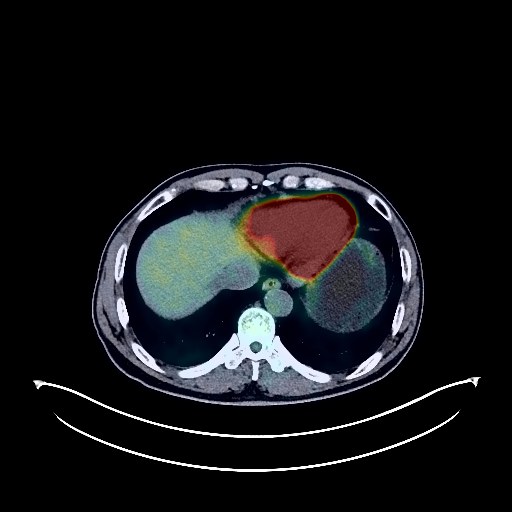
Lung Cancer PET/CT (case 983827-000152 from PETWB-REP)
0 views10 days agoWhole-body 18F-FDG PET/CT scan in a patient with Lung Cancer taken from the PETWB-REP dataset. The following English report (translated from original Chinese) is taken verbatim from the public dataset and has not been modified or otherwise checked for accuracy (see the end for citation). Impression a. A mass in the posterior segment of the right upper lobe, with increased FDG metabolism, suggestive of lung cancer, accompanied by distal obstructive changes. Possible right hilar lymph node metastasis. b. Scattered inflammation and remnants in both lungs; CT follow-up is recommended after anti-inflammatory treatment. Emphysema, bullae. Calcification of some arterial walls (including coronary arteries). Slight thickening of part of the gastric body wall, with increased FDG metabolism, suggestive of gastritis; gastroscopy is recommended to rule out other possibilities. A low-density nodule in the left inner lobe of the liver, with normal FDG metabolism, suggestive of hemangioma; contrast-enhanced MRI is recommended. Liver cyst. Increased FDG metabolism in part of the intestinal tract, likely due to inflammatory uptake; hemorrhoidal changes. Degenerative changes in the spine; lumbar vertebral instability, mild wedging of the L1 and L2 vertebral bodies. L4/5 and L5/S1 intervertebral disc bulges; L1-3 intervertebral disc pneumoconiosis and degeneration. Post-fracture changes of the left 5th and 6th ribs. Cranial scintigraphy showed no abnormalities. Bilateral submandibular gland physiological uptake is highly probable. This case is from PETWB-REP, a curated dataset of whole-body 18F-FDG PET/CT scans and corresponding radiology reports from 490 patients with a broad spectrum of malignancies. The data were retrospectively collected from patients who underwent clinically indicated whole-body 18F-FDG PET/CT scans at the Shanghai Universal Medical Imaging Diagnostic Center between 2021 and 2024. License: Creative Commons Attribution 4.0 International (CC BY 4.0) Citation: Xue, L., Feng, G., Wenbo, Z., Zhang, Y., Li, L., Wang, S., Peng, L., Peng, S., & Gao, X. (2026). PETWB-REP: A Multi-Cancer Whole-Body FDG PET/CT Dataset with Corresponding Radiology Reports [Data set]. Zenodo. https://doi.org/10.5281/zenodo.18670487
Whole BodyPET/CT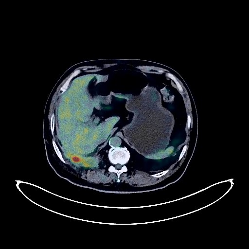
Lung Cancer PET/CT (case 983827-000034 from PETWB-REP)
0 views10 days agoWhole-body 18F-FDG PET/CT scan in a patient with Lung Cancer taken from the PETWB-REP dataset. The following English report (translated from original Chinese) is taken verbatim from the public dataset and has not been modified or otherwise checked for accuracy (see the end for citation). Impression a. Postoperative right lower lobe lung cancer, postoperative changes in the right chest wall, no signs of tumor recurrence in the surgical area. b. Right upper lobe bronchus is patent, no obvious space-occupying lesion, FDG metabolism is normal. c. Right upper lobe infection, anti-inflammatory treatment and follow-up examination are recommended to rule out other causes. Scattered post-inflammatory lesions in both lungs. d. Right pleural effusion and pneumothorax; right pleural thickening with possible inflammatory uptake, pleural metastasis to be ruled out, close observation is recommended. e. Chronic inflammatory lymph nodes in the right hilum and mediastinum. Slightly enlarged cardiac silhouette. Partial arteriosclerosis, linear high-density shadows seen in the coronary artery course. Chronic inflammatory changes in the cardia, antrum of the stomach and part of the intestine, hemorrhoids are possible, please follow up with endoscopy. Right inguinal hernia is possible. Left testis not visualized. Degenerative changes in the spine, L4/5 and L5/S1 intervertebral disc bulge. In elderly patients with lacunar infarcts deep within the brain, please combine with MRI examination. This case is from PETWB-REP, a curated dataset of whole-body 18F-FDG PET/CT scans and corresponding radiology reports from 490 patients with a broad spectrum of malignancies. The data were retrospectively collected from patients who underwent clinically indicated whole-body 18F-FDG PET/CT scans at the Shanghai Universal Medical Imaging Diagnostic Center between 2021 and 2024. License: Creative Commons Attribution 4.0 International (CC BY 4.0) Citation: Xue, L., Feng, G., Wenbo, Z., Zhang, Y., Li, L., Wang, S., Peng, L., Peng, S., & Gao, X. (2026). PETWB-REP: A Multi-Cancer Whole-Body FDG PET/CT Dataset with Corresponding Radiology Reports [Data set]. Zenodo. https://doi.org/10.5281/zenodo.18670487
Whole BodyPET/CT