Loading...
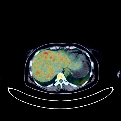
Lung Cancer PET/CT (case 983827-000048 from PETWB-REP)
0 views10 days agoWhole-body 18F-FDG PET/CT scan in a patient with Lung Cancer taken from the PETWB-REP dataset. The following English report (translated from original Chinese) is taken verbatim from the public dataset and has not been modified or otherwise checked for accuracy (see the end for citation). Impression a. A mass in the posterior segment of the left lower lobe with increased FDG uptake, suggestive of lung cancer. An irregular nodular lesion in the posterior basal segment of the left lower lobe with increased FDG metabolism, suggestive of possible lung cancer. b. Multiple metastases in both lungs. Multiple lymph node metastases in the left hilum, mediastinum, bilateral supraclavicular fossa, bilateral neck, and retroperitoneum. c. Metastasis in the left frontal lobe. Metastases in the T7 and L5 adnexa. Bilateral adrenal metastases. Chronic inflammation and post-inflammatory remnants in both lungs. Left pleural thickening, pericardial thickening with effusion. Minor arteriosclerosis in some arteries. A low-density mass in the left adnexal region with locally increased FDG metabolism at the periphery, suggestive of ovarian neoplastic lesions or cysts; please combine with enhanced MRI for comprehensive analysis. Nabothian cysts of the cervix. Low-density nodules in both lobes of the thyroid gland, with calcification on the right side and increased FDG uptake in some areas, suggestive of possible nodular goiter; malignancy in some areas needs to be ruled out; ultrasound follow-up is recommended. Chronic inflammatory changes in the cardia and antrum of the stomach. Mild osteophyte formation in the spine, with a bony island in the left iliac bone. Chronic inflammation of both ethmoid and maxillary sinuses. This case is from PETWB-REP, a curated dataset of whole-body 18F-FDG PET/CT scans and corresponding radiology reports from 490 patients with a broad spectrum of malignancies. The data were retrospectively collected from patients who underwent clinically indicated whole-body 18F-FDG PET/CT scans at the Shanghai Universal Medical Imaging Diagnostic Center between 2021 and 2024. License: Creative Commons Attribution 4.0 International (CC BY 4.0) Citation: Xue, L., Feng, G., Wenbo, Z., Zhang, Y., Li, L., Wang, S., Peng, L., Peng, S., & Gao, X. (2026). PETWB-REP: A Multi-Cancer Whole-Body FDG PET/CT Dataset with Corresponding Radiology Reports [Data set]. Zenodo. https://doi.org/10.5281/zenodo.18670487
Whole BodyPET/CT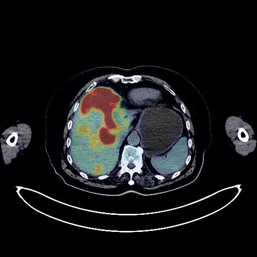
Liver Cancer PET/CT (case 983827-000122 from PETWB-REP)
0 views10 days agoWhole-body 18F-FDG PET/CT scan in a patient with Liver Cancer taken from the PETWB-REP dataset. The following English report (translated from original Chinese) is taken verbatim from the public dataset and has not been modified or otherwise checked for accuracy (see the end for citation). Impression a. Diffuse mass in the liver, elevated FDG metabolism, suggestive of malignancy, possibly liver cancer with intrahepatic metastasis; metastatic tumors cannot be ruled out. Please correlate with clinical findings and tumor markers. b. Slightly enlarged lymph nodes beside the right aorta, mildly elevated FDG metabolism, suggestive of reactive lymph node hyperplasia; metastasis to be ruled out. Several small chronic inflammatory nodules in the right lung. Scattered chronic inflammation and old lesions in both lungs. Tracheal diverticulum. Enlarged cardiac silhouette. Calcification of some arterial walls (including coronary arteries). Left renal cyst. Uterus in elderly patients. Degenerative changes in the spine. L5/S1 vertebral endplate inflammation. Osteoporosis. L4/5, L5/S1 intervertebral disc bulge. A few ischemic foci in the deep cerebral regions bilaterally; age-related brain. Chronic inflammation of the bilateral maxillary sinuses. This case is from PETWB-REP, a curated dataset of whole-body 18F-FDG PET/CT scans and corresponding radiology reports from 490 patients with a broad spectrum of malignancies. The data were retrospectively collected from patients who underwent clinically indicated whole-body 18F-FDG PET/CT scans at the Shanghai Universal Medical Imaging Diagnostic Center between 2021 and 2024. License: Creative Commons Attribution 4.0 International (CC BY 4.0) Citation: Xue, L., Feng, G., Wenbo, Z., Zhang, Y., Li, L., Wang, S., Peng, L., Peng, S., & Gao, X. (2026). PETWB-REP: A Multi-Cancer Whole-Body FDG PET/CT Dataset with Corresponding Radiology Reports [Data set]. Zenodo. https://doi.org/10.5281/zenodo.18670487
Whole BodyPET/CT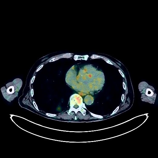
Lung Cancer PET/CT (case 983827-000224 from PETWB-REP)
0 views10 days agoWhole-body 18F-FDG PET/CT scan in a patient with Lung Cancer taken from the PETWB-REP dataset. The following English report (translated from original Chinese) is taken verbatim from the public dataset and has not been modified or otherwise checked for accuracy (see the end for citation). Impression a. Masses in the apical-posterior segment of the left upper lobe and the posterior-basal segment of the right lower lobe with increased FDG metabolism, highly suggestive of lung cancer; please confirm with pathology. b. Multiple solid nodules in both lungs, FDG uptake normal, suggestive of chronic inflammatory nodules; please follow up with CT to rule out other confounding nodules. Scattered chronic inflammation and remnants in both lungs. Emphysema and bullae in both lungs. c. Reactive hyperplasia of hilar and mediastinal lymph nodes in both lungs. Calcification of some arterial walls (including coronary arteries). Postoperative rectal cancer surgery, no obvious signs of tumor recurrence in the surgical area. Cyst in the right lobe of the liver. Bilateral hydrocele. Localized enlargement of the abdominal aorta, suggestive of aneurysm; CTA follow-up is recommended. Partial vertebral osteophyte formation. L3/4, L4/5, L5/S1 intervertebral disc bulge. Age-related brain changes. This case is from PETWB-REP, a curated dataset of whole-body 18F-FDG PET/CT scans and corresponding radiology reports from 490 patients with a broad spectrum of malignancies. The data were retrospectively collected from patients who underwent clinically indicated whole-body 18F-FDG PET/CT scans at the Shanghai Universal Medical Imaging Diagnostic Center between 2021 and 2024. License: Creative Commons Attribution 4.0 International (CC BY 4.0) Citation: Xue, L., Feng, G., Wenbo, Z., Zhang, Y., Li, L., Wang, S., Peng, L., Peng, S., & Gao, X. (2026). PETWB-REP: A Multi-Cancer Whole-Body FDG PET/CT Dataset with Corresponding Radiology Reports [Data set]. Zenodo. https://doi.org/10.5281/zenodo.18670487
Whole BodyPET/CT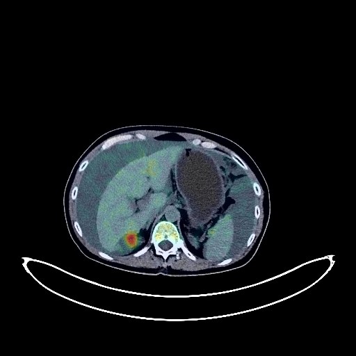
Ovarian Cancer PET/CT (case 983827-000189 from PETWB-REP)
0 views10 days agoWhole-body 18F-FDG PET/CT scan in a patient with Ovarian Cancer taken from the PETWB-REP dataset. The following English report (translated from original Chinese) is taken verbatim from the public dataset and has not been modified or otherwise checked for accuracy (see the end for citation). Impression Bilateral adnexal lesions with elevated FDG metabolism, suggestive of malignancy, most likely ovarian cancer; please correlate with clinicopathology. Extensive peritoneal and pelvic metastases. Significant abdominal and pelvic effusion. Scattered chronic inflammatory micronodules in the left lower lobe and right middle lobe, calcification in the right upper lobe. Small amount of bilateral pleural effusion. Calcification of some arterial walls. Bilateral breast proliferative changes; specialist and ultrasound examination recommended. Mild osteophyte formation in some vertebrae. No obvious abnormalities on cranial FDG scintigraphy. Minor chronic inflammation of the left maxillary sinus. This case is from PETWB-REP, a curated dataset of whole-body 18F-FDG PET/CT scans and corresponding radiology reports from 490 patients with a broad spectrum of malignancies. The data were retrospectively collected from patients who underwent clinically indicated whole-body 18F-FDG PET/CT scans at the Shanghai Universal Medical Imaging Diagnostic Center between 2021 and 2024. License: Creative Commons Attribution 4.0 International (CC BY 4.0) Citation: Xue, L., Feng, G., Wenbo, Z., Zhang, Y., Li, L., Wang, S., Peng, L., Peng, S., & Gao, X. (2026). PETWB-REP: A Multi-Cancer Whole-Body FDG PET/CT Dataset with Corresponding Radiology Reports [Data set]. Zenodo. https://doi.org/10.5281/zenodo.18670487
Whole BodyPET/CT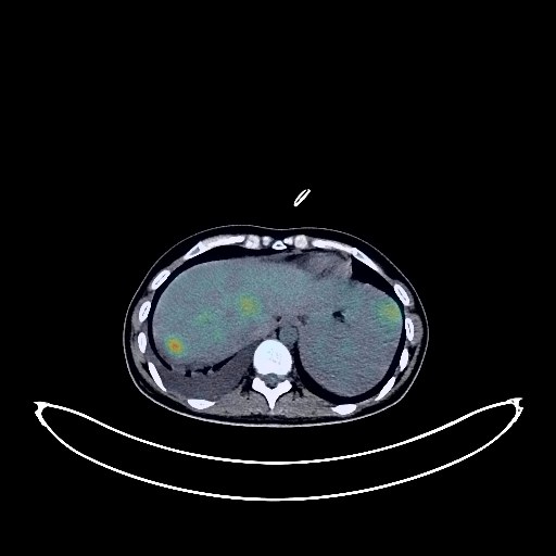
Liver Cancer PET/CT (case 983827-000118 from PETWB-REP)
0 views10 days agoWhole-body 18F-FDG PET/CT scan in a patient with Liver Cancer taken from the PETWB-REP dataset. The following English report (translated from original Chinese) is taken verbatim from the public dataset and has not been modified or otherwise checked for accuracy (see the end for citation). Impression a. Postoperative changes in the abdominal wall after liver cancer surgery; multiple space-occupying lesions in the liver with increased FDG metabolism, suggestive of tumor recurrence and metastasis, portal vein tumor thrombus formation. b. Peritoneal seeding metastasis, abdominopelvic effusion; multiple lymph node metastases in the hepatic hilum and retroperitoneum. c. Multiple lung metastases, right pleural metastasis, right pleural effusion. d. Multiple bone metastases throughout the body (see description for details). Chronic inflammation in the lower lobe of the right lung. Anemia. Small kidney stone in the right kidney. Chronic inflammatory changes or physiological uptake in some intestinal segments; please follow up with endoscopy. No obvious abnormalities were found on cranial scintigraphy. Chronic inflammation of the right maxillary sinus. Large inflammatory lymph nodes in the right deep cervical space. This case is from PETWB-REP, a curated dataset of whole-body 18F-FDG PET/CT scans and corresponding radiology reports from 490 patients with a broad spectrum of malignancies. The data were retrospectively collected from patients who underwent clinically indicated whole-body 18F-FDG PET/CT scans at the Shanghai Universal Medical Imaging Diagnostic Center between 2021 and 2024. License: Creative Commons Attribution 4.0 International (CC BY 4.0) Citation: Xue, L., Feng, G., Wenbo, Z., Zhang, Y., Li, L., Wang, S., Peng, L., Peng, S., & Gao, X. (2026). PETWB-REP: A Multi-Cancer Whole-Body FDG PET/CT Dataset with Corresponding Radiology Reports [Data set]. Zenodo. https://doi.org/10.5281/zenodo.18670487
Whole BodyPET/CT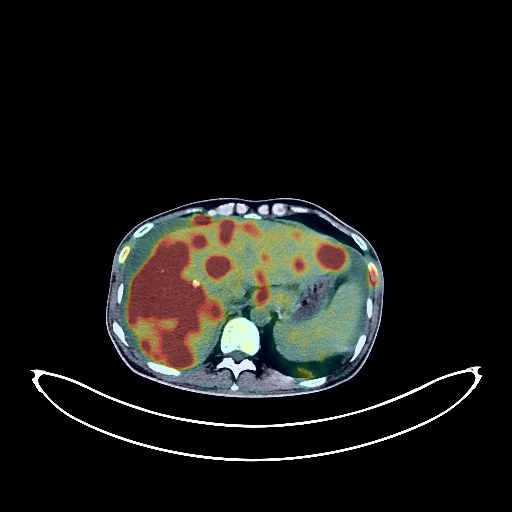
Liver Cancer PET/CT (case 983827-000114 from PETWB-REP)
0 views10 days agoWhole-body 18F-FDG PET/CT scan in a patient with Liver Cancer taken from the PETWB-REP dataset. The following English report (translated from original Chinese) is taken verbatim from the public dataset and has not been modified or otherwise checked for accuracy (see the end for citation). Impression a. After interventional treatment for liver cancer: Tumor activity in the right lobe of the liver is suppressed; multiple space-occupying lesions in the remaining liver with significantly increased FDG metabolism are considered metastatic tumors with tumor activity. b. Multiple lymph node metastases throughout the body (as mentioned above). Multiple bone metastases throughout the body (as mentioned above). Multiple lung metastases. Left adrenal gland metastasis. High probability of metastasis in the penile root region. c. Slight thickening of the peritoneum in some areas, likely due to edema; metastasis to be ruled out. Liver cirrhosis, splenomegaly, and large amounts of effusion in the abdominal and pelvic cavities. Poor gastric filling; possible inflammatory uptake of part of the gastric wall; gastroscopy re-examination is necessary if needed. Prostatic calcification. Right testicular hydrocele. A few chronic lesions and sequelae in both lungs. Right pleural effusion with partial atelectasis in the lower lobe of the right lung. Scoliosis, osteophyte formation at the margins of some vertebral bodies, grade I anterior slippage of the L5 vertebral body with bilateral pars interarticularis fractures. Post-fracture changes of multiple posterior ribs on the right side. Cranial scintigraphy showed no obvious abnormalities. Minor chronic inflammation of the left ethmoid and maxillary sinuses. This case is from PETWB-REP, a curated dataset of whole-body 18F-FDG PET/CT scans and corresponding radiology reports from 490 patients with a broad spectrum of malignancies. The data were retrospectively collected from patients who underwent clinically indicated whole-body 18F-FDG PET/CT scans at the Shanghai Universal Medical Imaging Diagnostic Center between 2021 and 2024. License: Creative Commons Attribution 4.0 International (CC BY 4.0) Citation: Xue, L., Feng, G., Wenbo, Z., Zhang, Y., Li, L., Wang, S., Peng, L., Peng, S., & Gao, X. (2026). PETWB-REP: A Multi-Cancer Whole-Body FDG PET/CT Dataset with Corresponding Radiology Reports [Data set]. Zenodo. https://doi.org/10.5281/zenodo.18670487
Whole BodyPET/CT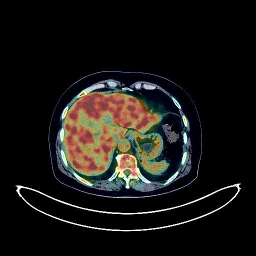
Lung Cancer PET/CT (case 983827-000249 from PETWB-REP)
0 views10 days agoWhole-body 18F-FDG PET/CT scan in a patient with Lung Cancer taken from the PETWB-REP dataset. The following English report (translated from original Chinese) is taken verbatim from the public dataset and has not been modified or otherwise checked for accuracy (see the end for citation). Impression a. A mass near the hilum in the apical-posterior segment of the left upper lobe, with increased FDG metabolism, suggestive of lung cancer. Left hilar lymph node metastasis cannot be ruled out. b. Multiple ground-glass opacities in both lungs, some with mildly increased FDG metabolism, highly suggestive of atypical adenomatous hyperplasia, with the largest nodules in the apical segment of the right upper lobe, the apical-posterior segment of the left upper lobe, and the right middle lobe. Early-stage tumors need to be ruled out; regular CT scans for comparison are recommended. c. Several small chronic inflammatory nodules (solid) in the right middle lobe and both lower lobes; mild inflammatory changes in the left lower lobe; follow-up CT scan is recommended. Some arteriosclerosis. Left breast hyperplasia. d. Slightly blurred local bone structure in the right iliac bone, with slightly increased FDG metabolism; follow-up MRI is recommended to rule out metastasis. Calcification at the fundus of the uterus. Slightly low-density lesion in the right lobe of the liver; FDG metabolism normal; suggestive of cyst or hemangioma; MRI recommended. Chronic gastritis. Mild scoliosis; degenerative changes in the spine. L4/5 and L5/S1 intervertebral disc bulge. Age-related brain changes; deep lacunar infarcts in the brain; MRI recommended. Minor inflammation of bilateral ethmoid and maxillary sinuses. This case is from PETWB-REP, a curated dataset of whole-body 18F-FDG PET/CT scans and corresponding radiology reports from 490 patients with a broad spectrum of malignancies. The data were retrospectively collected from patients who underwent clinically indicated whole-body 18F-FDG PET/CT scans at the Shanghai Universal Medical Imaging Diagnostic Center between 2021 and 2024. License: Creative Commons Attribution 4.0 International (CC BY 4.0) Citation: Xue, L., Feng, G., Wenbo, Z., Zhang, Y., Li, L., Wang, S., Peng, L., Peng, S., & Gao, X. (2026). PETWB-REP: A Multi-Cancer Whole-Body FDG PET/CT Dataset with Corresponding Radiology Reports [Data set]. Zenodo. https://doi.org/10.5281/zenodo.18670487
Whole BodyPET/CT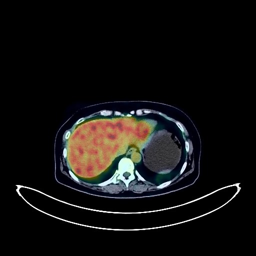
Cervical Cancer PET/CT (case 983827-000096 from PETWB-REP)
0 views10 days agoWhole-body 18F-FDG PET/CT scan in a patient with Cervical Cancer taken from the PETWB-REP dataset. The following English report (translated from original Chinese) is taken verbatim from the public dataset and has not been modified or otherwise checked for accuracy (see the end for citation). Impression a. Uterine mass with elevated FDG metabolism, suggestive of malignancy; please correlate with clinicopathology; uterine cavity effusion, small amount of pelvic effusion. b. Multiple lymph node metastases in the right parauterine region, bilateral iliac vessels, retroperitoneum, bilateral supraclavicular fossa, and superior mediastinal vascular space. c. Multiple bone metastases in the right iliac bone, right hip body, left pubic tubercle, and bilateral inferior pubic rami. d. Multiple solid nodules in both lungs, with clear borders, some showing mild uptake, suggesting possible partial metastasis; CT follow-up is recommended. A few post-inflammatory lesions in both lungs. Tracheal diverticulum. Calcification in the left breast; ultrasound follow-up is recommended. Splenomegaly. Chronic cholecystitis. Possible chronic inflammatory changes in some intestinal segments; please correlate with endoscopy. Mild vertebral osteophyte formation, L5/S1 intervertebral disc bulge. Bilateral subcutaneous calcifications in the buttocks. Sacral canal cyst. No obvious abnormalities were found on cranial scintigraphy. This case is from PETWB-REP, a curated dataset of whole-body 18F-FDG PET/CT scans and corresponding radiology reports from 490 patients with a broad spectrum of malignancies. The data were retrospectively collected from patients who underwent clinically indicated whole-body 18F-FDG PET/CT scans at the Shanghai Universal Medical Imaging Diagnostic Center between 2021 and 2024. License: Creative Commons Attribution 4.0 International (CC BY 4.0) Citation: Xue, L., Feng, G., Wenbo, Z., Zhang, Y., Li, L., Wang, S., Peng, L., Peng, S., & Gao, X. (2026). PETWB-REP: A Multi-Cancer Whole-Body FDG PET/CT Dataset with Corresponding Radiology Reports [Data set]. Zenodo. https://doi.org/10.5281/zenodo.18670487
Whole BodyPET/CT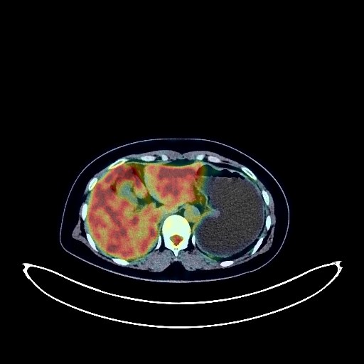
Lung Cancer PET/CT (case 983827-000060 from PETWB-REP)
0 views10 days agoWhole-body 18F-FDG PET/CT scan in a patient with Lung Cancer taken from the PETWB-REP dataset. The following English report (translated from original Chinese) is taken verbatim from the public dataset and has not been modified or otherwise checked for accuracy (see the end for citation). Impression a. A mass in the posterior basal segment of the right lower lobe, with increased FDG metabolism, suggestive of lung cancer. b. Multiple pure ground-glass nodules in both lungs, with normal FDG metabolism, suggestive of atypical adenomatous hyperplasia or inflammatory nodules; annual HRCT follow-up is recommended. c. Inflammatory nodules (solid) in the left and right middle lobes; calcification in the left lower lobe. Multiple focal emphysema in both lungs; bullae in the apical segment of the right upper lobe. Increased FDG metabolism in the left tip of the tongue and bilateral laryngopharynx, suggestive of physiological uptake. Minor inflammation of the right maxillary sinus. Malrotation of the left kidney. High probability of uterine fibroids; left ovarian cyst; ultrasound follow-up is recommended. Mild osteophyte formation in the spine. No obvious abnormalities were found on cranial imaging; MRI follow-up is recommended. This case is from PETWB-REP, a curated dataset of whole-body 18F-FDG PET/CT scans and corresponding radiology reports from 490 patients with a broad spectrum of malignancies. The data were retrospectively collected from patients who underwent clinically indicated whole-body 18F-FDG PET/CT scans at the Shanghai Universal Medical Imaging Diagnostic Center between 2021 and 2024. License: Creative Commons Attribution 4.0 International (CC BY 4.0) Citation: Xue, L., Feng, G., Wenbo, Z., Zhang, Y., Li, L., Wang, S., Peng, L., Peng, S., & Gao, X. (2026). PETWB-REP: A Multi-Cancer Whole-Body FDG PET/CT Dataset with Corresponding Radiology Reports [Data set]. Zenodo. https://doi.org/10.5281/zenodo.18670487
Whole BodyPET/CT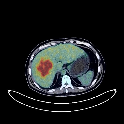
Liver Cancer PET/CT (case 983827-000053 from PETWB-REP)
0 views10 days agoWhole-body 18F-FDG PET/CT scan in a patient with Liver Cancer taken from the PETWB-REP dataset. The following English report (translated from original Chinese) is taken verbatim from the public dataset and has not been modified or otherwise checked for accuracy (see the end for citation). Impression a. Liver cirrhosis with a large mass in the right lobe of the liver, elevated FDG metabolism, suggestive of primary liver cancer with intrahepatic dissemination and right portal vein thrombus formation. b. Multiple lymph node metastases in the hepatic hilum, para-aortic region, left para-gastric cardia, bilateral posterior diaphragmatic crura, right cardiophrenic angle, and left cervical root. a. Multiple inflammatory nodules in the right middle lobe and both lower lobes are highly probable; follow-up is recommended to rule out mixed metastases. Bilateral emphysema. A few fibrotic lesions in the right lower lobe. b. Partial calcification of the aortic and coronary artery walls. Chronic cholecystitis. Seminal vesicle cyst. L3 vertebral instability. Spinal osteophyte formation. Partial intervertebral disc degeneration. No obvious abnormalities seen on cranial imaging. Bilateral maxillary sinusitis. This case is from PETWB-REP, a curated dataset of whole-body 18F-FDG PET/CT scans and corresponding radiology reports from 490 patients with a broad spectrum of malignancies. The data were retrospectively collected from patients who underwent clinically indicated whole-body 18F-FDG PET/CT scans at the Shanghai Universal Medical Imaging Diagnostic Center between 2021 and 2024. License: Creative Commons Attribution 4.0 International (CC BY 4.0) Citation: Xue, L., Feng, G., Wenbo, Z., Zhang, Y., Li, L., Wang, S., Peng, L., Peng, S., & Gao, X. (2026). PETWB-REP: A Multi-Cancer Whole-Body FDG PET/CT Dataset with Corresponding Radiology Reports [Data set]. Zenodo. https://doi.org/10.5281/zenodo.18670487
Whole BodyPET/CT