Loading...
Breast Cancer PET/CT (case 983827-000164 from PETWB-REP)
0 views10 days agoWhole-body 18F-FDG PET/CT scan in a patient with Breast Cancer taken from the PETWB-REP dataset. The following English report (translated from original Chinese) is taken verbatim from the public dataset and has not been modified or otherwise checked for accuracy (see the end for citation). Impression a. Left breast mass with increased FDG metabolism, suggestive of breast cancer; please correlate with clinicopathology. b. Multiple lymph node metastases in the left axilla, retroperitoneum, and root of the mesenteric cavity. c. Cerebellar metastasis; MRI follow-up recommended. Small softening lesion in the left frontal lobe. Uterine fibroid; uneven endometrial density with patchy increased FDG metabolism, endometrial cancer to be ruled out; enhanced MRI recommended for further examination. Thyroid calcifications; enlarged thyroid gland with multiple nodules and masses, increased FDG metabolism, suggestive of nodular goiter; please correlate with ultrasound. Inflammatory manifestations in the antrum of the stomach and duodenum; please correlate with endoscopy to rule out other possibilities. Chronic miliary lesions in the anterior segment of the right upper lobe; calcifications in the anterior medial basal segment of the left lower lobe. A few post-inflammatory lesions in both lungs. Anemia. Changes after cholecystectomy. Degenerative changes in the spine, L2/3 vertebral endplate inflammation. Multiple lumbar disc bulges. Chronic subcutaneous inflammation in the right groin. Chronic inflammation of the nasopharynx. This case is from PETWB-REP, a curated dataset of whole-body 18F-FDG PET/CT scans and corresponding radiology reports from 490 patients with a broad spectrum of malignancies. The data were retrospectively collected from patients who underwent clinically indicated whole-body 18F-FDG PET/CT scans at the Shanghai Universal Medical Imaging Diagnostic Center between 2021 and 2024. License: Creative Commons Attribution 4.0 International (CC BY 4.0) Citation: Xue, L., Feng, G., Wenbo, Z., Zhang, Y., Li, L., Wang, S., Peng, L., Peng, S., & Gao, X. (2026). PETWB-REP: A Multi-Cancer Whole-Body FDG PET/CT Dataset with Corresponding Radiology Reports [Data set]. Zenodo. https://doi.org/10.5281/zenodo.18670487
Whole BodyPET/CT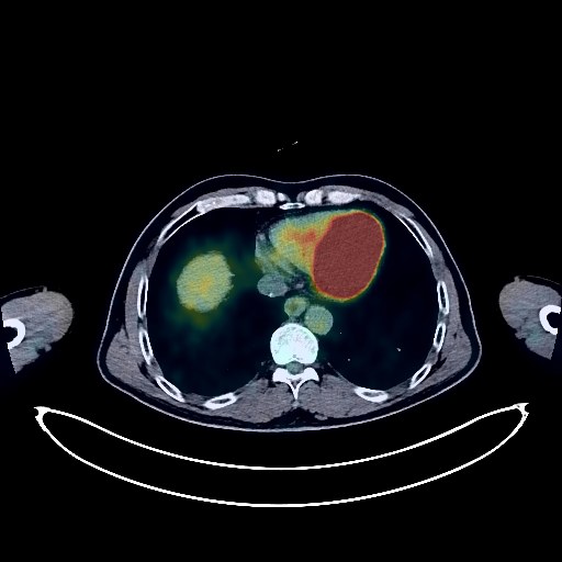
Lung Cancer PET/CT (case 983827-000143 from PETWB-REP)
0 views10 days agoWhole-body 18F-FDG PET/CT scan in a patient with Lung Cancer taken from the PETWB-REP dataset. The following English report (translated from original Chinese) is taken verbatim from the public dataset and has not been modified or otherwise checked for accuracy (see the end for citation). Impression a. Right upper lobe posterior segment mass, elevated FDG metabolism, suggestive of lung cancer. b. Multiple intracranial metastases. Metastasis to the left 9th posterior rib. c. Ground-glass nodules in both upper lobes, normal FDG metabolism, suggestive of inflammatory nodules or atypical adenomatous hyperplasia; annual HRCT follow-up recommended. d. Possible chronic inflammatory nodules in the right middle lobe and interlobar pleura, metastasis to be ruled out; follow-up recommended. Bilateral emphysema. A few chronic inflammations and old lesions in both lungs. e. Reactive hyperplasia of bilateral hilar and mediastinal lymph nodes. Calcification of some arterial walls. Liver cyst. Degenerative changes in the spine. L4/5, L5/S1 intervertebral disc bulge. Bilateral frozen shoulder. Right maxillary sinus submucosal cyst. Cyst in the midline of the maxilla. This case is from PETWB-REP, a curated dataset of whole-body 18F-FDG PET/CT scans and corresponding radiology reports from 490 patients with a broad spectrum of malignancies. The data were retrospectively collected from patients who underwent clinically indicated whole-body 18F-FDG PET/CT scans at the Shanghai Universal Medical Imaging Diagnostic Center between 2021 and 2024. License: Creative Commons Attribution 4.0 International (CC BY 4.0) Citation: Xue, L., Feng, G., Wenbo, Z., Zhang, Y., Li, L., Wang, S., Peng, L., Peng, S., & Gao, X. (2026). PETWB-REP: A Multi-Cancer Whole-Body FDG PET/CT Dataset with Corresponding Radiology Reports [Data set]. Zenodo. https://doi.org/10.5281/zenodo.18670487
Whole BodyPET/CT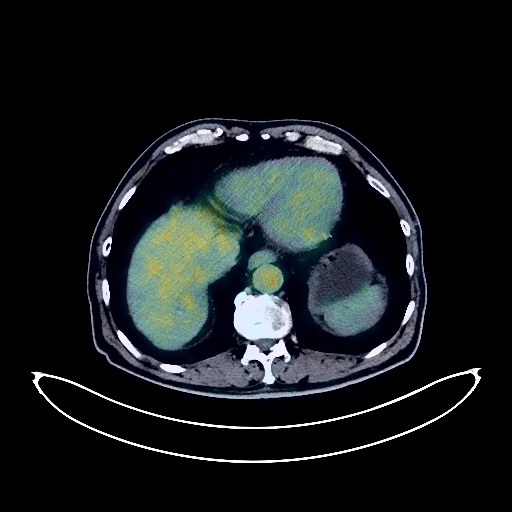
Esophageal Cancer PET/CT (case 983827-000012 from PETWB-REP)
0 views10 days agoWhole-body 18F-FDG PET/CT scan in a patient with Esophageal Cancer taken from the PETWB-REP dataset. The following English report (translated from original Chinese) is taken verbatim from the public dataset and has not been modified or otherwise checked for accuracy (see the end for citation). Impression Thickening of the mid-thoracic esophagus with soft tissue mass, increased FDG metabolism, consistent with esophageal cancer. Metastasis to the right paratracheal and para-aortic lymph nodes in the superior mediastinum and posterior mediastinum. Mixed ground-glass nodules in the posterior basal segment of the left lower lobe and ground-glass nodules in the posterior segment of the left lower lobe, with no increased FDG metabolism, both suggestive of chronic inflammatory nodules. Several solid chronic inflammatory nodules in both lungs, please follow up with CT scan. Changes in chronic bronchitis and emphysema in both lungs. Calcification of some arterial walls (including coronary arteries). Cyst in the right posterior lobe of the liver. Calcification in the right lobe of the liver. Cholestasis in the gallbladder. Slight thickening of the gastric wall in the antrum, with mildly increased FDG metabolism, suggestive of inflammatory uptake, please follow up with gastroscopy. a. Localized low-density lesion in the right lower abdomen above the mesentery, with normal FDG metabolism, suggestive of benign lesion, please follow up. b. Increased fat density and multiple small nodules around the root of the mesentery, with no abnormal FDG uptake, suggest panniculitis is highly likely; please correlate with clinical findings and conduct follow-up examinations. Small kidney stones in both kidneys. Benign prostatic hyperplasia, with unevenly increased FDG uptake, suggests chronic inflammation is highly likely; PSA testing is recommended. Spinal degenerative changes. L4/5 and L5/S1 intervertebral disc bulges. L2/3 and L4/5 intervertebral space narrowing. Multiple pneumothorax in the lumbar intervertebral discs. Lipoma on the medial side of the right scapula. A few ischemic foci deep in the brain. Submucosal cyst of the left maxillary sinus. Calcifications in the right lobe and isthmus of the thyroid gland; please follow up with ultrasound. This case is from PETWB-REP, a curated dataset of whole-body 18F-FDG PET/CT scans and corresponding radiology reports from 490 patients with a broad spectrum of malignancies. The data were retrospectively collected from patients who underwent clinically indicated whole-body 18F-FDG PET/CT scans at the Shanghai Universal Medical Imaging Diagnostic Center between 2021 and 2024. License: Creative Commons Attribution 4.0 International (CC BY 4.0) Citation: Xue, L., Feng, G., Wenbo, Z., Zhang, Y., Li, L., Wang, S., Peng, L., Peng, S., & Gao, X. (2026). PETWB-REP: A Multi-Cancer Whole-Body FDG PET/CT Dataset with Corresponding Radiology Reports [Data set]. Zenodo. https://doi.org/10.5281/zenodo.18670487
Whole BodyPET/CT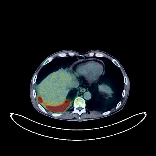
Lung Cancer PET/CT (case 983827-000031 from PETWB-REP)
0 views10 days agoWhole-body 18F-FDG PET/CT scan in a patient with Lung Cancer taken from the PETWB-REP dataset. The following English report (translated from original Chinese) is taken verbatim from the public dataset and has not been modified or otherwise checked for accuracy (see the end for citation). Impression a. Soft tissue nodules near the hilum of the right lower lobe with increased FDG metabolism, consistent with central lung cancer presentation, distal obstructive inflammation, and atelectasis; please correlate with clinical findings. b. Right hilar and mediastinal lymph node metastasis is highly probable; reactive hyperplasia of left hilar lymph nodes is possible. c. Bilateral adrenal gland enlargement with increased FDG uptake suggests possible hyperplasia; metastasis needs to be ruled out. Multiple solid small nodules in both lungs, FDG metabolism normal; chronic inflammatory nodules are highly probable; follow-up CT scan recommended to rule out metastasis. A few linear and punctate lesions are also seen in both lungs, FDG metabolism normal. Interstitial changes in both lungs with scattered chronic inflammation and remnants. Emphysema in both lungs. Small amount of pleural effusion on the right side. Pericardial effusion. Partial arteriosclerosis (including coronary arteries). a. Soft tissue density shadows in the head of the pancreas and between the duodenum, FDG uptake normal; physiological changes in the head of the pancreas are possible; space-occupying lesion needs to be ruled out; contrast-enhanced examination recommended. b. Splenic calcifications. Left renal cyst. Increased FDG metabolism in part of the gastric wall, suggesting possible chronic inflammation; please follow up with gastroscopy. Spinal degenerative changes. Mild wedge-shaped L1 vertebral body. L4/5 and L5/S1 intervertebral disc bulges. Blurred right sacroiliac joint space. Possible paravertebral inflammatory changes at the L2/3 intervertebral space level. Bilateral deep lacunar infarcts, age-related brain; MRI follow-up is recommended to rule out occult lesions. Chronic inflammation of the nasopharynx and bilateral palatine tonsils. This case is from PETWB-REP, a curated dataset of whole-body 18F-FDG PET/CT scans and corresponding radiology reports from 490 patients with a broad spectrum of malignancies. The data were retrospectively collected from patients who underwent clinically indicated whole-body 18F-FDG PET/CT scans at the Shanghai Universal Medical Imaging Diagnostic Center between 2021 and 2024. License: Creative Commons Attribution 4.0 International (CC BY 4.0) Citation: Xue, L., Feng, G., Wenbo, Z., Zhang, Y., Li, L., Wang, S., Peng, L., Peng, S., & Gao, X. (2026). PETWB-REP: A Multi-Cancer Whole-Body FDG PET/CT Dataset with Corresponding Radiology Reports [Data set]. Zenodo. https://doi.org/10.5281/zenodo.18670487
Whole BodyPET/CT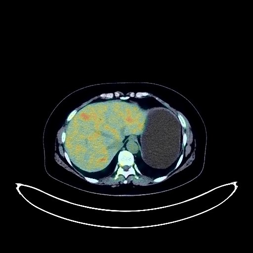
Renal Cancer PET/CT (case 983827-000104 from PETWB-REP)
0 views10 days agoWhole-body 18F-FDG PET/CT scan in a patient with Renal Cancer taken from the PETWB-REP dataset. The following English report (translated from original Chinese) is taken verbatim from the public dataset and has not been modified or otherwise checked for accuracy (see the end for citation). Impression A mass with small cystic changes in the lower pole of the left kidney, with mild FDG uptake, suggests a neoplastic lesion, possibly renal cell carcinoma. Please combine with enhanced MRI for comprehensive analysis. Small renal calculus in the left kidney. Multiple cysts in the liver. Possible cholestasis in the gallbladder; ultrasound examination is recommended. Chronic inflammatory nodules in the apical-posterior segment of the left upper lobe and beside the oblique fissure of the right lung. A few old lesions in the middle lobe of the right lung. Spinal degenerative changes, L4/5 intervertebral disc bulge. A slightly high-density nodule in the posterior part of the falx cerebri, with absent FDG uptake, suggests a possible meningioma; enhanced MRI is recommended. This case is from PETWB-REP, a curated dataset of whole-body 18F-FDG PET/CT scans and corresponding radiology reports from 490 patients with a broad spectrum of malignancies. The data were retrospectively collected from patients who underwent clinically indicated whole-body 18F-FDG PET/CT scans at the Shanghai Universal Medical Imaging Diagnostic Center between 2021 and 2024. License: Creative Commons Attribution 4.0 International (CC BY 4.0) Citation: Xue, L., Feng, G., Wenbo, Z., Zhang, Y., Li, L., Wang, S., Peng, L., Peng, S., & Gao, X. (2026). PETWB-REP: A Multi-Cancer Whole-Body FDG PET/CT Dataset with Corresponding Radiology Reports [Data set]. Zenodo. https://doi.org/10.5281/zenodo.18670487
Whole BodyPET/CT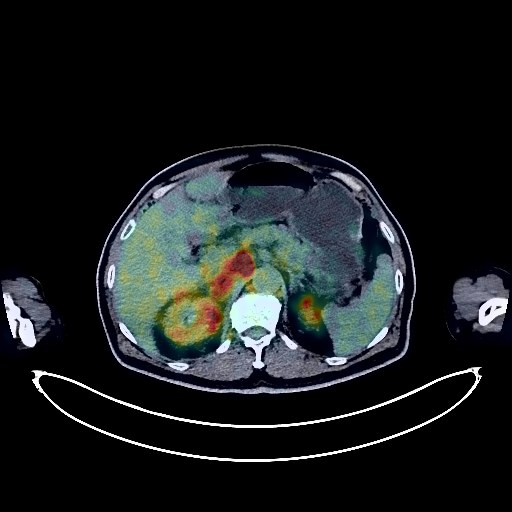
Renal Cancer PET/CT (case 983827-000254 from PETWB-REP)
0 views10 days agoWhole-body 18F-FDG PET/CT scan in a patient with Renal Cancer taken from the PETWB-REP dataset. The following English report (translated from original Chinese) is taken verbatim from the public dataset and has not been modified or otherwise checked for accuracy (see the end for citation). Impression a. A mass in the lower pole of the right kidney with increased FDG metabolism; multiple enlarged lymph nodes throughout the body with increased FDG metabolism (see description for details; some lesions are poorly demarcated from the middle and lower segments of the right ureter); focal increase in FDG uptake in the L4 vertebral body. All of these suggest malignancy, but lymphoma cannot be ruled out. Please correlate with clinicopathology. b. An irregular patchy shadow in the posterior basal segment of the right lower lobe, adjacent to the mediastinum, with increased FDG metabolism. Tumor infiltration is highly probable; inflammation needs to be ruled out. Please correlate with clinicopathology. c. Extensive thickening of the right peritoneum and pelvic floor fascia, with a small amount of pelvic effusion. d. Localized thickening of the rectal wall; tumor needs to be ruled out. Colonoscopy is recommended. Localized nodular FDG metabolism in the left lobe of the liver; please correlate with contrast-enhanced MRI to rule out tumor. Chronic inflammatory micronodules in both lungs; follow-up CT is recommended. Bilateral emphysema, bilateral interstitial lung inflammation. Tracheal diverticulum. Calcification of some arterial walls (including coronary arteries). Small gallstones. Accessory spleen. Possible splenic angioma. Benign prostatic hyperplasia with calcification. Small amount of hydrocele in the right testis. Chronic inflammatory changes in part of the gastric wall; please confirm with gastroscopy. Degenerative changes in the spine, anterior slippage of the L4 vertebral body, L4/5 and L5/S1 intervertebral disc bulging with pneumodegenerative changes. Subcutaneous inflammation in the left buttock. Age-related brain, deep lacunar infarcts; please confirm with MRI. Chronic inflammation of the left ethmoid sinus. This case is from PETWB-REP, a curated dataset of whole-body 18F-FDG PET/CT scans and corresponding radiology reports from 490 patients with a broad spectrum of malignancies. The data were retrospectively collected from patients who underwent clinically indicated whole-body 18F-FDG PET/CT scans at the Shanghai Universal Medical Imaging Diagnostic Center between 2021 and 2024. License: Creative Commons Attribution 4.0 International (CC BY 4.0) Citation: Xue, L., Feng, G., Wenbo, Z., Zhang, Y., Li, L., Wang, S., Peng, L., Peng, S., & Gao, X. (2026). PETWB-REP: A Multi-Cancer Whole-Body FDG PET/CT Dataset with Corresponding Radiology Reports [Data set]. Zenodo. https://doi.org/10.5281/zenodo.18670487
Whole BodyPET/CT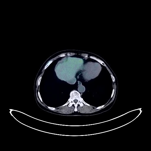
Lung Cancer PET/CT (case 983827-000009 from PETWB-REP)
0 views10 days agoWhole-body 18F-FDG PET/CT scan in a patient with Lung Cancer taken from the PETWB-REP dataset. The following English report (translated from original Chinese) is taken verbatim from the public dataset and has not been modified or otherwise checked for accuracy (see the end for citation). Impression a. Solid nodule in the posterior segment of the left upper lobe with increased FDG metabolism, suggestive of peripheral lung cancer. Left upper hilar lymph node metastasis. b. Small solid nodules in the pleura of the left upper lobe and right oblique fissure, without increased FDG metabolism, suggestive of chronic inflammatory nodules; follow-up with CT is recommended. Scattered emphysema, chronic inflammation, and remnants in both upper lobes. c. Possible reactive hyperplasia of mediastinal and bilateral axillary lymph nodes; partial lymph node metastasis at the aortic window needs further investigation; follow-up is recommended. Calcification of some arterial walls (including coronary arteries). d. Small nodule in the anterior mediastinal thymic region, without abnormal FDG uptake, likely a small thymoma; follow-up is recommended. A slightly high-density lesion with uneven distribution under the capsule of the right kidney, appearing crescent-shaped in the sagittal plane. FDG uptake at the lesion's edge is physiological, while FDG metabolism is absent internally. This suggests a possible subcapsular hematoma in the absorption phase, but space-occupying lesion cannot be ruled out. Further diagnosis requires clinical examination and enhanced MRI. Right renal angiomyolipoma. Right lobe hepatic cyst. Cholestasis in the gallbladder. Linear high-density shadows in part of the intestinal wall, suggestive of schistosomiasis. Scattered calcifications in the left breast; follow-up ultrasound is needed. Possible uterine fibroid; follow-up ultrasound is needed. Fractured changes in the right 6th-10th ribs; clinical correlation is needed. Partial vertebral osteophyte formation. L4/5 and L5/S1 intervertebral disc bulge. Multiple subcutaneous calcifications in the left buttock. No obvious abnormalities were found on cranial scintigraphy. Localized inflammatory uptake in the right inferior alveolar ridge. This case is from PETWB-REP, a curated dataset of whole-body 18F-FDG PET/CT scans and corresponding radiology reports from 490 patients with a broad spectrum of malignancies. The data were retrospectively collected from patients who underwent clinically indicated whole-body 18F-FDG PET/CT scans at the Shanghai Universal Medical Imaging Diagnostic Center between 2021 and 2024. License: Creative Commons Attribution 4.0 International (CC BY 4.0) Citation: Xue, L., Feng, G., Wenbo, Z., Zhang, Y., Li, L., Wang, S., Peng, L., Peng, S., & Gao, X. (2026). PETWB-REP: A Multi-Cancer Whole-Body FDG PET/CT Dataset with Corresponding Radiology Reports [Data set]. Zenodo. https://doi.org/10.5281/zenodo.18670487
Whole BodyPET/CT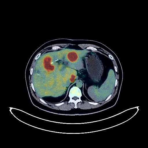
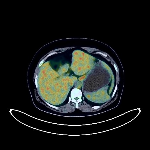
Colon Cancer PET/CT (case 983827-000187 from PETWB-REP)
0 views10 days agoWhole-body 18F-FDG PET/CT scan in a patient with Colon Cancer taken from the PETWB-REP dataset. The following English report (translated from original Chinese) is taken verbatim from the public dataset and has not been modified or otherwise checked for accuracy (see the end for citation). Impression a. Postoperative colon cancer surgery, no clear signs of tumor recurrence were observed at the anastomosis site. Please follow up with colonoscopy. ? b. Postoperative changes in liver metastases with a small amount of local effusion; multiple metastases in the remaining liver. ? c. Multiple lymph node metastases in the hepatic hilum, hepatogastric space, lower esophageal segment of the posterior mediastinum, and left lower hilum. Metastasis to the left side of the lower abdominal aorta. ? d. Soft tissue lesions in the left lower pulmonary ligament and posterior segment of the right upper lobe with significantly increased FDG metabolism; multiple solid miliary lesions and small nodules in both lungs, with a larger one in the left lower lobe showing mildly increased FDG metabolism. All of the above lesions are considered metastases. Please compare with old films and follow up with CT scan. Cystic low-density lesion with increased FDG metabolism in the common bile duct (upper pancreatic segment) area, suggestive of possible lymph node metastasis with internal necrosis. Please compare with old films and perform enhanced MRI. Left renal calculus. Benign prostatic hyperplasia. Manifestations of chronic gastritis. Duodenal diverticulum in the descending part of the duodenum. A few chronic pulmonary lesions and sequelae, a small amount of pleural effusion on the right side with partial atelectasis in the lower lobe of the right lung. Mild osteophyte formation in some vertebral bodies, multiple Schmorl's nodes in the lumbar vertebrae. Benign osteopathy in the left iliac bone. No obvious abnormalities were found on cranial scintigraphy; enhanced MRI may be necessary. Small submucosal cysts in both maxillary sinuses. This case is from PETWB-REP, a curated dataset of whole-body 18F-FDG PET/CT scans and corresponding radiology reports from 490 patients with a broad spectrum of malignancies. The data were retrospectively collected from patients who underwent clinically indicated whole-body 18F-FDG PET/CT scans at the Shanghai Universal Medical Imaging Diagnostic Center between 2021 and 2024. License: Creative Commons Attribution 4.0 International (CC BY 4.0) Citation: Xue, L., Feng, G., Wenbo, Z., Zhang, Y., Li, L., Wang, S., Peng, L., Peng, S., & Gao, X. (2026). PETWB-REP: A Multi-Cancer Whole-Body FDG PET/CT Dataset with Corresponding Radiology Reports [Data set]. Zenodo. https://doi.org/10.5281/zenodo.18670487
Whole BodyPET/CT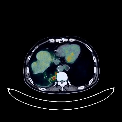
Lung Cancer PET/CT (case 983827-000004 from PETWB-REP)
0 views10 days agoWhole-body 18F-FDG PET/CT scan in a patient with Lung Cancer taken from the PETWB-REP dataset. The following English report (translated from original Chinese) is taken verbatim from the public dataset and has not been modified or otherwise checked for accuracy (see the end for citation). Impression a. After lung cancer treatment, a mass in the lower lobe of the right lung with increased FDG metabolism suggests continued high tumor activity. Right hilar and mediastinal lymph node metastasis. Right pleural metastasis, small amount of right pleural effusion. b. Patchy lesion in the apical segment of the upper lobe of the right lung, FDG metabolism normal; comparison with previous imaging data and follow-up are recommended. c. Bilateral chronic inflammatory nodules are highly probable; comparison with old images and re-examination are recommended. Scattered post-inflammatory lesions in both lungs. d. Pericardial thickening with a small amount of effusion, arteriosclerosis, changes after coronary stent placement. Right clavicular port placement. Mass in the pancreatic tail region with increased FDG metabolism, suggestive of metastasis; please combine with MRI. Postoperative changes in gastric cardia cancer; no signs of tumor recurrence in the surgical area; please combine with gastroscopy for follow-up. Splenic capsule calcification. Residual contrast agent in the cystostomy. Possible chronic inflammatory changes in some intestinal segments; please combine with endoscopy for follow-up. Osteoporosis, degenerative changes in the spine, multiple lumbar disc bulges, and wedge-shaped deformity of the L3 vertebral body. Some changes are post-radiotherapy changes in the cervical and thoracic spine. Elderly brain, deep lacunar infarcts and softening lesions in the brain; MRI follow-up is recommended. This case is from PETWB-REP, a curated dataset of whole-body 18F-FDG PET/CT scans and corresponding radiology reports from 490 patients with a broad spectrum of malignancies. The data were retrospectively collected from patients who underwent clinically indicated whole-body 18F-FDG PET/CT scans at the Shanghai Universal Medical Imaging Diagnostic Center between 2021 and 2024. License: Creative Commons Attribution 4.0 International (CC BY 4.0) Citation: Xue, L., Feng, G., Wenbo, Z., Zhang, Y., Li, L., Wang, S., Peng, L., Peng, S., & Gao, X. (2026). PETWB-REP: A Multi-Cancer Whole-Body FDG PET/CT Dataset with Corresponding Radiology Reports [Data set]. Zenodo. https://doi.org/10.5281/zenodo.18670487
Whole BodyPET/CT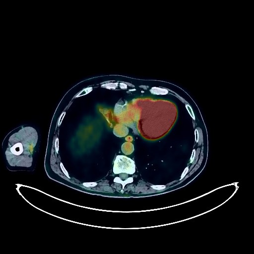
Lung Cancer PET/CT (case 983827-000181 from PETWB-REP)
0 views10 days agoWhole-body 18F-FDG PET/CT scan in a patient with Lung Cancer taken from the PETWB-REP dataset. The following English report (translated from original Chinese) is taken verbatim from the public dataset and has not been modified or otherwise checked for accuracy (see the end for citation). Impression a. A mass in the right middle lobe of the lung with increased FDG metabolism, suggestive of peripheral lung cancer; please correlate with clinical and pathological findings. A soft tissue mass in the right back muscles with bone destruction of the adjacent scapula suggests a high probability of metastasis. b. Multiple solid nodules in both lungs with normal FDG metabolism, suggestive of chronic inflammatory nodules; follow-up CT is recommended. Scattered chronic inflammation and sequelae in both lungs. High probability of reactive hyperplasia of the hilar and mediastinal lymph nodes. Calcification of some arterial walls (including coronary arteries). A soft tissue nodule in the right parotid gland with increased FDG metabolism, suggestive of adenolymphoma or mixed tumor; ultrasound follow-up is recommended. A few ischemic lesions in the deep bilateral brain; senile cerebral hemorrhage; please correlate with MRI for follow-up. Inflammation of the left ethmoid sinus. Right renal cyst, complex cyst. Prostatic calcification. Increased FDG metabolism in some intestinal segments, possibly due to inflammatory or physiological uptake; colonoscopy is recommended. Degenerative changes in the spine. Wedge-shaped changes in the T12 and L1 vertebral bodies. Schmorl's nodes in some thoracic vertebrae. Mild bulging of the L4/5 and L5/S1 intervertebral discs; pneumoconiosis and degeneration in some thoracic and lumbar vertebrae. This case is from PETWB-REP, a curated dataset of whole-body 18F-FDG PET/CT scans and corresponding radiology reports from 490 patients with a broad spectrum of malignancies. The data were retrospectively collected from patients who underwent clinically indicated whole-body 18F-FDG PET/CT scans at the Shanghai Universal Medical Imaging Diagnostic Center between 2021 and 2024. License: Creative Commons Attribution 4.0 International (CC BY 4.0) Citation: Xue, L., Feng, G., Wenbo, Z., Zhang, Y., Li, L., Wang, S., Peng, L., Peng, S., & Gao, X. (2026). PETWB-REP: A Multi-Cancer Whole-Body FDG PET/CT Dataset with Corresponding Radiology Reports [Data set]. Zenodo. https://doi.org/10.5281/zenodo.18670487
Whole BodyPET/CT