Loading...
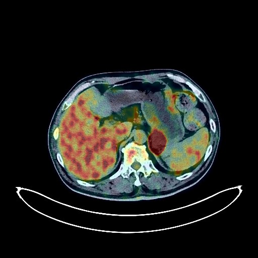
Lung Cancer PET/CT (case 983827-000073 from PETWB-REP)
0 views10 days agoWhole-body 18F-FDG PET/CT scan in a patient with Lung Cancer taken from the PETWB-REP dataset. The following English report (translated from original Chinese) is taken verbatim from the public dataset and has not been modified or otherwise checked for accuracy (see the end for citation). Impression a. A mass in the lingular segment of the left upper lobe of the lung, with increased FDG metabolism, suggestive of lung cancer. Left adrenal metastasis. b. Metastasis to the pulmonary artery window and the left anterior lymph nodes of the abdominal aorta. Left hilar lymph node metastasis is highly probable. c. Several small, solid, chronic inflammatory nodules in both lungs. Bilateral emphysema, a few chronic inflammations, and old lesions. Calcification of some arterial walls (including the coronary arteries). d. Softening lesions in the right basal ganglia region, bilateral deep cerebral ischemia, leukoencephalopathy, and age-related brain changes; no obvious space-occupying lesions seen intracranially. Enhanced intracranial MRI is recommended. Slight thickening of the walls of part of the gastric body and antrum, with mildly increased FDG uptake, suggestive of chronic gastritis; increased FDG metabolism in part of the colon and rectum, suggestive of inflammatory or physiological uptake. Follow-up with gastroscopy and colonoscopy is recommended. Chronic cholecystitis. Gallstones. Degenerative changes in the spine. L4/5 and L5/S1 intervertebral disc bulges. Several low-density nodules in both lobes of the thyroid gland, with calcification in the right lobe. FDG metabolism was normal. Nodular goiter is highly suspected, and ultrasound re-examination is recommended. This case is from PETWB-REP, a curated dataset of whole-body 18F-FDG PET/CT scans and corresponding radiology reports from 490 patients with a broad spectrum of malignancies. The data were retrospectively collected from patients who underwent clinically indicated whole-body 18F-FDG PET/CT scans at the Shanghai Universal Medical Imaging Diagnostic Center between 2021 and 2024. License: Creative Commons Attribution 4.0 International (CC BY 4.0) Citation: Xue, L., Feng, G., Wenbo, Z., Zhang, Y., Li, L., Wang, S., Peng, L., Peng, S., & Gao, X. (2026). PETWB-REP: A Multi-Cancer Whole-Body FDG PET/CT Dataset with Corresponding Radiology Reports [Data set]. Zenodo. https://doi.org/10.5281/zenodo.18670487
Whole BodyPET/CT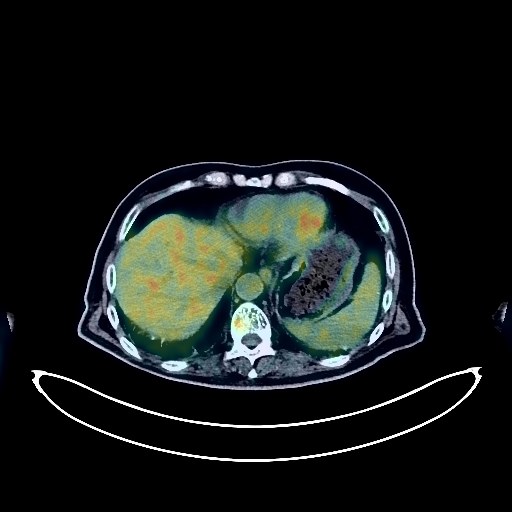
Liver Cancer PET/CT (case 983827-000253 from PETWB-REP)
0 views10 days agoWhole-body 18F-FDG PET/CT scan in a patient with Liver Cancer taken from the PETWB-REP dataset. The following English report (translated from original Chinese) is taken verbatim from the public dataset and has not been modified or otherwise checked for accuracy (see the end for citation). Impression a. "Post-hepatocellular carcinoma surgery changes": No obvious space-occupying lesions were seen in the remaining liver; FDG metabolism was normal. Follow-up MRI is recommended. Liver cirrhosis, slightly enlarged spleen. b. Irregular thickening of the duodenal bulb wall with mass formation; increased FDG metabolism, consistent with metastatic tumor presentation based on medical history. Scattered chronic inflammation and remnants in both lungs. Pleural thickening bilaterally. Anemia. Small hepatic cysts. Post-cholecystectomy changes. Bilateral renal cysts. Bilateral adrenal hyperplasia. Prostatic hyperplasia with calcification. "Post-bladder cancer surgery" changes; no signs of tumor recurrence were observed. Chronic gastritis; continuous increased FDG uptake in the remaining intestinal tract, suggestive of chronic enteritis or physiological uptake. Duodenal diverticulum. 6.a. The previously observed increased FDG uptake lesions on the left side of the L5 vertebral body and the left erector spinae muscle were not visualized. Increased FDG uptake strips within the spinal canal at the T11-T12 vertebral body level are similar to the previous findings, suggesting a physiological change. b. T10 vertebral hemangioma. Degenerative changes in the spine. Mild posterior slippage of the L2 vertebral body. Multiple intervertebral disc bulges with pneumoconiosis and degeneration. Bilateral frozen shoulder. Bilateral deep cerebral ischemic lesions, age-related brain changes. Chronic inflammation of the left maxillary sinus. This case is from PETWB-REP, a curated dataset of whole-body 18F-FDG PET/CT scans and corresponding radiology reports from 490 patients with a broad spectrum of malignancies. The data were retrospectively collected from patients who underwent clinically indicated whole-body 18F-FDG PET/CT scans at the Shanghai Universal Medical Imaging Diagnostic Center between 2021 and 2024. License: Creative Commons Attribution 4.0 International (CC BY 4.0) Citation: Xue, L., Feng, G., Wenbo, Z., Zhang, Y., Li, L., Wang, S., Peng, L., Peng, S., & Gao, X. (2026). PETWB-REP: A Multi-Cancer Whole-Body FDG PET/CT Dataset with Corresponding Radiology Reports [Data set]. Zenodo. https://doi.org/10.5281/zenodo.18670487
Whole BodyPET/CT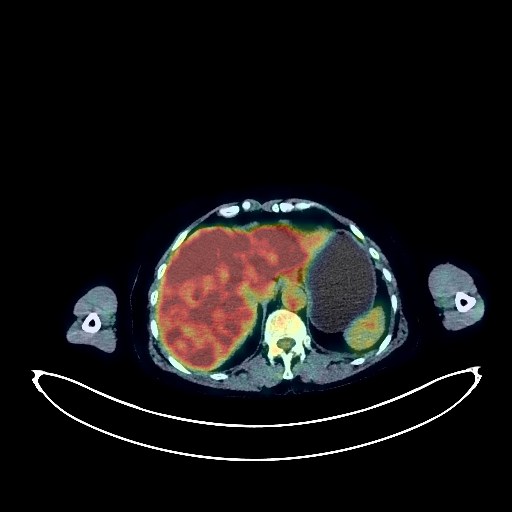
Lung Cancer PET/CT (case 983827-000044 from PETWB-REP)
0 views10 days agoWhole-body 18F-FDG PET/CT scan in a patient with Lung Cancer taken from the PETWB-REP dataset. The following English report (translated from original Chinese) is taken verbatim from the public dataset and has not been modified or otherwise checked for accuracy (see the end for citation). Impression a. Solid nodule in the anterior basal segment of the right lower lobe with slightly elevated FDG metabolism, suggestive of lung cancer; atypical granulomatous lesion to be ruled out. Pathological examination is required for confirmation. b. Several chronic inflammatory miliary nodules in the upper lobes of both lungs and the right lower lobe. Atelectasis in the left lower lobe, no definite space-occupying lesion seen. A few fibrotic lesions in both lungs. Localized soft tissue lesions in the rectum with elevated FDG metabolism, suggestive of polyps; local malignancy to be ruled out. Colonoscopy is recommended for further examination. Partial vertebral osteophyte formation. Partial lumbar vertebral island formation. Subcutaneous calcification and inflammatory remnants in the right buttock. No obvious abnormalities were seen on cranial scintigraphy. This case is from PETWB-REP, a curated dataset of whole-body 18F-FDG PET/CT scans and corresponding radiology reports from 490 patients with a broad spectrum of malignancies. The data were retrospectively collected from patients who underwent clinically indicated whole-body 18F-FDG PET/CT scans at the Shanghai Universal Medical Imaging Diagnostic Center between 2021 and 2024. License: Creative Commons Attribution 4.0 International (CC BY 4.0) Citation: Xue, L., Feng, G., Wenbo, Z., Zhang, Y., Li, L., Wang, S., Peng, L., Peng, S., & Gao, X. (2026). PETWB-REP: A Multi-Cancer Whole-Body FDG PET/CT Dataset with Corresponding Radiology Reports [Data set]. Zenodo. https://doi.org/10.5281/zenodo.18670487
Whole BodyPET/CT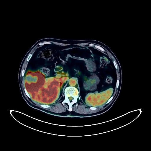
Liver Cancer PET/CT (case 983827-000040 from PETWB-REP)
0 views10 days agoWhole-body 18F-FDG PET/CT scan in a patient with Liver Cancer taken from the PETWB-REP dataset. The following English report (translated from original Chinese) is taken verbatim from the public dataset and has not been modified or otherwise checked for accuracy (see the end for citation). Impression a. Mass lesion in the lower segment of the right anterior lobe of the liver, with increased FDG metabolism, suggestive of hepatocellular carcinoma. b. Diffuse metastases in both lungs. Multiple intracranial metastases. c. Cirrhosis. Scattered chronic inflammation and old lesions in both lungs (most prominent in the upper lobe of the right lung). Slight thickening of the pleura bilaterally. Enlarged chronic inflammatory lymph nodes in the hilar and mediastinal regions. Calcification of some arterial walls. Slight pericardial thickening. Postoperative changes after intestinal polyp surgery. Intervertebral colon. Increased FDG metabolism in part of the colon, suggestive of physiological or inflammatory uptake. Degenerative changes in the spine. L4/5 and L5/S1 intervertebral disc bulges. This case is from PETWB-REP, a curated dataset of whole-body 18F-FDG PET/CT scans and corresponding radiology reports from 490 patients with a broad spectrum of malignancies. The data were retrospectively collected from patients who underwent clinically indicated whole-body 18F-FDG PET/CT scans at the Shanghai Universal Medical Imaging Diagnostic Center between 2021 and 2024. License: Creative Commons Attribution 4.0 International (CC BY 4.0) Citation: Xue, L., Feng, G., Wenbo, Z., Zhang, Y., Li, L., Wang, S., Peng, L., Peng, S., & Gao, X. (2026). PETWB-REP: A Multi-Cancer Whole-Body FDG PET/CT Dataset with Corresponding Radiology Reports [Data set]. Zenodo. https://doi.org/10.5281/zenodo.18670487
Whole BodyPET/CT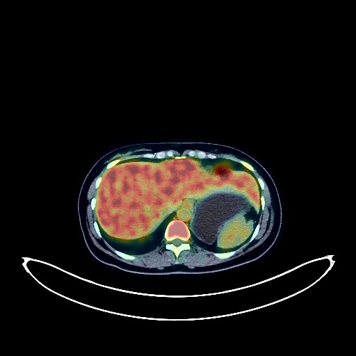
Breast Cancer PET/CT (case 983827-000080 from PETWB-REP)
0 views10 days agoWhole-body 18F-FDG PET/CT scan in a patient with Breast Cancer taken from the PETWB-REP dataset. The following English report (translated from original Chinese) is taken verbatim from the public dataset and has not been modified or otherwise checked for accuracy (see the end for citation). Impression a. Left breast mass with elevated FDG metabolism, suggestive of breast cancer. b. Multiple lymph node metastases in the left axilla, left interpectoral space, and left supraclavicular fossa. c. Multiple bone metastases throughout the body (see description for details). Right upper lobe containing an air sac. A few post-inflammatory remnants in both lungs. Mild anemia. Chronic cholecystitis. Intrauterine device insertion, Nabothian cyst in the cervix. L4/5 intervertebral disc herniation with posterior calcification. Cranial scintigraphy showed no obvious abnormalities. Chronic inflammation of the right maxillary sinus. This case is from PETWB-REP, a curated dataset of whole-body 18F-FDG PET/CT scans and corresponding radiology reports from 490 patients with a broad spectrum of malignancies. The data were retrospectively collected from patients who underwent clinically indicated whole-body 18F-FDG PET/CT scans at the Shanghai Universal Medical Imaging Diagnostic Center between 2021 and 2024. License: Creative Commons Attribution 4.0 International (CC BY 4.0) Citation: Xue, L., Feng, G., Wenbo, Z., Zhang, Y., Li, L., Wang, S., Peng, L., Peng, S., & Gao, X. (2026). PETWB-REP: A Multi-Cancer Whole-Body FDG PET/CT Dataset with Corresponding Radiology Reports [Data set]. Zenodo. https://doi.org/10.5281/zenodo.18670487
Whole BodyPET/CT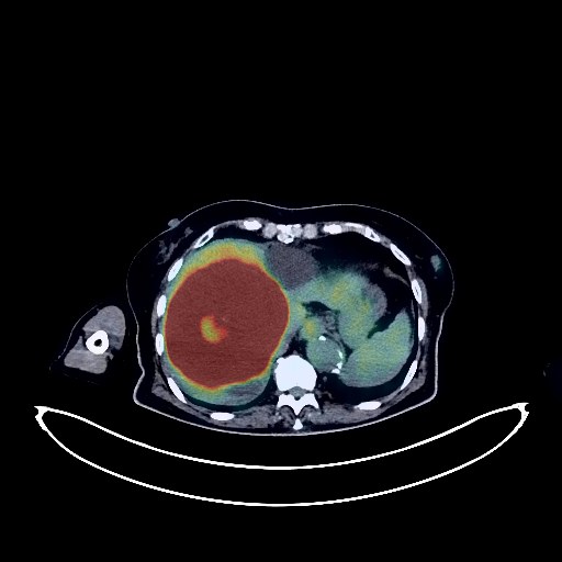
Colon Cancer PET/CT (case 983827-000209 from PETWB-REP)
0 views10 days agoWhole-body 18F-FDG PET/CT scan in a patient with Colon Cancer taken from the PETWB-REP dataset. The following English report (translated from original Chinese) is taken verbatim from the public dataset and has not been modified or otherwise checked for accuracy (see the end for citation). Impression a. Increased FDG metabolism in the surgical area after sigmoid colon cancer surgery, likely due to inflammatory uptake. Colonoscopy is recommended to rule out recurrence. Increased FDG metabolism in parts of the stomach wall and intestines, possibly due to physiological uptake or chronic inflammation. Hemorrhoids. b. A cystic-solid lesion in the right posterior parietal lobe with increased FDG metabolism in the solid portion, likely due to brain metastasis with edema; enhanced MRI is recommended. Metastasis to the right parietal and occipital bones is also possible. c. Liver metastasis. Possible left adrenal metastasis. High FDG metabolism in the pancreatic tail, metastasis to be ruled out. Slightly dense nodule in the lower outer quadrant of the left breast, with increased FDG metabolism, possibly a fibroadenoma, but malignancy to be ruled out; enhanced MRI is recommended. Dense lesion in the right breast. Reactive hyperplasia of the left axillary lymph nodes. A small, chronic inflammatory nodule in the posterior segment of the right lower lobe is likely large; CT follow-up is recommended to rule out other possibilities. Old lesions in the posterior segment of the left upper lobe. Localized pleural thickening with calcification in the left apex. Scattered chronic inflammation and remnants in both lungs. Reactive hyperplasia of the hilar and mediastinal lymph nodes bilaterally. Calcified hilar lymph nodes bilaterally. Partial arteriosclerosis (including coronary arteries). Liver cysts. Possible gallbladder fundus adenomyoma, bile concentration. Bilateral renal cysts, bilateral renal stones. Reactive hyperplasia of the bilateral inguinal lymph nodes. Degenerative changes in the spine. L4/5 and L5/S1 intervertebral disc bulges. L2 vertebral instability. Mild anterior slippage of the L3-4 vertebral bodies. Pneumatosis and degeneration of the L1/2, L2/3, and L4/5 intervertebral discs. Right iliac bone island. The density of both lobes of the thyroid gland is uneven, suggesting nodular goiter. Ultrasound follow-up is recommended. Age-related brain changes: deep lacunar infarcts. Localized calcification of the falx cerebri. Minor inflammation of the right maxillary sinus, bilateral ethmoid sinus submucosal cysts. Right superior alveolar ulceritis. This case is from PETWB-REP, a curated dataset of whole-body 18F-FDG PET/CT scans and corresponding radiology reports from 490 patients with a broad spectrum of malignancies. The data were retrospectively collected from patients who underwent clinically indicated whole-body 18F-FDG PET/CT scans at the Shanghai Universal Medical Imaging Diagnostic Center between 2021 and 2024. License: Creative Commons Attribution 4.0 International (CC BY 4.0) Citation: Xue, L., Feng, G., Wenbo, Z., Zhang, Y., Li, L., Wang, S., Peng, L., Peng, S., & Gao, X. (2026). PETWB-REP: A Multi-Cancer Whole-Body FDG PET/CT Dataset with Corresponding Radiology Reports [Data set]. Zenodo. https://doi.org/10.5281/zenodo.18670487
Whole BodyPET/CT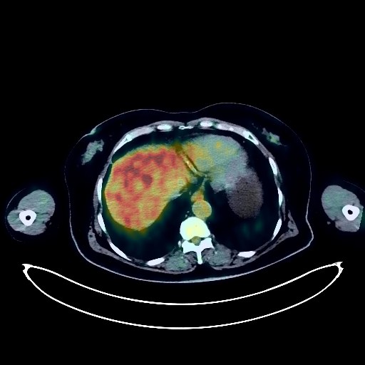
Cervical Cancer PET/CT (case 983827-000025 from PETWB-REP)
0 views10 days agoWhole-body 18F-FDG PET/CT scan in a patient with Cervical Cancer taken from the PETWB-REP dataset. The following English report (translated from original Chinese) is taken verbatim from the public dataset and has not been modified or otherwise checked for accuracy (see the end for citation). Impression a. A slightly low-density mass in the cervix with increased FDG metabolism, consistent with cervical cancer, involving the upper vagina and uterine cavity effusion; please confirm with MRI. b. Left pelvic wall lymph node metastasis is highly probable. Multiple chronic inflammatory nodules in both lungs; please follow up with CT. Bilateral pulmonary fibrosis. Reactive hyperplasia of hilar and mediastinal lymph nodes in both lungs. Partial arteriosclerosis (including coronary arteries). Bilateral breast hyperplasia. Cyst or hemangioma in the left lobe of the liver. Bilateral renal cysts. Nodular FDG hypermetabolic foci in the lower rectum, possibly due to inflammatory uptake; local space-occupying lesion to be ruled out; continuous FDG metabolism increase in the remaining intestinal segments, possibly due to physiological uptake or chronic inflammation; please confirm with endoscopy. Osteoporosis. Slight scoliosis with degenerative changes. Schmorl's nodes at the L2-4 vertebral margins. L3/4 and L4/5 intervertebral disc herniation; partial cervical, thoracic, and lumbar intervertebral disc degeneration with pneumoconiosis. Bilateral frozen shoulder. A low-density nodule with calcification in the right lobe of the thyroid gland; FDG metabolism was normal, suggesting possible nodular goiter. Ultrasound follow-up is recommended to rule out other possibilities. Bilateral reactive hyperplasia of cervical lymph nodes. A few lacunar ischemic foci in the deep brain regions of both sides, indicative of age-related brain disorders. This case is from PETWB-REP, a curated dataset of whole-body 18F-FDG PET/CT scans and corresponding radiology reports from 490 patients with a broad spectrum of malignancies. The data were retrospectively collected from patients who underwent clinically indicated whole-body 18F-FDG PET/CT scans at the Shanghai Universal Medical Imaging Diagnostic Center between 2021 and 2024. License: Creative Commons Attribution 4.0 International (CC BY 4.0) Citation: Xue, L., Feng, G., Wenbo, Z., Zhang, Y., Li, L., Wang, S., Peng, L., Peng, S., & Gao, X. (2026). PETWB-REP: A Multi-Cancer Whole-Body FDG PET/CT Dataset with Corresponding Radiology Reports [Data set]. Zenodo. https://doi.org/10.5281/zenodo.18670487
Whole BodyPET/CT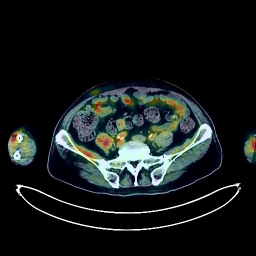
Lung Cancer PET/CT (case 983827-000125 from PETWB-REP)
0 views10 days agoWhole-body 18F-FDG PET/CT scan in a patient with Lung Cancer taken from the PETWB-REP dataset. The following English report (translated from original Chinese) is taken verbatim from the public dataset and has not been modified or otherwise checked for accuracy (see the end for citation). Impression a. A mass in the right middle lobe of the lung with increased FDG metabolism, suggestive of lung cancer. b. Multiple muscle metastases throughout the body (see description for details); linear FDG increase in the right plantar fossa, suggesting possible tonic uptake; please correlate with clinical findings and follow up. c. Right hilar lymph node metastasis. Left hilar and mediastinal lymph node metastasis to be ruled out. Symmetrical increased FDG metabolism in the bilateral lateral pterygoid muscles, suggestive of tonic uptake. Several small, solid, chronic inflammatory nodules in both lungs. Minor chronic inflammation and old lesions in both lungs. Calcification of some arterial walls (including coronary arteries). Mild anterior slippage of the L4 vertebral body and mild posterior slippage of the L5 vertebral body. Degenerative changes in the spine. L4/5 and L5/S1 intervertebral disc bulges. A few ischemic lesions in the deep bilateral brain regions, indicative of age-related brain disorders. This case is from PETWB-REP, a curated dataset of whole-body 18F-FDG PET/CT scans and corresponding radiology reports from 490 patients with a broad spectrum of malignancies. The data were retrospectively collected from patients who underwent clinically indicated whole-body 18F-FDG PET/CT scans at the Shanghai Universal Medical Imaging Diagnostic Center between 2021 and 2024. License: Creative Commons Attribution 4.0 International (CC BY 4.0) Citation: Xue, L., Feng, G., Wenbo, Z., Zhang, Y., Li, L., Wang, S., Peng, L., Peng, S., & Gao, X. (2026). PETWB-REP: A Multi-Cancer Whole-Body FDG PET/CT Dataset with Corresponding Radiology Reports [Data set]. Zenodo. https://doi.org/10.5281/zenodo.18670487
Whole BodyPET/CT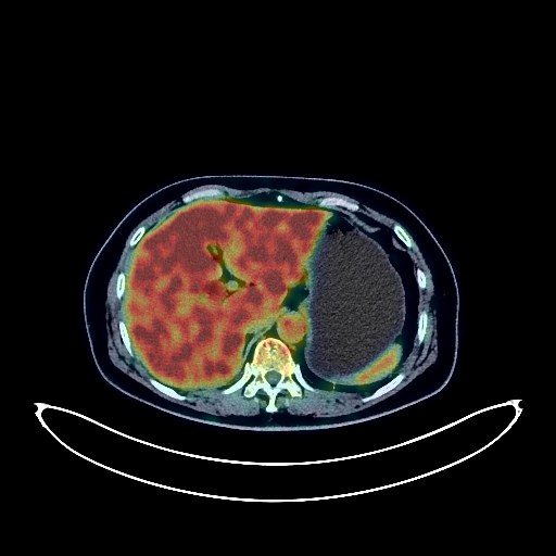
Lung Cancer PET/CT (case 983827-000016 from PETWB-REP)
0 views10 days agoWhole-body 18F-FDG PET/CT scan in a patient with Lung Cancer taken from the PETWB-REP dataset. The following English report (translated from original Chinese) is taken verbatim from the public dataset and has not been modified or otherwise checked for accuracy (see the end for citation). Impression a. An irregular soft tissue density mass with increased FDG metabolism in the right upper deep cervical space, suggestive of malignancy, more likely primary than metastatic. The lesion involves the adjacent submandibular gland; please correlate with clinical findings and tissue biopsy. b. Full morphology of the bilateral oropharyngeal lateral walls with increased FDG uptake, suggesting possible chronic inflammation or physiological changes; specialist examination recommended. a. Mixed ground-glass nodules with slight FDG uptake in the apical segment of the right upper lobe and the posterior segment of the right lower lobe, highly suggestive of invasive adenocarcinoma (AIC). b. Small inflammatory nodules in the right upper lobe; please follow up with CT scan. Scattered chronic inflammation and remnants in both lungs. Reactive hyperplasia of bilateral hilar and mediastinal lymph nodes. Partial arteriosclerosis (including coronary arteries). Thickening of the intestinal wall in the hepatic flexure of the ascending colon, protruding into the lumen in a nodular manner with increased FDG metabolism, suggesting possible polyps; colonoscopy recommended to rule out malignancy. Post-cholecystectomy, the common bile duct is widened, and the lower segment appears to be cup-shaped. FDG showed no abnormal uptake, suggesting possible post-operative changes. MRI is recommended to rule out space-occupying lesions. Multiple small cysts in the liver. Osteoporosis. Degenerative changes in the spine. Mild L5/S1 intervertebral disc bulge. Cranial scintigraphy showed no obvious abnormalities. A small amount of chronic inflammation in the right maxillary sinus. This case is from PETWB-REP, a curated dataset of whole-body 18F-FDG PET/CT scans and corresponding radiology reports from 490 patients with a broad spectrum of malignancies. The data were retrospectively collected from patients who underwent clinically indicated whole-body 18F-FDG PET/CT scans at the Shanghai Universal Medical Imaging Diagnostic Center between 2021 and 2024. License: Creative Commons Attribution 4.0 International (CC BY 4.0) Citation: Xue, L., Feng, G., Wenbo, Z., Zhang, Y., Li, L., Wang, S., Peng, L., Peng, S., & Gao, X. (2026). PETWB-REP: A Multi-Cancer Whole-Body FDG PET/CT Dataset with Corresponding Radiology Reports [Data set]. Zenodo. https://doi.org/10.5281/zenodo.18670487
Whole BodyPET/CT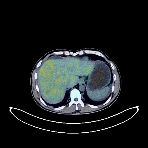
Liver Cancer PET/CT (case 983827-000110 from PETWB-REP)
0 views10 days agoWhole-body 18F-FDG PET/CT scan in a patient with Liver Cancer taken from the PETWB-REP dataset. The following English report (translated from original Chinese) is taken verbatim from the public dataset and has not been modified or otherwise checked for accuracy (see the end for citation). Impression a. Multiple space-occupying lesions in the liver with increased FDG metabolism, highly suggestive of primary liver cancer with portal vein tumor thrombus formation. Please combine clinical findings with enhanced MRI for comprehensive analysis. b. Changes in liver cirrhosis, slightly enlarged spleen, portal hypertension with collateral circulation formation; possible reactive hyperplasia of the hilar and retroperitoneal lymph nodes, follow-up recommended; ascites and pelvic effusion. Small solid nodules in the lower lobes of both lungs, FDG metabolism normal, suggestive of inflammatory nodules, CT follow-up recommended to rule out other possibilities. A few post-inflammatory lesions in both lungs. Mild anemia. Chronic cholecystitis. Left adrenal hyperplasia. Bilateral renal calculi. Prostatic calcification. Possible chronic inflammatory changes in part of the gastric wall and intestinal tract, please combine endoscopic follow-up. Mild vertebral osteophyte formation. Bilateral sacroiliac joint condensing osteitis. Cranial scintigraphy showed no obvious abnormalities. This case is from PETWB-REP, a curated dataset of whole-body 18F-FDG PET/CT scans and corresponding radiology reports from 490 patients with a broad spectrum of malignancies. The data were retrospectively collected from patients who underwent clinically indicated whole-body 18F-FDG PET/CT scans at the Shanghai Universal Medical Imaging Diagnostic Center between 2021 and 2024. License: Creative Commons Attribution 4.0 International (CC BY 4.0) Citation: Xue, L., Feng, G., Wenbo, Z., Zhang, Y., Li, L., Wang, S., Peng, L., Peng, S., & Gao, X. (2026). PETWB-REP: A Multi-Cancer Whole-Body FDG PET/CT Dataset with Corresponding Radiology Reports [Data set]. Zenodo. https://doi.org/10.5281/zenodo.18670487
Whole BodyPET/CT