Loading...
Lung Cancer PET/CT (case 983827-000144 from PETWB-REP)
0 views9 days agoWhole-body 18F-FDG PET/CT scan in a patient with Lung Cancer taken from the PETWB-REP dataset. The following English report (translated from original Chinese) is taken verbatim from the public dataset and has not been modified or otherwise checked for accuracy (see the end for citation). Impression a. A mass in the left upper lobe of the lung, with increased FDG metabolism, suggestive of lung cancer with obstructive atelectasis. Metastasis to lymph nodes in the bilateral hilar, mediastinal, right lower lobe basal segment, peribronchial, and pancreatic head regions. b. Several small, solid, chronic inflammatory nodules in both lungs. Scattered chronic inflammation and old lesions in both lungs. Slight thickening of the pleura bilaterally. Calcification of some arterial walls (including coronary arteries). Multiple cysts in the liver. Chronic cholecystitis; gallstones in the neck of the gallbladder. Bilateral adrenal hyperplasia is possible; follow-up CT is recommended. Slight thickening of the lower esophageal wall near the cardia, with increased FDG metabolism, suggestive of inflammation; slight thickening of the ascending colon wall with increased FDG metabolism, suggestive of inflammatory or physiological uptake. Please follow up with endoscopy. Uniform density in both lobes of the thyroid gland, increased FDG metabolism, suggestive of nodular goiter; ultrasound and thyroid function follow-up are recommended. Degenerative changes in the spine. L4/5 and L5/S1 intervertebral disc bulge. Right shoulder periarthritis. Symmetrical increased FDG metabolism in the bilateral costovertebral joint areas, suggestive of brown adipose tissue uptake. A few ischemic lesions in the deep bilateral brain regions, suggestive of age-related brain changes. This case is from PETWB-REP, a curated dataset of whole-body 18F-FDG PET/CT scans and corresponding radiology reports from 490 patients with a broad spectrum of malignancies. The data were retrospectively collected from patients who underwent clinically indicated whole-body 18F-FDG PET/CT scans at the Shanghai Universal Medical Imaging Diagnostic Center between 2021 and 2024. License: Creative Commons Attribution 4.0 International (CC BY 4.0) Citation: Xue, L., Feng, G., Wenbo, Z., Zhang, Y., Li, L., Wang, S., Peng, L., Peng, S., & Gao, X. (2026). PETWB-REP: A Multi-Cancer Whole-Body FDG PET/CT Dataset with Corresponding Radiology Reports [Data set]. Zenodo. https://doi.org/10.5281/zenodo.18670487
Whole BodyPET/CT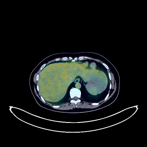
Lung Cancer PET/CT (case 983827-000243 from PETWB-REP)
0 views9 days agoWhole-body 18F-FDG PET/CT scan in a patient with Lung Cancer taken from the PETWB-REP dataset. The following English report (translated from original Chinese) is taken verbatim from the public dataset and has not been modified or otherwise checked for accuracy (see the end for citation). Impression a. A mass in the posterior segment of the right lower lobe, with increased FDG metabolism, suggestive of lung cancer, accompanied by distal obstructive changes. Please confirm the diagnosis with pathological examination. Right hilar lymph node metastasis. b. Ground-glass nodule in the posterior segment of the left lower lobe, with normal FDG metabolism, likely atypical adenomatous hyperplasia. HRCT follow-up at 6-12 months is recommended to rule out other possibilities. c. Solid micronodules in both lungs, with normal FDG metabolism, likely chronic inflammatory micronodules. Please follow up with CT. Sequelae of pneumonia in both lungs. Small amount of pleural effusion on the right side. Bilateral breast hyperplasia; please follow up with ultrasound. Increased FDG metabolism in parts of the stomach wall and intestines, suggestive of inflammatory or physiological uptake. Colonoscopy follow-up is recommended. Liver calcifications. Fatty liver. Small uterine fibroids may be large. Increased FDG metabolism in the left adnexal region is likely a physiological change; please follow up with ultrasound. Small amount of pelvic effusion. Cranial FDG imaging showed no abnormalities; MRI follow-up is recommended. This case is from PETWB-REP, a curated dataset of whole-body 18F-FDG PET/CT scans and corresponding radiology reports from 490 patients with a broad spectrum of malignancies. The data were retrospectively collected from patients who underwent clinically indicated whole-body 18F-FDG PET/CT scans at the Shanghai Universal Medical Imaging Diagnostic Center between 2021 and 2024. License: Creative Commons Attribution 4.0 International (CC BY 4.0) Citation: Xue, L., Feng, G., Wenbo, Z., Zhang, Y., Li, L., Wang, S., Peng, L., Peng, S., & Gao, X. (2026). PETWB-REP: A Multi-Cancer Whole-Body FDG PET/CT Dataset with Corresponding Radiology Reports [Data set]. Zenodo. https://doi.org/10.5281/zenodo.18670487
Whole BodyPET/CT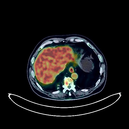
Lung Cancer PET/CT (case 983827-000180 from PETWB-REP)
0 views9 days agoWhole-body 18F-FDG PET/CT scan in a patient with Lung Cancer taken from the PETWB-REP dataset. The following English report (translated from original Chinese) is taken verbatim from the public dataset and has not been modified or otherwise checked for accuracy (see the end for citation). Impression a. Space-occupying lesion in the lower lobe of the right lung, with significantly increased FDG metabolism, consistent with lung cancer with obstructive atelectasis based on pathology. b. Old fibrosis and calcifications in the upper and posterior segments of the lower lobe of the left lung. Partial arteriosclerosis (including coronary arteries). Chronic inflammatory changes in the lower esophagus and stomach; gastroscopy follow-up may be necessary. Hiatal hernia. Liver cyst. Prostatic calcifications. Degenerative changes in the spine, instability of the L1, L2, and L5 vertebral bodies with bilateral pars interarticularis fractures at L5. Right-sided frozen shoulder. No obvious abnormalities were found on cranial scintigraphy. This case is from PETWB-REP, a curated dataset of whole-body 18F-FDG PET/CT scans and corresponding radiology reports from 490 patients with a broad spectrum of malignancies. The data were retrospectively collected from patients who underwent clinically indicated whole-body 18F-FDG PET/CT scans at the Shanghai Universal Medical Imaging Diagnostic Center between 2021 and 2024. License: Creative Commons Attribution 4.0 International (CC BY 4.0) Citation: Xue, L., Feng, G., Wenbo, Z., Zhang, Y., Li, L., Wang, S., Peng, L., Peng, S., & Gao, X. (2026). PETWB-REP: A Multi-Cancer Whole-Body FDG PET/CT Dataset with Corresponding Radiology Reports [Data set]. Zenodo. https://doi.org/10.5281/zenodo.18670487
Whole BodyPET/CT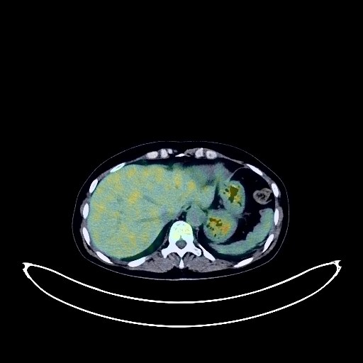
Ovarian Cancer PET/CT (case 983827-000111 from PETWB-REP)
0 views9 days agoWhole-body 18F-FDG PET/CT scan in a patient with Ovarian Cancer taken from the PETWB-REP dataset. The following English report (translated from original Chinese) is taken verbatim from the public dataset and has not been modified or otherwise checked for accuracy (see the end for citation). Impression Bilateral adnexal soft tissue lesions with increased FDG metabolism, increased FDG metabolism in the endometrial area and upper cervix, all suggestive of malignancy, with a high probability of cancer. Please confirm with pathology. Reactive hyperplasia of retroperitoneal and mesenteric lymph nodes is highly probable; please follow up to rule out other possibilities. a. Ground-glass nodule in the apical-posterior segment of the left upper lobe is suggestive of atypical adenomatous hyperplasia, early lung cancer to be ruled out; HRCT follow-up every six months is recommended. Ground-glass nodules in the apical segment of the right upper lobe and the posterior segment of the right lower lobe are suggestive of chronic inflammatory nodules or atypical adenomatous hyperplasia; please confirm with annual HRCT follow-up. b. Chronic inflammatory nodules (solid) in the right upper lobe and both lower lobes. Bilateral pulmonary fibrosis. Reactive hyperplasia of both hilar and mediastinal lymph nodes. Micropleural effusion bilaterally. Bilateral breast hyperplasia; ultrasound follow-up is recommended. Liver cysts, liver calcifications. Cholestasis of the gallbladder. Increased FDG metabolism in parts of the stomach wall and intestines, possibly due to physiological uptake or chronic inflammation; please follow up with endoscopy. Spinal degeneration. Lumbar instability. L5 and S1 vertebral endplate inflammation. L3/4 and L4/5 intervertebral disc bulging, L5/S1 intervertebral disc pneumatosis and degeneration. No obvious abnormalities seen on cranial scintigraphy. Reactive hyperplasia of bilateral deep cervical interspaces, submandibular, and left supraclavicular lymph nodes. This case is from PETWB-REP, a curated dataset of whole-body 18F-FDG PET/CT scans and corresponding radiology reports from 490 patients with a broad spectrum of malignancies. The data were retrospectively collected from patients who underwent clinically indicated whole-body 18F-FDG PET/CT scans at the Shanghai Universal Medical Imaging Diagnostic Center between 2021 and 2024. License: Creative Commons Attribution 4.0 International (CC BY 4.0) Citation: Xue, L., Feng, G., Wenbo, Z., Zhang, Y., Li, L., Wang, S., Peng, L., Peng, S., & Gao, X. (2026). PETWB-REP: A Multi-Cancer Whole-Body FDG PET/CT Dataset with Corresponding Radiology Reports [Data set]. Zenodo. https://doi.org/10.5281/zenodo.18670487
Whole BodyPET/CT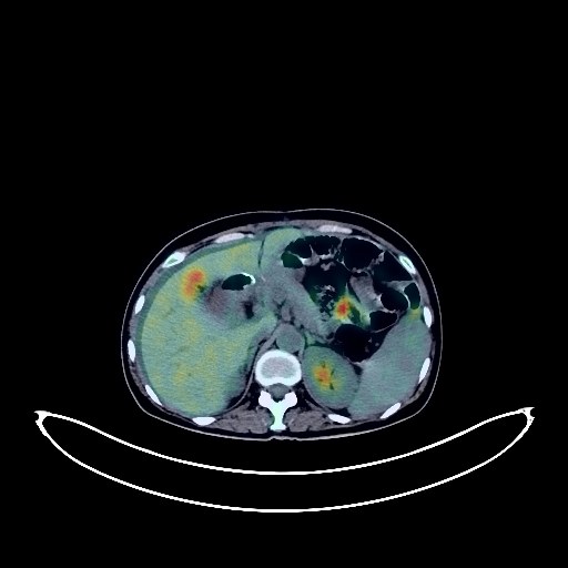
Gastric Cancer PET/CT (case 983827-000177 from PETWB-REP)
0 views9 days agoWhole-body 18F-FDG PET/CT scan in a patient with Gastric Cancer taken from the PETWB-REP dataset. The following English report (translated from original Chinese) is taken verbatim from the public dataset and has not been modified or otherwise checked for accuracy (see the end for citation). Impression Post-gastric cancer surgery, slight thickening of the intestinal wall at the anastomosis site and increased FDG metabolism suggest anastomotic inflammation or physiological uptake; follow-up gastroscopy is recommended. Large cystic-solid mass in the abdominopelvic cavity with increased FDG metabolism in the cyst wall, suggestive of ovarian cystadenoma, ovarian cancer to be ruled out; please correlate with clinicopathology. Small amount of fluid in the abdominopelvic cavity. Several small chronic inflammatory nodules in the right lung; follow-up is recommended to rule out other mixed nodules. A small amount of chronic inflammation and old lesions in both lungs. Calcification of some arterial walls (including coronary arteries). Liver cyst. Chronic cholecystitis; gallstones. Continuous increased FDG metabolism in the colon and rectum, suggestive of inflammatory or physiological uptake; colonoscopy is recommended. Mild degenerative changes in the spine. L4/5, L5/S1 intervertebral disc bulge. Increased FDG uptake throughout the bone marrow suggests reactive bone marrow hyperplasia. Cranial scintigraphy showed no abnormalities. This case is from PETWB-REP, a curated dataset of whole-body 18F-FDG PET/CT scans and corresponding radiology reports from 490 patients with a broad spectrum of malignancies. The data were retrospectively collected from patients who underwent clinically indicated whole-body 18F-FDG PET/CT scans at the Shanghai Universal Medical Imaging Diagnostic Center between 2021 and 2024. License: Creative Commons Attribution 4.0 International (CC BY 4.0) Citation: Xue, L., Feng, G., Wenbo, Z., Zhang, Y., Li, L., Wang, S., Peng, L., Peng, S., & Gao, X. (2026). PETWB-REP: A Multi-Cancer Whole-Body FDG PET/CT Dataset with Corresponding Radiology Reports [Data set]. Zenodo. https://doi.org/10.5281/zenodo.18670487
Whole BodyPET/CT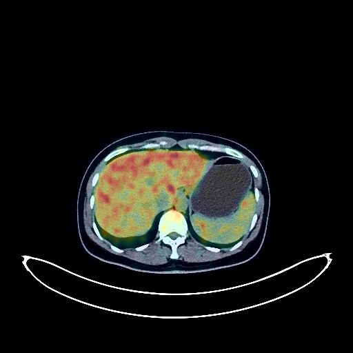
Glioma PET/CT (case 983827-000024 from PETWB-REP)
0 views9 days agoWhole-body 18F-FDG PET/CT scan in a patient with Glioma taken from the PETWB-REP dataset. The following English report (translated from original Chinese) is taken verbatim from the public dataset and has not been modified or otherwise checked for accuracy (see the end for citation). Impression A mass in the left hippocampus with increased FDG metabolism, suggestive of malignancy, accompanied by peritumoral edema and slight rightward midline deviation. Please combine with enhanced MRI for comprehensive assessment. Ground-glass nodule in the apical segment of the right upper lobe, with normal FDG metabolism, suggestive of chronic inflammatory nodule or atypical adenomatous hyperplasia. Please combine with annual follow-up via HRCT. Bilateral breast hyperplasia. Reactive hyperplasia of bilateral axillary lymph nodes. Increased FDG metabolism in the endometrial area after intrauterine device insertion, likely a physiological change. Please combine with ultrasound follow-up. L4/5 and L5/S1 intervertebral disc bulge. Chronic inflammation of the nasopharynx and bilateral palatine tonsils is highly probable. Please combine with specialist examination. This case is from PETWB-REP, a curated dataset of whole-body 18F-FDG PET/CT scans and corresponding radiology reports from 490 patients with a broad spectrum of malignancies. The data were retrospectively collected from patients who underwent clinically indicated whole-body 18F-FDG PET/CT scans at the Shanghai Universal Medical Imaging Diagnostic Center between 2021 and 2024. License: Creative Commons Attribution 4.0 International (CC BY 4.0) Citation: Xue, L., Feng, G., Wenbo, Z., Zhang, Y., Li, L., Wang, S., Peng, L., Peng, S., & Gao, X. (2026). PETWB-REP: A Multi-Cancer Whole-Body FDG PET/CT Dataset with Corresponding Radiology Reports [Data set]. Zenodo. https://doi.org/10.5281/zenodo.18670487
Whole BodyPET/CT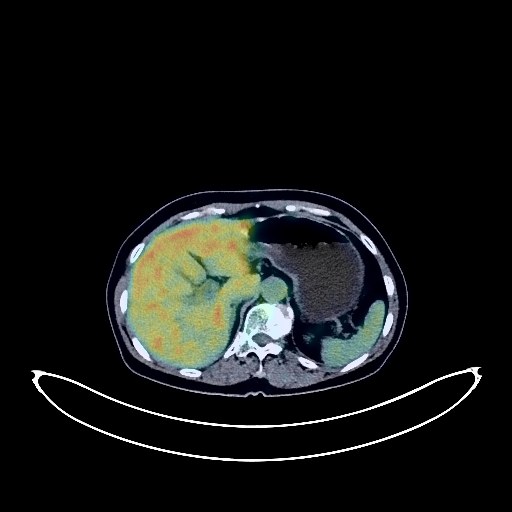
Lung Cancer PET/CT (case 983827-000020 from PETWB-REP)
0 views9 days agoWhole-body 18F-FDG PET/CT scan in a patient with Lung Cancer taken from the PETWB-REP dataset. The following English report (translated from original Chinese) is taken verbatim from the public dataset and has not been modified or otherwise checked for accuracy (see the end for citation). Impression a. Soft tissue mass in the medial segment of the right middle lobe with increased FDG metabolism, highly suggestive of peripheral lung cancer; please confirm with pathology. b. Scattered ground-glass nodules in both lungs, mixed ground-glass nodule in the posterior basal segment of the left lower lobe, no increased FDG metabolism, suggestive of atypical adenomatous hyperplasia or chronic inflammatory nodule; bilateral chronic inflammatory nodules (solid); please follow up with CT. Scattered chronic inflammation and sequelae in both lungs. Calcification in the left upper lobe. A few ischemic lesions deep in the brain; please confirm with MRI. Low-density nodule in the right lobe of the thyroid gland, no increased FDG metabolism, suggestive of benign; please follow up with ultrasound. Cyst in the right lobe of the liver. Intrahepatic calcification. Scattered subcutaneous calcifications in the abdominopelvic cavity and bilateral buttocks. Subcutaneous inflammatory nodule in the right buttock. Scoliosis with degeneration. L2-S1 intervertebral disc bulge, L4/5 intervertebral disc posterior margin calcification, L5/S1 intervertebral disc gas accumulation. This case is from PETWB-REP, a curated dataset of whole-body 18F-FDG PET/CT scans and corresponding radiology reports from 490 patients with a broad spectrum of malignancies. The data were retrospectively collected from patients who underwent clinically indicated whole-body 18F-FDG PET/CT scans at the Shanghai Universal Medical Imaging Diagnostic Center between 2021 and 2024. License: Creative Commons Attribution 4.0 International (CC BY 4.0) Citation: Xue, L., Feng, G., Wenbo, Z., Zhang, Y., Li, L., Wang, S., Peng, L., Peng, S., & Gao, X. (2026). PETWB-REP: A Multi-Cancer Whole-Body FDG PET/CT Dataset with Corresponding Radiology Reports [Data set]. Zenodo. https://doi.org/10.5281/zenodo.18670487
Whole BodyPET/CT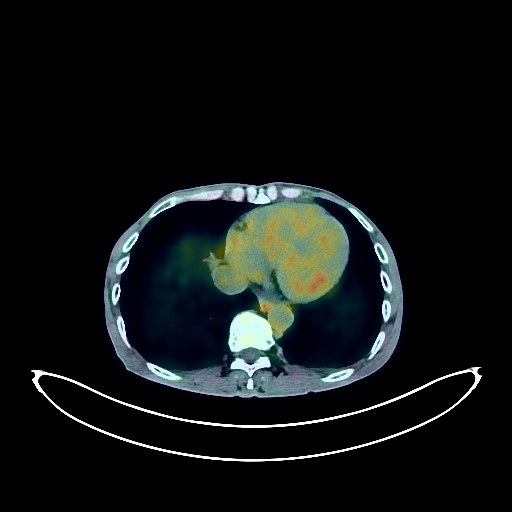
Renal Cancer PET/CT (case 983827-000052 from PETWB-REP)
0 views9 days agoWhole-body 18F-FDG PET/CT scan in a patient with Renal Cancer taken from the PETWB-REP dataset. The following English report (translated from original Chinese) is taken verbatim from the public dataset and has not been modified or otherwise checked for accuracy (see the end for citation). Impression Left renal mass with elevated FDG metabolism, suggestive of renal cell carcinoma; please correlate with clinicopathology; left renal vein and inferior vena cava tumor thrombus formation. Chronic inflammatory nodules in both lungs. Bilateral emphysema, scattered post-inflammatory lesions in both lungs. Minor bilateral pleural reaction. Anemia changes, partial arterial wall calcification (including coronary arteries). Small cyst in the right lobe of the liver. Chronic cholecystitis. Accessory spleen. Right renal cyst. Benign prostatic hyperplasia with calcification. Small amount of hydrocele in both testes, with calcification on the right side. Chronic inflammatory changes in part of the gastric wall and intestines. Osteoporosis, degenerative changes in the spine, multiple intervertebral disc bulges, L4/5 and L5/S1 intervertebral disc pneumodegenerative changes. Bilateral pars interarticularis fracture of the L4 vertebral body, anterior slippage of the vertebral body. Right shoulder periarthritis. Age-related brain, deep lacunar infarcts. Chronic inflammation of both ethmoid sinuses and the left maxillary sinus; submucosal cyst of the left maxillary sinus; right maxillary alveolar ulcer. This case is from PETWB-REP, a curated dataset of whole-body 18F-FDG PET/CT scans and corresponding radiology reports from 490 patients with a broad spectrum of malignancies. The data were retrospectively collected from patients who underwent clinically indicated whole-body 18F-FDG PET/CT scans at the Shanghai Universal Medical Imaging Diagnostic Center between 2021 and 2024. License: Creative Commons Attribution 4.0 International (CC BY 4.0) Citation: Xue, L., Feng, G., Wenbo, Z., Zhang, Y., Li, L., Wang, S., Peng, L., Peng, S., & Gao, X. (2026). PETWB-REP: A Multi-Cancer Whole-Body FDG PET/CT Dataset with Corresponding Radiology Reports [Data set]. Zenodo. https://doi.org/10.5281/zenodo.18670487
Whole BodyPET/CT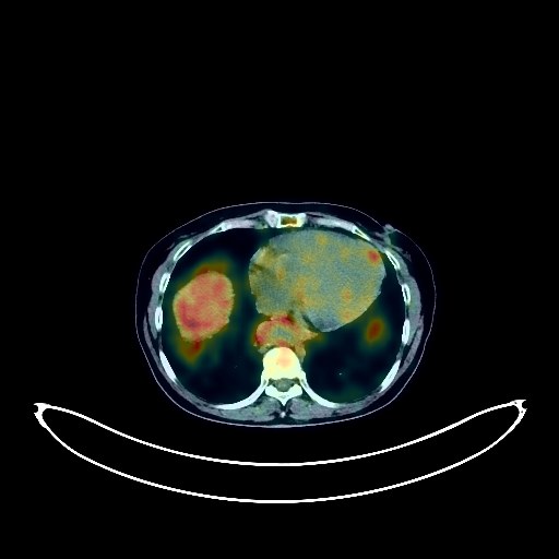
Ovarian Cancer PET/CT (case 983827-000235 from PETWB-REP)
0 views9 days agoWhole-body 18F-FDG PET/CT scan in a patient with Ovarian Cancer taken from the PETWB-REP dataset. The following English report (translated from original Chinese) is taken verbatim from the public dataset and has not been modified or otherwise checked for accuracy (see the end for citation). Impression a. Extensive peritoneal thickening and increased density with nodules and patchy shadows, elevated FDG metabolism, implantation metastasis is the primary consideration; please correlate with clinicopathology. Abdominal and pelvic effusion. b. Right adnexal region mass with elevated FDG metabolism, considered malignant tumor, primary tumor is the primary consideration, metastasis to be ruled out; enhanced MRI is recommended for further examination. Chronic inflammatory micronodules in both lungs. A few post-inflammatory lesions in both lungs. Reactive hyperplasia of hilar, mediastinal, and left axillary lymph nodes. Widening of the ascending aorta, with partial calcification of the arterial wall (including coronary arteries). Left renal cyst. Uterine fibroid to be ruled out. Reactive hyperplasia of right anterior diaphragmatic and right cardiophrenic angle lymph nodes. Partial chronic inflammatory changes in the gastric wall. Degenerative changes in the spine, L4/5 and L5/S1 intervertebral disc bulges. Age-related brain abnormalities, deep lacunar infarcts. Inflammation of the base of the tongue and bilateral palatine tonsils. Reactive hyperplasia of bilateral cervical lymph nodes. This case is from PETWB-REP, a curated dataset of whole-body 18F-FDG PET/CT scans and corresponding radiology reports from 490 patients with a broad spectrum of malignancies. The data were retrospectively collected from patients who underwent clinically indicated whole-body 18F-FDG PET/CT scans at the Shanghai Universal Medical Imaging Diagnostic Center between 2021 and 2024. License: Creative Commons Attribution 4.0 International (CC BY 4.0) Citation: Xue, L., Feng, G., Wenbo, Z., Zhang, Y., Li, L., Wang, S., Peng, L., Peng, S., & Gao, X. (2026). PETWB-REP: A Multi-Cancer Whole-Body FDG PET/CT Dataset with Corresponding Radiology Reports [Data set]. Zenodo. https://doi.org/10.5281/zenodo.18670487
Whole BodyPET/CT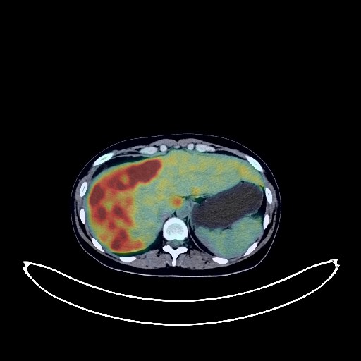
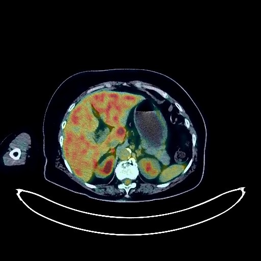
Liver Cancer PET/CT (case 983827-000190 from PETWB-REP)
0 views9 days agoWhole-body 18F-FDG PET/CT scan in a patient with Liver Cancer taken from the PETWB-REP dataset. The following English report (translated from original Chinese) is taken verbatim from the public dataset and has not been modified or otherwise checked for accuracy (see the end for citation). Impression a. Multiple space-occupying lesions in the liver with increased FDG metabolism. Combined with the enhanced MRI images from another hospital on June 7, 2023, malignancy is highly likely, possibly epithelioid hemangioendothelioma or sarcoma. A biopsy is recommended. b. A few punctate and linear shadows in the fat space adjacent to the right lobe of the liver and the right paracolic gutter, peritoneal seeding metastasis should be suspected. Follow-up CT scan is recommended. Abdominal and pelvic effusion. c. Solid nodules in the bilateral adnexa, irregular in shape, with slightly increased FDG metabolism. Neoplasticity needs to be ruled out. Further enhanced MRI is recommended. Physiological uptake in the uterine cavity is highly likely. d. Reactive hyperplasia of retroperitoneal, mesenteric, and bilateral inguinal lymph nodes. e. Scattered multiple solid nodules of varying sizes in both lungs, with smooth margins, are highly suggestive of metastatic tumors. Follow-up CT scan is recommended. a. Multiple soft tissue lesions in both breasts, with slightly increased FDG metabolism. Based on the ultrasound report and MRI from another hospital, fibroadenoma is suspected. Please correlate with clinical findings and perform a biopsy if necessary to confirm the diagnosis. b. Reactive hyperplasia of bilateral axillary lymph nodes, including one enlarged lymph node in the left axilla with increased FDG metabolism. Please follow up with ultrasound to rule out metastasis. A few fibrotic lesions in the right middle lobe of the lung. Incomplete thymic regression. Gastric antrum in a contracted state, no increased FDG metabolism observed. Physiological uptake of some intestinal segments is highly likely. A linear shadow with increased FDG metabolism in the muscle layer of the left upper wall, considered to be physiological or inflammatory uptake. Please correlate with clinical findings. L5/S1 disc herniation. No obvious abnormalities were found on cranial scintigraphy. This case is from PETWB-REP, a curated dataset of whole-body 18F-FDG PET/CT scans and corresponding radiology reports from 490 patients with a broad spectrum of malignancies. The data were retrospectively collected from patients who underwent clinically indicated whole-body 18F-FDG PET/CT scans at the Shanghai Universal Medical Imaging Diagnostic Center between 2021 and 2024. License: Creative Commons Attribution 4.0 International (CC BY 4.0) Citation: Xue, L., Feng, G., Wenbo, Z., Zhang, Y., Li, L., Wang, S., Peng, L., Peng, S., & Gao, X. (2026). PETWB-REP: A Multi-Cancer Whole-Body FDG PET/CT Dataset with Corresponding Radiology Reports [Data set]. Zenodo. https://doi.org/10.5281/zenodo.18670487
Whole BodyPET/CT