Loading...
Lung Cancer PET/CT (case 983827-000214 from PETWB-REP)
0 views9 days agoWhole-body 18F-FDG PET/CT scan in a patient with Lung Cancer taken from the PETWB-REP dataset. The following English report (translated from original Chinese) is taken verbatim from the public dataset and has not been modified or otherwise checked for accuracy (see the end for citation). Impression a. A mass in the anterior segment of the left upper lobe with increased FDG metabolism, suggestive of lung cancer with obstructive changes; please correlate with clinicopathology. Reactive hyperplasia of mediastinal lymph nodes is highly probable, but mixed metastasis needs to be ruled out. b. Chronic inflammatory micronodules or atypical adenomatous hyperplasia in both lungs; annual HRCT follow-up is recommended. A few post-inflammatory lesions in both lungs. Tracheal diverticulum. Calcification of some arterial walls (including coronary arteries). Liver cyst. Post-cholecystectomy changes. Right kidney stone. Small cyst in the left kidney. Benign prostatic hyperplasia with calcification. Small amount of hydrocele in the left testis. Chronic inflammatory changes in the gastric antrum; please correlate with endoscopic follow-up. Degenerative changes in the spine, L4/5 and L5/S1 intervertebral disc bulges. Right ischial tuberosity inflammation. Lipoma on the right anterior chest wall. No obvious abnormalities were found on cranial scintigraphy. Calcification lesion in the right palatine tonsil. This case is from PETWB-REP, a curated dataset of whole-body 18F-FDG PET/CT scans and corresponding radiology reports from 490 patients with a broad spectrum of malignancies. The data were retrospectively collected from patients who underwent clinically indicated whole-body 18F-FDG PET/CT scans at the Shanghai Universal Medical Imaging Diagnostic Center between 2021 and 2024. License: Creative Commons Attribution 4.0 International (CC BY 4.0) Citation: Xue, L., Feng, G., Wenbo, Z., Zhang, Y., Li, L., Wang, S., Peng, L., Peng, S., & Gao, X. (2026). PETWB-REP: A Multi-Cancer Whole-Body FDG PET/CT Dataset with Corresponding Radiology Reports [Data set]. Zenodo. https://doi.org/10.5281/zenodo.18670487
Whole BodyPET/CT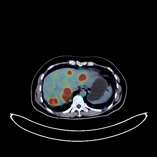
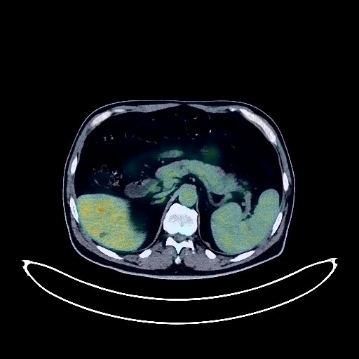
Gallbladder Cancer PET/CT (case 983827-000139 from PETWB-REP)
0 views9 days agoWhole-body 18F-FDG PET/CT scan in a patient with Gallbladder Cancer taken from the PETWB-REP dataset. The following English report (translated from original Chinese) is taken verbatim from the public dataset and has not been modified or otherwise checked for accuracy (see the end for citation). Impression a. Irregular low-density mass in the gallbladder area, multiple intrahepatic lesions, elevated FDG metabolism, suggestive of malignancy, likely gallbladder cancer with multiple intrahepatic metastases; please correlate with clinicopathology. b. Multiple metastatic tumors in both lungs. Multiple lymph node metastases in the left hilum, mediastinum, hepatic hilum, hepatogastric space, retroperitoneum, and local superior mesenteric region. Multiple implantation metastases in the pelvic cavity. Bilateral ovarian metastases are highly probable. Dilatation of some intrahepatic bile ducts. Dilatation of the pancreatic duct in the head of the pancreas. Liver cysts. Elevated FDG metabolism in the rectum and ileocecal region, suggestive of physiological or inflammatory uptake; please correlate with colonoscopy follow-up. Uterine fibroids, intrauterine device visible. Subcutaneous inflammatory nodule in the right buttock. No obvious abnormalities were found on cranial scintigraphy. This case is from PETWB-REP, a curated dataset of whole-body 18F-FDG PET/CT scans and corresponding radiology reports from 490 patients with a broad spectrum of malignancies. The data were retrospectively collected from patients who underwent clinically indicated whole-body 18F-FDG PET/CT scans at the Shanghai Universal Medical Imaging Diagnostic Center between 2021 and 2024. License: Creative Commons Attribution 4.0 International (CC BY 4.0) Citation: Xue, L., Feng, G., Wenbo, Z., Zhang, Y., Li, L., Wang, S., Peng, L., Peng, S., & Gao, X. (2026). PETWB-REP: A Multi-Cancer Whole-Body FDG PET/CT Dataset with Corresponding Radiology Reports [Data set]. Zenodo. https://doi.org/10.5281/zenodo.18670487
Whole BodyPET/CT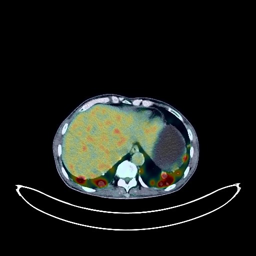
Renal Cancer PET/CT (case 983827-000051 from PETWB-REP)
0 views9 days agoWhole-body 18F-FDG PET/CT scan in a patient with Renal Cancer taken from the PETWB-REP dataset. The following English report (translated from original Chinese) is taken verbatim from the public dataset and has not been modified or otherwise checked for accuracy (see the end for citation). Impression a. Bone destruction and soft tissue mass formation in the right mandibular ramus with increased FDG uptake, suggestive of a metastatic tumor. Multiple bone metastases throughout the body. b. Multiple lymph node metastases in the right submandibular region, bilateral supraclavicular fossa, mediastinum, hepatogastric space, hilar space, and retroperitoneum. Multiple lung metastases. c. Enlarged left adrenal gland with increased FDG metabolism, suggestive of a metastatic tumor. A large mass in the right kidney, suggestive of a malignant tumor, most likely renal cell carcinoma. The right adrenal gland is not clearly visualized. Benign prostatic hyperplasia with calcification. Osteophyte formation in some vertebrae. L4/5 and L5/S1 intervertebral disc bulges. No obvious abnormalities were found on cranial scintigraphy. This case is from PETWB-REP, a curated dataset of whole-body 18F-FDG PET/CT scans and corresponding radiology reports from 490 patients with a broad spectrum of malignancies. The data were retrospectively collected from patients who underwent clinically indicated whole-body 18F-FDG PET/CT scans at the Shanghai Universal Medical Imaging Diagnostic Center between 2021 and 2024. License: Creative Commons Attribution 4.0 International (CC BY 4.0) Citation: Xue, L., Feng, G., Wenbo, Z., Zhang, Y., Li, L., Wang, S., Peng, L., Peng, S., & Gao, X. (2026). PETWB-REP: A Multi-Cancer Whole-Body FDG PET/CT Dataset with Corresponding Radiology Reports [Data set]. Zenodo. https://doi.org/10.5281/zenodo.18670487
Whole BodyPET/CT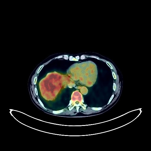
Prostate Cancer PET/CT (case 983827-000185 from PETWB-REP)
0 views9 days agoWhole-body 18F-FDG PET/CT scan in a patient with Prostate Cancer taken from the PETWB-REP dataset. The following English report (translated from original Chinese) is taken verbatim from the public dataset and has not been modified or otherwise checked for accuracy (see the end for citation). Impression a. Benign prostatic hyperplasia (BPH), with uneven glandular density and increased FDG metabolism, consistent with prostate cancer, involving the left seminal vesicle and adjacent bladder wall, and the distal left ureter, leading to hydronephrosis of the ureter and renal pelvis. b. Possible reactive hyperplasia of left pelvic wall lymph nodes, metastasis to be ruled out. c. Uneven density of the right intertrochanteric region of the femur, increased FDG metabolism; follow-up is recommended to rule out metastasis. Pituitary lesion, increased FDG uptake, suggestive of pituitary tumor; enhanced MRI is recommended for further examination to rule out other possibilities. a. Possible several chronic inflammatory micronodules in both lungs, some metastasis to be ruled out; regular HRCT follow-up is recommended for comparison. A few chronic inflammations and old lesions in both lungs. b. Reactive hyperplasia of hilar and mediastinal lymph nodes in both lungs. Some arterial wall calcification (including coronary arteries). Cervical, thoracic, and lumbar spondylosis. L4/5 and L5/S1 intervertebral disc bulges. A few ischemic lesions in the deep bilateral cerebral regions, indicative of age-related encephalopathy. This case is from PETWB-REP, a curated dataset of whole-body 18F-FDG PET/CT scans and corresponding radiology reports from 490 patients with a broad spectrum of malignancies. The data were retrospectively collected from patients who underwent clinically indicated whole-body 18F-FDG PET/CT scans at the Shanghai Universal Medical Imaging Diagnostic Center between 2021 and 2024. License: Creative Commons Attribution 4.0 International (CC BY 4.0) Citation: Xue, L., Feng, G., Wenbo, Z., Zhang, Y., Li, L., Wang, S., Peng, L., Peng, S., & Gao, X. (2026). PETWB-REP: A Multi-Cancer Whole-Body FDG PET/CT Dataset with Corresponding Radiology Reports [Data set]. Zenodo. https://doi.org/10.5281/zenodo.18670487
Whole BodyPET/CT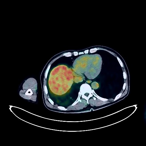
Lung Cancer PET/CT (case 983827-000106 from PETWB-REP)
0 views9 days agoWhole-body 18F-FDG PET/CT scan in a patient with Lung Cancer taken from the PETWB-REP dataset. The following English report (translated from original Chinese) is taken verbatim from the public dataset and has not been modified or otherwise checked for accuracy (see the end for citation). Impression a. Irregular soft tissue nodules with increased FDG metabolism in the apical-posterior segment of the left upper lobe, highly suggestive of lung cancer; please correlate with clinicopathology. Possible metastasis to the right upper tracheal lymph nodes; reactive hyperplasia of the hilar and remaining mediastinal lymph nodes is highly likely, pending further investigation of mixed metastases. b. Chronic inflammatory micronodules in the remaining two lungs; CT follow-up is recommended to rule out other mixed metastases. Emphysema in both lungs. Scattered post-inflammatory lesions in both lungs. Anemia changes, post-coronary stent placement changes, partial arteriosclerosis. Low-density masses in the right lateral ventricle trigone and frontoparietal lobe, with absent FDG uptake, suggestive of malignancy; metastasis is highly probable. Cystic mass in the head of the pancreas, possibly a cyst; MRI with contrast enhancement is recommended for comprehensive analysis. Irregular kidney shape in both kidneys. Benign prostatic hyperplasia. Chronic inflammatory changes or physiological uptake in some intestinal segments; endoscopic follow-up is recommended. Degenerative changes in the spine, multiple bulging lumbar intervertebral discs. Bilateral hip periarthritis. This case is from PETWB-REP, a curated dataset of whole-body 18F-FDG PET/CT scans and corresponding radiology reports from 490 patients with a broad spectrum of malignancies. The data were retrospectively collected from patients who underwent clinically indicated whole-body 18F-FDG PET/CT scans at the Shanghai Universal Medical Imaging Diagnostic Center between 2021 and 2024. License: Creative Commons Attribution 4.0 International (CC BY 4.0) Citation: Xue, L., Feng, G., Wenbo, Z., Zhang, Y., Li, L., Wang, S., Peng, L., Peng, S., & Gao, X. (2026). PETWB-REP: A Multi-Cancer Whole-Body FDG PET/CT Dataset with Corresponding Radiology Reports [Data set]. Zenodo. https://doi.org/10.5281/zenodo.18670487
Whole BodyPET/CT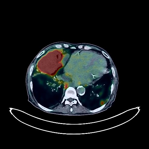
Lung Cancer PET/CT (case 983827-000148 from PETWB-REP)
0 views9 days agoWhole-body 18F-FDG PET/CT scan in a patient with Lung Cancer taken from the PETWB-REP dataset. The following English report (translated from original Chinese) is taken verbatim from the public dataset and has not been modified or otherwise checked for accuracy (see the end for citation). Impression a. Irregular mass in the right middle lobe with increased FDG metabolism, suggestive of lung cancer with obstructive changes; multiple lymph node metastases in the bilateral hilar, mediastinal, right supraclavicular fossa, right supradiaphragmatic, and right posterior diaphragmatic angle spaces. b. Multiple metastatic tumors in both lungs; bilateral pleural metastases, bilateral pleural effusions (partially loculated). c. Multiple bone metastases throughout the body (see description for details). Multiple bronchial wall thickenings in the upper and lower lobes of the right lung, multiple bronchiectasis in both lungs. Emphysema in both lungs, bullae in the left upper lobe. Chronic inflammation and post-inflammatory remnants in both lungs. Pericardial thickening with a small amount of effusion, anemia. Calcification of some arterial walls (including coronary arteries). Small liver cysts. Chronic cholecystitis, gallstones. Left renal cyst, left renal calculus. Prostatic calcifications. Left inguinal hernia. Right testicular tunica vaginalis calcifications. Partial chronic inflammatory changes in the gastric wall. Degenerative changes in the spine, bilateral L4 spondylolysis with anterior vertebral slippage, multiple intervertebral disc bulges. Age-related brain, deep lacunar infarcts. Chronic inflammation of the left sphenoid sinus, bilateral ethmoid sinuses, and bilateral maxillary sinuses; left maxillary sinus submucosal cyst. This case is from PETWB-REP, a curated dataset of whole-body 18F-FDG PET/CT scans and corresponding radiology reports from 490 patients with a broad spectrum of malignancies. The data were retrospectively collected from patients who underwent clinically indicated whole-body 18F-FDG PET/CT scans at the Shanghai Universal Medical Imaging Diagnostic Center between 2021 and 2024. License: Creative Commons Attribution 4.0 International (CC BY 4.0) Citation: Xue, L., Feng, G., Wenbo, Z., Zhang, Y., Li, L., Wang, S., Peng, L., Peng, S., & Gao, X. (2026). PETWB-REP: A Multi-Cancer Whole-Body FDG PET/CT Dataset with Corresponding Radiology Reports [Data set]. Zenodo. https://doi.org/10.5281/zenodo.18670487
Whole BodyPET/CT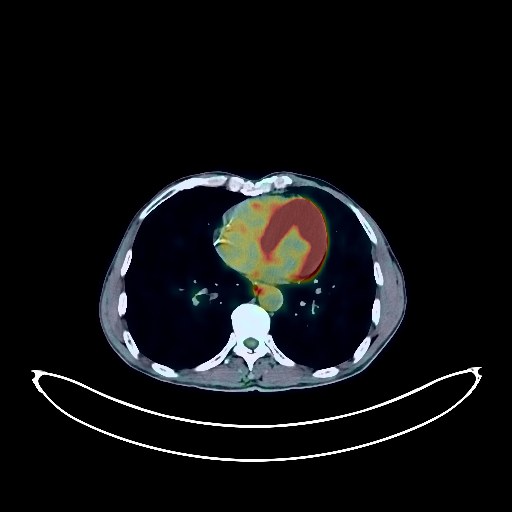
Lung Cancer PET/CT (case 983827-000261 from PETWB-REP)
0 views9 days agoWhole-body 18F-FDG PET/CT scan in a patient with Lung Cancer taken from the PETWB-REP dataset. The following English report (translated from original Chinese) is taken verbatim from the public dataset and has not been modified or otherwise checked for accuracy (see the end for citation). Impression a. Following chemotherapy for squamous cell carcinoma of the left upper lung, a mass is observed in the apical-posterior segment and lingular segment of the left upper lobe, with unevenly increased FDG metabolism in the solid component, suggesting continued tumor activity. There is slight inflammation around the lesion. b. Increased FDG metabolism is observed in the hilar lymph nodes of both lungs; no enlarged lymph nodes are seen in the mediastinum. Follow-up is recommended. c. Bilateral emphysema and scattered chronic inflammatory nodules (solid) in both lungs are present. Follow-up with CT scan is recommended. Calcification in the basal segment of the right lower lobe. A few post-inflammatory remnants in both lungs. Partial arteriosclerosis (including coronary arteries). Anemia is present. Slightly eccentric thickening of the rectal wall in the middle and lower segments, with increased FDG metabolism, suggests inflammation is highly likely; neoplastic lesions should be ruled out. Colonoscopy is recommended for clarification. Increased FDG metabolism in the remaining colon and rectum suggests physiological uptake or chronic inflammatory changes. Chronic gastritis. Left adrenal hyperplasia is highly probable; follow-up CT scan is recommended. Liver cyst. Left kidney cyst. Benign prostatic hyperplasia with calcification. Degenerative changes in the spine. Schmorl's node at the upper margin of the L5 vertebral body; L4/5 and L5/S1 intervertebral disc bulge with pneumoconiosis and degeneration. Age-related brain changes; deep lacunar infarcts in the brain; MRI is recommended. Minor inflammation of both ethmoid sinuses. This case is from PETWB-REP, a curated dataset of whole-body 18F-FDG PET/CT scans and corresponding radiology reports from 490 patients with a broad spectrum of malignancies. The data were retrospectively collected from patients who underwent clinically indicated whole-body 18F-FDG PET/CT scans at the Shanghai Universal Medical Imaging Diagnostic Center between 2021 and 2024. License: Creative Commons Attribution 4.0 International (CC BY 4.0) Citation: Xue, L., Feng, G., Wenbo, Z., Zhang, Y., Li, L., Wang, S., Peng, L., Peng, S., & Gao, X. (2026). PETWB-REP: A Multi-Cancer Whole-Body FDG PET/CT Dataset with Corresponding Radiology Reports [Data set]. Zenodo. https://doi.org/10.5281/zenodo.18670487
Whole BodyPET/CT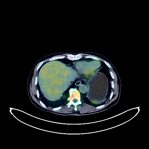
Prostate Cancer PET/CT (case 983827-000234 from PETWB-REP)
0 views9 days agoWhole-body 18F-FDG PET/CT scan in a patient with Prostate Cancer taken from the PETWB-REP dataset. The following English report (translated from original Chinese) is taken verbatim from the public dataset and has not been modified or otherwise checked for accuracy (see the end for citation). Impression a. Benign prostatic hyperplasia with calcification, prostatic mass with increased FDG metabolism, suggestive of prostate cancer. b. Multiple lymph node metastases in the bilateral pelvic walls, bilateral iliac vessels, presacral region, and retroperitoneum. c. Multiple bone metastases throughout the body (see description), involvement of the spinal canal and sacral canal at the T7 and T11 levels, pathological fracture of the T11 vertebra. Multiple lung metastases. Scattered post-inflammatory lesions in both lungs. Reactive hyperplasia of mediastinal lymph nodes. Calcification of some arterial walls (including coronary arteries). Mild fatty liver, calcification in the right lobe of the liver, hepatic cysts. Right renal calculus, small renal cysts in both kidneys. Bladder calculus, catheter in place. Right inguinal hernia. Calcification of the tunica vaginalis in the left testis. Chronic inflammatory changes or physiological uptake in the gastric antrum and part of the intestine; please follow up with endoscopy. Degenerative changes in the spine, multiple intervertebral disc bulges. Multiple thyroid nodules, some with calcification, increased FDG metabolism, suggestive of nodular goiter. Age-related brain, deep lacunar infarcts. Bilateral chronic ethmoid sinusitis. This case is from PETWB-REP, a curated dataset of whole-body 18F-FDG PET/CT scans and corresponding radiology reports from 490 patients with a broad spectrum of malignancies. The data were retrospectively collected from patients who underwent clinically indicated whole-body 18F-FDG PET/CT scans at the Shanghai Universal Medical Imaging Diagnostic Center between 2021 and 2024. License: Creative Commons Attribution 4.0 International (CC BY 4.0) Citation: Xue, L., Feng, G., Wenbo, Z., Zhang, Y., Li, L., Wang, S., Peng, L., Peng, S., & Gao, X. (2026). PETWB-REP: A Multi-Cancer Whole-Body FDG PET/CT Dataset with Corresponding Radiology Reports [Data set]. Zenodo. https://doi.org/10.5281/zenodo.18670487
Whole BodyPET/CT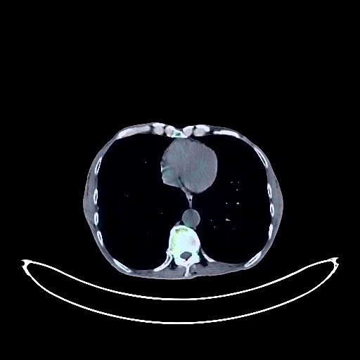
Pancreatic Cancer PET/CT (case 983827-000168 from PETWB-REP)
0 views9 days agoWhole-body 18F-FDG PET/CT scan in a patient with Pancreatic Cancer taken from the PETWB-REP dataset. The following English report (translated from original Chinese) is taken verbatim from the public dataset and has not been modified or otherwise checked for accuracy (see the end for citation). Impression Pancreatic mass with increased FDG metabolism, suggestive of pancreatic cancer with biliary obstruction and obstructive pancreatitis. Please correlate with clinicopathology. Peripancreatic and mesenteric lymph nodes show increased FDG metabolism, possibly indicating metastasis. Please correlate with clinical findings. a. Multiple solid nodules, plaque-like lesions, calcifications, and linear lesions in both lungs, some with increased FDG metabolism, suggestive of pulmonary tuberculosis. Some are slightly mobile; follow-up CT is recommended. b. Chronic bronchitis with bilateral emphysema. Scattered post-inflammatory lesions in both lungs. Partial pleural thickening bilaterally. Reactive hyperplasia of hilar and mediastinal lymph nodes bilaterally. Calcification of some arterial walls (including coronary arteries). Left adrenal hyperplasia. Bilateral renal calculi. Benign prostatic hyperplasia with calcification. Possible chronic inflammatory changes in part of the gastric wall; please follow up with endoscopy. Osteoporosis, degenerative changes in the spine, L4/5 and L5/S1 intervertebral disc bulge. Left pubic tubercle bony island. Unevenly increased FDG metabolism throughout the medullary cavity, likely due to reactive proliferative changes; please correlate with clinical findings. Age-related brain, deep lacunar infarcts; please correlate with MRI. Right inferior alveolar alveolitis. This case is from PETWB-REP, a curated dataset of whole-body 18F-FDG PET/CT scans and corresponding radiology reports from 490 patients with a broad spectrum of malignancies. The data were retrospectively collected from patients who underwent clinically indicated whole-body 18F-FDG PET/CT scans at the Shanghai Universal Medical Imaging Diagnostic Center between 2021 and 2024. License: Creative Commons Attribution 4.0 International (CC BY 4.0) Citation: Xue, L., Feng, G., Wenbo, Z., Zhang, Y., Li, L., Wang, S., Peng, L., Peng, S., & Gao, X. (2026). PETWB-REP: A Multi-Cancer Whole-Body FDG PET/CT Dataset with Corresponding Radiology Reports [Data set]. Zenodo. https://doi.org/10.5281/zenodo.18670487
Whole BodyPET/CT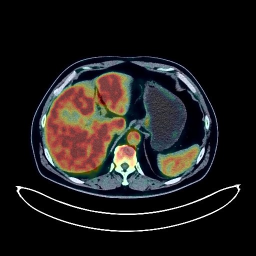
Nasopharyngeal Cancer PET/CT (case 983827-000267 from PETWB-REP)
0 views9 days agoWhole-body 18F-FDG PET/CT scan in a patient with Nasopharyngeal Cancer taken from the PETWB-REP dataset. The following English report (translated from original Chinese) is taken verbatim from the public dataset and has not been modified or otherwise checked for accuracy (see the end for citation). Impression a. Soft tissue lesions on the posterior and right lateral walls of the nasopharynx with increased FDG uptake, involving both posterior nasal apertures, consistent with nasopharyngeal carcinoma. b. Right retropharyngeal lymph node metastasis. Metastasis to the bilateral deep cervical spaces, left posterior cervical triangle, and part of the left supraclavicular lymph nodes needs to be ruled out; please follow up. c. Bilateral ethmoid and maxillary sinusitis. Nasal cavity inflammation. Right otitis media and mastoiditis. Multiple lesions in the left anterior and middle mediastinum with increased FDG uptake; malignancy is the primary consideration, with high-risk thymoma and lymph node metastasis being possible. Comprehensive analysis with contrast-enhanced MRI is recommended. Multiple solid micronodules in both lungs with normal FDG uptake, suggestive of chronic inflammatory nodules; please follow up with CT. Scattered chronic inflammation and remnants in both lungs. Calcification of some arterial walls (including coronary arteries). Fatty liver, punctate calcifications in the right lobe of the liver. Mild fatty infiltration of the pancreas. Bilateral renal cysts. Prostatic calcification. Physiological changes or inflammatory lesions in the gastric antrum; follow-up gastroscopy is recommended. Spinal degenerative changes. No obvious abnormalities were found on cranial FDG imaging. This case is from PETWB-REP, a curated dataset of whole-body 18F-FDG PET/CT scans and corresponding radiology reports from 490 patients with a broad spectrum of malignancies. The data were retrospectively collected from patients who underwent clinically indicated whole-body 18F-FDG PET/CT scans at the Shanghai Universal Medical Imaging Diagnostic Center between 2021 and 2024. License: Creative Commons Attribution 4.0 International (CC BY 4.0) Citation: Xue, L., Feng, G., Wenbo, Z., Zhang, Y., Li, L., Wang, S., Peng, L., Peng, S., & Gao, X. (2026). PETWB-REP: A Multi-Cancer Whole-Body FDG PET/CT Dataset with Corresponding Radiology Reports [Data set]. Zenodo. https://doi.org/10.5281/zenodo.18670487
Whole BodyPET/CT