Loading...
Lymphoma PET/CT (case 983827-000015 from PETWB-REP)
1 views9 days agoWhole-body 18F-FDG PET/CT scan in a patient with Lymphoma taken from the PETWB-REP dataset. The following English report (translated from original Chinese) is taken verbatim from the public dataset and has not been modified or otherwise checked for accuracy (see the end for citation). Impression Post-lymphoma treatment: a. Multiple lymph nodes throughout the body showing increased FDG metabolism, suggestive of lymphoma infiltration; comparison with previous imaging data and follow-up are recommended. b. No space-occupying lesions were seen in the nasal cavity; FDG metabolism was normal, suggesting suppressed tumor activity. Old tuberculosis in both lungs; CT follow-up is recommended. Scattered post-inflammatory lesions in both lungs. Bilateral breast hyperplasia. Physiological uptake in the endometrial area is possible; please combine with specialist examination. Chronic inflammatory changes in the lower esophagus, gastric antrum, and part of the intestine; please combine with endoscopic follow-up. Mild osteophyte formation in the spine. Uneven thyroid density and increased FDG metabolism suggest possible inflammation; ultrasound follow-up is recommended. No obvious abnormalities were seen on cranial scintigraphy. This case is from PETWB-REP, a curated dataset of whole-body 18F-FDG PET/CT scans and corresponding radiology reports from 490 patients with a broad spectrum of malignancies. The data were retrospectively collected from patients who underwent clinically indicated whole-body 18F-FDG PET/CT scans at the Shanghai Universal Medical Imaging Diagnostic Center between 2021 and 2024. License: Creative Commons Attribution 4.0 International (CC BY 4.0) Citation: Xue, L., Feng, G., Wenbo, Z., Zhang, Y., Li, L., Wang, S., Peng, L., Peng, S., & Gao, X. (2026). PETWB-REP: A Multi-Cancer Whole-Body FDG PET/CT Dataset with Corresponding Radiology Reports [Data set]. Zenodo. https://doi.org/10.5281/zenodo.18670487
Whole BodyPET/CT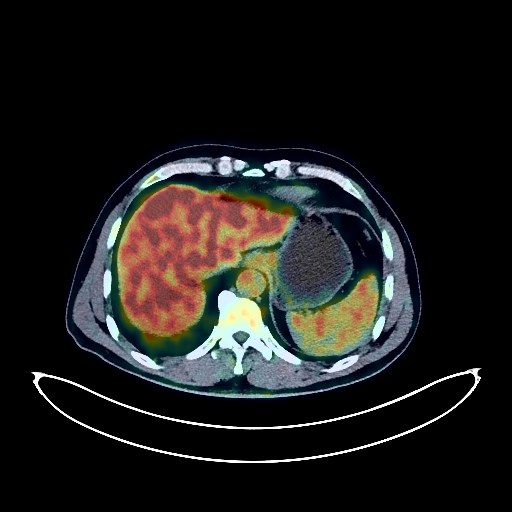
Lung Cancer PET/CT (case 983827-000027 from PETWB-REP)
0 views9 days agoWhole-body 18F-FDG PET/CT scan in a patient with Lung Cancer taken from the PETWB-REP dataset. The following English report (translated from original Chinese) is taken verbatim from the public dataset and has not been modified or otherwise checked for accuracy (see the end for citation). Impression a. A cavitary lesion with increased FDG metabolism in the posterior segment of the left upper lobe, highly suggestive of lung cancer, but atypical inflammatory granuloma cannot be ruled out. Please combine clinical examinations and pathology. b. Scattered ground-glass nodules in the anterior segment of the left upper lobe and the apical segment of the right upper lobe, without increased FDG metabolism, suggestive of atypical adenomatous hyperplasia or chronic inflammatory nodules. Please follow up with CT. Chronic inflammatory nodule in the posterior segment of the left upper lobe. Possible right adrenal adenoma. Please compare with CT to rule out metastasis. Right renal calculus. Prostatic calcification. Calcification of the tunica vaginalis in the left testis. Partial vertebral osteophyte formation. L4/5 and L5/S1 intervertebral disc bulge. Increased FDG metabolism in the bone marrow of the left upper femur. MRI follow-up is recommended. A few ischemic lesions in the deep brain. Bilateral maxillary sinusitis. Chronic inflammation of the right mastoid process. This case is from PETWB-REP, a curated dataset of whole-body 18F-FDG PET/CT scans and corresponding radiology reports from 490 patients with a broad spectrum of malignancies. The data were retrospectively collected from patients who underwent clinically indicated whole-body 18F-FDG PET/CT scans at the Shanghai Universal Medical Imaging Diagnostic Center between 2021 and 2024. License: Creative Commons Attribution 4.0 International (CC BY 4.0) Citation: Xue, L., Feng, G., Wenbo, Z., Zhang, Y., Li, L., Wang, S., Peng, L., Peng, S., & Gao, X. (2026). PETWB-REP: A Multi-Cancer Whole-Body FDG PET/CT Dataset with Corresponding Radiology Reports [Data set]. Zenodo. https://doi.org/10.5281/zenodo.18670487
Whole BodyPET/CT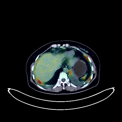
Ovarian Cancer PET/CT (case 983827-000162 from PETWB-REP)
1 views9 days agoWhole-body 18F-FDG PET/CT scan in a patient with Ovarian Cancer taken from the PETWB-REP dataset. The following English report (translated from original Chinese) is taken verbatim from the public dataset and has not been modified or otherwise checked for accuracy (see the end for citation). Impression a. Bilateral adnexal region cystic-solid lesions, with increased FDG metabolism in the solid portion, suggestive of malignancy, most likely ovarian cancer; please correlate with clinicopathology. b. Multiple peritoneal seeding metastases in the abdominopelvic cavity. Multiple lymph node metastases in the bilateral pelvic walls, retroperitoneum, cardiophrenic angle, and left internal mammary chain. Bilateral pleural metastases. Small amount of effusion in the abdomen and pelvis. c. Soft tissue nodule near the diaphragm in the left lower lobe, with increased FDG metabolism, highly suggestive of metastasis. Several small chronic inflammatory nodules (solid) in both lungs are possible; CT scan is recommended to rule out metastasis. A few chronic inflammations and old lesions in both lungs. Reactive hyperplasia of the left hilar lymph nodes is possible; follow-up is recommended. Small amount of pleural effusion in both cavities, with partial atelectasis in the left lower lobe. Bilateral breast proliferative changes. Mild osteophyte formation in the cervical, thoracic, and lumbar spine. L4/5 and L5/S1 intervertebral disc bulge. No abnormalities found on cranial scintigraphy. Submucosal cyst of the left maxillary sinus. This case is from PETWB-REP, a curated dataset of whole-body 18F-FDG PET/CT scans and corresponding radiology reports from 490 patients with a broad spectrum of malignancies. The data were retrospectively collected from patients who underwent clinically indicated whole-body 18F-FDG PET/CT scans at the Shanghai Universal Medical Imaging Diagnostic Center between 2021 and 2024. License: Creative Commons Attribution 4.0 International (CC BY 4.0) Citation: Xue, L., Feng, G., Wenbo, Z., Zhang, Y., Li, L., Wang, S., Peng, L., Peng, S., & Gao, X. (2026). PETWB-REP: A Multi-Cancer Whole-Body FDG PET/CT Dataset with Corresponding Radiology Reports [Data set]. Zenodo. https://doi.org/10.5281/zenodo.18670487
Whole BodyPET/CT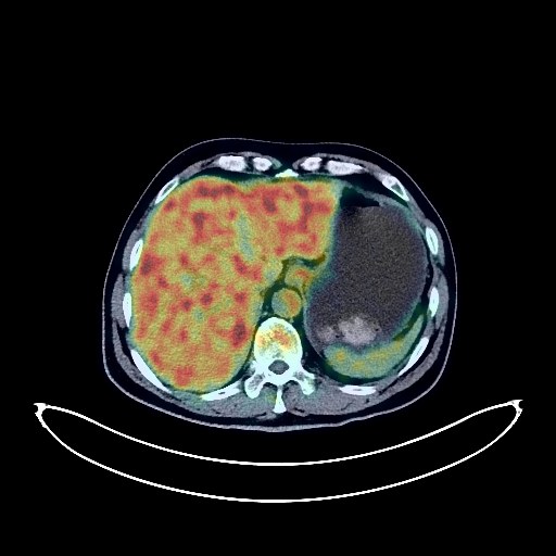
Lung Cancer PET/CT (case 983827-000002 from PETWB-REP)
0 views9 days agoWhole-body 18F-FDG PET/CT scan in a patient with Lung Cancer taken from the PETWB-REP dataset. The following English report (translated from original Chinese) is taken verbatim from the public dataset and has not been modified or otherwise checked for accuracy (see the end for citation). Impression Comparison of PET/CT scan of left lung cancer after chemotherapy with the scan taken on December 17, 2021 at our center: a. Soft tissue patchy lesions in the left lower hilum: The lesions have significantly shrunk compared to the previous scan, and the FDG metabolic value has significantly decreased compared to the previous scan, suggesting post-treatment changes and suppression of tumor activity. Bronchiectasis in the left lower lobe, with a small amount of distal inflammation. b. Lymph nodes in the left hilum and below the carina: The lesions have shrunk in size compared to the previous scan, and the FDG metabolic value has increased, suggesting partial suppression of tumor activity. c. Lymph nodes in the anterior superior mediastinum, the space between the great vessels, and the right lateral tracheal inlet: Some have higher density and calcification, with slightly increased FDG uptake, suggesting possible post-treatment changes (partial suppression) of metastatic lymph nodes. Please correlate with clinical findings. d. Slightly low-density shadow near the diaphragm in the right lobe of the liver: This is a newly added lesion compared to the previous scan. No abnormal FDG uptake was observed, suggesting possible artifact. Space-occupying lesions need to be ruled out. Further MRI examination is recommended. Calcification lesions in the anterior segment of the right upper lobe. Bilateral reactive adrenal hyperplasia, please follow up. Partial vertebral osteophyte formation. L3/4, L4/5, and L5/S1 intervertebral disc bulge. L5/S1 intervertebral disc pneumothorax. No abnormal FDG metabolism observed in the entire skeletal system. A few ischemic foci deep within the brain. Bilateral deep cervical lymph node reactive hyperplasia. This case is from PETWB-REP, a curated dataset of whole-body 18F-FDG PET/CT scans and corresponding radiology reports from 490 patients with a broad spectrum of malignancies. The data were retrospectively collected from patients who underwent clinically indicated whole-body 18F-FDG PET/CT scans at the Shanghai Universal Medical Imaging Diagnostic Center between 2021 and 2024. License: Creative Commons Attribution 4.0 International (CC BY 4.0) Citation: Xue, L., Feng, G., Wenbo, Z., Zhang, Y., Li, L., Wang, S., Peng, L., Peng, S., & Gao, X. (2026). PETWB-REP: A Multi-Cancer Whole-Body FDG PET/CT Dataset with Corresponding Radiology Reports [Data set]. Zenodo. https://doi.org/10.5281/zenodo.18670487
Whole BodyPET/CT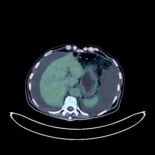
Bladder Cancer PET/CT (case 983827-000226 from PETWB-REP)
0 views9 days agoWhole-body 18F-FDG PET/CT scan in a patient with Bladder Cancer taken from the PETWB-REP dataset. The following English report (translated from original Chinese) is taken verbatim from the public dataset and has not been modified or otherwise checked for accuracy (see the end for citation). Impression Postoperative changes after bladder cancer surgery: a. A large soft tissue mass in the pelvic cavity with unevenly increased FDG metabolism, suggesting tumor recurrence or metastasis. The prostate is poorly visualized; please confirm with pathology. b. Partial uneven thickening of the peritoneum and mesentery in the upper and middle abdomen with mildly increased FDG metabolism, suggesting possible reactive hyperplasia; please follow up to rule out mixed metastases. c. Multiple dense bone shadows throughout the body (see description for details), FDG metabolism normal, suggesting benign bone disease; please follow up. d. Large amounts of fluid accumulation in the abdomen and pelvis, with some tissue structures poorly visualized. a. Chronic inflammatory nodules in both lungs. A few post-inflammatory remnants in both lungs, with air sacs in both lungs. Large amount of pleural effusion on the right side, with partial pulmonary edema in the right lung. Mild thickening of the left pleura. b. Calcification of some arterial walls (including coronary arteries). Mild bilateral gynecomastia. Mild fatty liver, multiple hepatic cysts, no definite space-occupying lesions. Chronic cholecystitis. Multiple renal cysts (some complex). Increased FDG metabolism in some esophageal and gastric walls, considered physiological uptake or chronic inflammatory changes; please follow up with endoscopy. Mild scoliosis with degenerative changes, L2 and L3 vertebral instability. L2-5 intervertebral disc bulge. Sacral canal cyst. A few lacunar infarcts in the deep bilateral brain, mild age-related brain changes. This case is from PETWB-REP, a curated dataset of whole-body 18F-FDG PET/CT scans and corresponding radiology reports from 490 patients with a broad spectrum of malignancies. The data were retrospectively collected from patients who underwent clinically indicated whole-body 18F-FDG PET/CT scans at the Shanghai Universal Medical Imaging Diagnostic Center between 2021 and 2024. License: Creative Commons Attribution 4.0 International (CC BY 4.0) Citation: Xue, L., Feng, G., Wenbo, Z., Zhang, Y., Li, L., Wang, S., Peng, L., Peng, S., & Gao, X. (2026). PETWB-REP: A Multi-Cancer Whole-Body FDG PET/CT Dataset with Corresponding Radiology Reports [Data set]. Zenodo. https://doi.org/10.5281/zenodo.18670487
Whole BodyPET/CT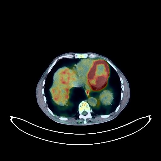
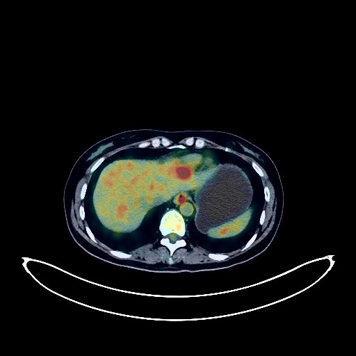
Liver Cancer PET/CT (case 983827-000090 from PETWB-REP)
0 views9 days agoWhole-body 18F-FDG PET/CT scan in a patient with Liver Cancer taken from the PETWB-REP dataset. The following English report (translated from original Chinese) is taken verbatim from the public dataset and has not been modified or otherwise checked for accuracy (see the end for citation). Impression a. Multiple intrahepatic lesions, some with increased FDG metabolism, suggesting possible hepatocellular carcinoma with multiple subfocal formation or intrahepatic metastasis. Some lesions show internal bleeding; please analyze in conjunction with MRI images from other hospitals. b. Changes consistent with cirrhosis, splenomegaly, portal hypertension with collateral circulation, and a cyst in the right lobe of the liver. Metastasis to some lymph nodes in the porta hepatis and retroperitoneum needs further investigation. Small amount of ascites. a. Ground-glass nodule in the apical segment of the right upper lobe, with normal FDG metabolism, suggesting inflammation or atypical adenomatous hyperplasia; follow-up CT is recommended. b. Chronic inflammatory micronodules in the upper lobes of both lungs. Emphysema in both lungs, calcification in the right upper lobe, and scattered post-inflammatory remnants in both lungs. c. Slight thickening of the pleura bilaterally. Anemia changes, and calcification of some arterial walls (including coronary arteries). Enlarged gallbladder. Bilateral renal cysts. Prostatic calcification. Chronic inflammatory changes or physiological uptake in the cardia, antrum of the stomach, and part of the intestine. Degenerative changes in the spine, with mild posterior slippage of the L1, L2, and L3 vertebral bodies, and multiple thoracolumbar disc bulges with pneumoconiosis and degeneration. Deep lacunar ischemic lesions in the brain, with mild age-related brain changes. Chronic inflammation of the left maxillary sinus. This case is from PETWB-REP, a curated dataset of whole-body 18F-FDG PET/CT scans and corresponding radiology reports from 490 patients with a broad spectrum of malignancies. The data were retrospectively collected from patients who underwent clinically indicated whole-body 18F-FDG PET/CT scans at the Shanghai Universal Medical Imaging Diagnostic Center between 2021 and 2024. License: Creative Commons Attribution 4.0 International (CC BY 4.0) Citation: Xue, L., Feng, G., Wenbo, Z., Zhang, Y., Li, L., Wang, S., Peng, L., Peng, S., & Gao, X. (2026). PETWB-REP: A Multi-Cancer Whole-Body FDG PET/CT Dataset with Corresponding Radiology Reports [Data set]. Zenodo. https://doi.org/10.5281/zenodo.18670487
Whole BodyPET/CT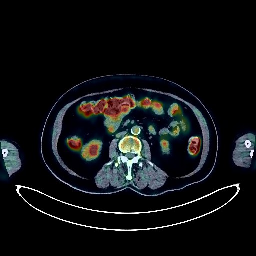
Lymphoma PET/CT (case 983827-000017 from PETWB-REP)
0 views9 days agoWhole-body 18F-FDG PET/CT scan in a patient with Lymphoma taken from the PETWB-REP dataset. The following English report (translated from original Chinese) is taken verbatim from the public dataset and has not been modified or otherwise checked for accuracy (see the end for citation). Impression a. Space-occupying lesions in the right anterior superior and middle mediastinum with increased FDG metabolism, suggestive of an invasive neoplastic lesion, possibly thymoma; please correlate with clinicopathology. b. Possible chronic lymphadenopathy in the left supraclavicular and right hilar regions; tumor infiltration to be ruled out; follow-up is recommended. Chronic inflammatory nodules in both lungs; CT follow-up is recommended. A few post-inflammatory lesions in both lungs. Partial pleural thickening on both sides. Calcification of some arterial walls (including coronary arteries). Uneven fatty liver; ultrasound follow-up is recommended. Bilateral renal cysts. Post-schistosomiasis sequelae, possibly chronic inflammatory changes in the gastric antrum and part of the intestine; please correlate with endoscopy. Osteoporosis, degenerative changes in the spine, post-lumbar spine surgery changes. Uneven thyroid density, calcification in the left lobe; ultrasound follow-up is recommended. Age-related brain, deep lacunar infarcts; please correlate with MRI. Right palatine tonsillitis. Reactive hyperplasia of bilateral cervical lymph nodes. This case is from PETWB-REP, a curated dataset of whole-body 18F-FDG PET/CT scans and corresponding radiology reports from 490 patients with a broad spectrum of malignancies. The data were retrospectively collected from patients who underwent clinically indicated whole-body 18F-FDG PET/CT scans at the Shanghai Universal Medical Imaging Diagnostic Center between 2021 and 2024. License: Creative Commons Attribution 4.0 International (CC BY 4.0) Citation: Xue, L., Feng, G., Wenbo, Z., Zhang, Y., Li, L., Wang, S., Peng, L., Peng, S., & Gao, X. (2026). PETWB-REP: A Multi-Cancer Whole-Body FDG PET/CT Dataset with Corresponding Radiology Reports [Data set]. Zenodo. https://doi.org/10.5281/zenodo.18670487
Whole BodyPET/CT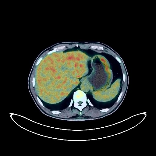
Lung Cancer PET/CT (case 983827-000021 from PETWB-REP)
0 views9 days agoWhole-body 18F-FDG PET/CT scan in a patient with Lung Cancer taken from the PETWB-REP dataset. The following English report (translated from original Chinese) is taken verbatim from the public dataset and has not been modified or otherwise checked for accuracy (see the end for citation). Impression a. Soft tissue mass near the hilum of the left upper lobe, with increased FDG uptake, suggesting a high probability of central lung cancer with distal atelectasis; bronchoscopy is recommended. b. Increased FDG metabolism in the left hilar and mediastinal lymph nodes, suggesting possible reactive proliferative lymph nodes, with partial metastasis not ruled out. Inflammatory nodules in the apical and posterior segments of the right upper lobe. Calcification of some arterial walls (including coronary arteries). Chronic cholecystitis. Chronic gastritis is possible; gastroscopy is recommended. Bilateral adrenal hyperplasia is possible; follow-up examination is recommended. Right renal cyst. Benign prostatic hyperplasia with multiple calcifications. Reactive hyperplasia of bilateral inguinal lymph nodes. Degenerative changes in the spine. Schmorl's node in the L5 vertebral body. L5-S1 intervertebral disc degeneration. Mild senile encephalopathy. Bilateral chronic maxillary sinusitis. This case is from PETWB-REP, a curated dataset of whole-body 18F-FDG PET/CT scans and corresponding radiology reports from 490 patients with a broad spectrum of malignancies. The data were retrospectively collected from patients who underwent clinically indicated whole-body 18F-FDG PET/CT scans at the Shanghai Universal Medical Imaging Diagnostic Center between 2021 and 2024. License: Creative Commons Attribution 4.0 International (CC BY 4.0) Citation: Xue, L., Feng, G., Wenbo, Z., Zhang, Y., Li, L., Wang, S., Peng, L., Peng, S., & Gao, X. (2026). PETWB-REP: A Multi-Cancer Whole-Body FDG PET/CT Dataset with Corresponding Radiology Reports [Data set]. Zenodo. https://doi.org/10.5281/zenodo.18670487
Whole BodyPET/CT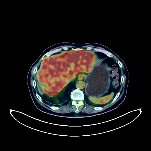
Lung Cancer PET/CT (case 983827-000081 from PETWB-REP)
0 views9 days agoWhole-body 18F-FDG PET/CT scan in a patient with Lung Cancer taken from the PETWB-REP dataset. The following English report (translated from original Chinese) is taken verbatim from the public dataset and has not been modified or otherwise checked for accuracy (see the end for citation). Impression a. Solid nodule in the right middle lobe with increased FDG metabolism, highly suggestive of peripheral lung cancer; please confirm the diagnosis with pathological examination. Possible metastasis to the right hilar and mediastinal lymph nodes. b. Large patchy high-density shadow in the right lower lobe with increased FDG metabolism, suggestive of infectious lesions; CT follow-up after regular anti-infective treatment is recommended to rule out other complications. c. Old pulmonary tuberculosis in both lungs. Chronic inflammatory nodules in both lungs. Pulmonary fibrosis and emphysema in both lungs. Mild pleural thickening bilaterally. Partial arteriosclerosis (including coronary arteries). Gallstones, chronic cholecystitis. Bilateral adrenal hyperplasia. Prostatic calcification. Spinal degeneration. L4 vertebral instability. L1 vertebral endplate inflammation. L5/S1 intervertebral disc bulge. Sellar region mass with increased FDG metabolism; enhanced MRI is recommended for further examination. Bilateral deep lacunar insufficiency lesions in the brain. Right maxillary sinusitis. This case is from PETWB-REP, a curated dataset of whole-body 18F-FDG PET/CT scans and corresponding radiology reports from 490 patients with a broad spectrum of malignancies. The data were retrospectively collected from patients who underwent clinically indicated whole-body 18F-FDG PET/CT scans at the Shanghai Universal Medical Imaging Diagnostic Center between 2021 and 2024. License: Creative Commons Attribution 4.0 International (CC BY 4.0) Citation: Xue, L., Feng, G., Wenbo, Z., Zhang, Y., Li, L., Wang, S., Peng, L., Peng, S., & Gao, X. (2026). PETWB-REP: A Multi-Cancer Whole-Body FDG PET/CT Dataset with Corresponding Radiology Reports [Data set]. Zenodo. https://doi.org/10.5281/zenodo.18670487
Whole BodyPET/CT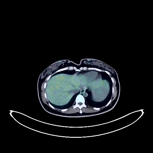
Gastric Cancer PET/CT (case 983827-000121 from PETWB-REP)
0 views9 days agoWhole-body 18F-FDG PET/CT scan in a patient with Gastric Cancer taken from the PETWB-REP dataset. The following English report (translated from original Chinese) is taken verbatim from the public dataset and has not been modified or otherwise checked for accuracy (see the end for citation). Impression a. Post-gastric cancer treatment: No abnormal FDG metabolic foci were found in the surgical area. Please combine clinical and endoscopic examinations. b. Cystic-solid mass with increased FDG metabolism in the solid component of the right adnexa, suggestive of metastasis; suspected metastasis in the left adnexa. Please combine enhanced MRI for comprehensive analysis. c. Abdominal and pelvic effusion. Reactive hyperplasia of retroperitoneal lymph nodes is highly likely; follow-up is recommended. Chronic inflammatory micronodules in the lateral basal segment of the left lower lobe. Mild thickening of the right pleura. Anemia. Bilateral breast hyperplasia. Liver calcification. Small cyst in the left lobe of the liver. Mild widening of the common bile duct; please follow up with ultrasound. Partial vertebral osteophyte formation. Slight bulging of the L4/5 and L5/S1 intervertebral discs. Cranial FDG imaging showed no obvious abnormalities, but there was a large occipital cisternaemagna or arachnoid cyst. There was also a small amount of chronic inflammation of the right maxillary sinus. This case is from PETWB-REP, a curated dataset of whole-body 18F-FDG PET/CT scans and corresponding radiology reports from 490 patients with a broad spectrum of malignancies. The data were retrospectively collected from patients who underwent clinically indicated whole-body 18F-FDG PET/CT scans at the Shanghai Universal Medical Imaging Diagnostic Center between 2021 and 2024. License: Creative Commons Attribution 4.0 International (CC BY 4.0) Citation: Xue, L., Feng, G., Wenbo, Z., Zhang, Y., Li, L., Wang, S., Peng, L., Peng, S., & Gao, X. (2026). PETWB-REP: A Multi-Cancer Whole-Body FDG PET/CT Dataset with Corresponding Radiology Reports [Data set]. Zenodo. https://doi.org/10.5281/zenodo.18670487
Whole BodyPET/CT