Loading...
Renal Cancer PET/CT (case 983827-000178 from PETWB-REP)
0 views9 days agoWhole-body 18F-FDG PET/CT scan in a patient with Renal Cancer taken from the PETWB-REP dataset. The following English report (translated from original Chinese) is taken verbatim from the public dataset and has not been modified or otherwise checked for accuracy (see the end for citation). Impression Mass in the left renal pelvis and calyces, with increased FDG metabolism, strongly suggestive of renal pelvis cancer; please correlate with clinicopathology. Dilatation and hydronephrosis of the left renal pelvis and upper ureter. Chronic inflammatory miliary nodules in both lungs; follow-up CT scan recommended. A few chronic inflammatory lesions and old lesions in both lungs. Calcification of some arterial walls. Benign prostatic hyperplasia with calcification, increased FDG metabolism in the gland; follow-up PSA and ultrasound recommended. Bilateral adrenal hyperplasia. Osteophyte formation in the cervical, thoracic, and lumbar vertebrae. L4/5 and L5/S1 intervertebral disc bulges. No abnormalities found on cranial scintigraphy. This case is from PETWB-REP, a curated dataset of whole-body 18F-FDG PET/CT scans and corresponding radiology reports from 490 patients with a broad spectrum of malignancies. The data were retrospectively collected from patients who underwent clinically indicated whole-body 18F-FDG PET/CT scans at the Shanghai Universal Medical Imaging Diagnostic Center between 2021 and 2024. License: Creative Commons Attribution 4.0 International (CC BY 4.0) Citation: Xue, L., Feng, G., Wenbo, Z., Zhang, Y., Li, L., Wang, S., Peng, L., Peng, S., & Gao, X. (2026). PETWB-REP: A Multi-Cancer Whole-Body FDG PET/CT Dataset with Corresponding Radiology Reports [Data set]. Zenodo. https://doi.org/10.5281/zenodo.18670487
Whole BodyPET/CT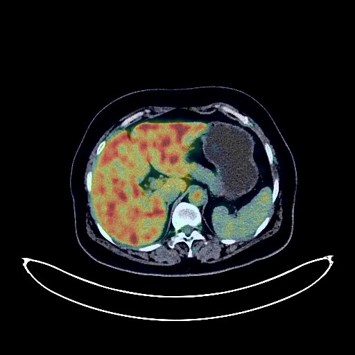
Lung Cancer PET/CT (case 983827-000049 from PETWB-REP)
0 views9 days agoWhole-body 18F-FDG PET/CT scan in a patient with Lung Cancer taken from the PETWB-REP dataset. The following English report (translated from original Chinese) is taken verbatim from the public dataset and has not been modified or otherwise checked for accuracy (see the end for citation). Impression a. Space-occupying lesion in the posterior segment of the left lower lobe, with increased FDG metabolism, suggestive of lung cancer. b. Metastatic tumor in the right frontoparietal lobe. Metastatic tumor in the left sacral region (S2). a. Ground-glass nodules in the apical-posterior segment of the left upper lobe and the right middle lobe, with normal FDG metabolism, suggestive of atypical adenomatous hyperplasia; annual HRCT follow-up is recommended. b. Chronic inflammatory nodules in the lateral basal segment and para-oblique fissure of the right lower lobe, calcification in the right lower lobe, and a few fibrotic lesions at the bases of both lungs. Reactive hyperplasia of bilateral deep cervical spaces, submandibular, and submental lymph nodes. Continuous increased FDG metabolism in the colon and rectum, suggestive of inflammation or physiological uptake; colonoscopy follow-up is recommended. Spinal osteophyte formation, L4/5 intervertebral disc bulge. Increased FDG metabolism in the right shoulder joint capsule, suggestive of frozen shoulder. Benign bone disease of the right frontal bone is highly probable. This case is from PETWB-REP, a curated dataset of whole-body 18F-FDG PET/CT scans and corresponding radiology reports from 490 patients with a broad spectrum of malignancies. The data were retrospectively collected from patients who underwent clinically indicated whole-body 18F-FDG PET/CT scans at the Shanghai Universal Medical Imaging Diagnostic Center between 2021 and 2024. License: Creative Commons Attribution 4.0 International (CC BY 4.0) Citation: Xue, L., Feng, G., Wenbo, Z., Zhang, Y., Li, L., Wang, S., Peng, L., Peng, S., & Gao, X. (2026). PETWB-REP: A Multi-Cancer Whole-Body FDG PET/CT Dataset with Corresponding Radiology Reports [Data set]. Zenodo. https://doi.org/10.5281/zenodo.18670487
Whole BodyPET/CT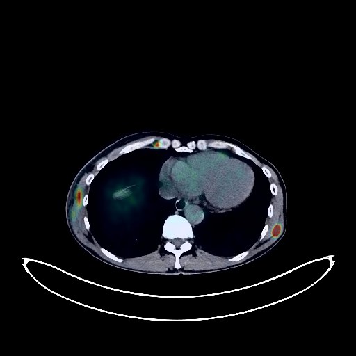
Lymphoma PET/CT (case 983827-000089 from PETWB-REP)
0 views9 days agoWhole-body 18F-FDG PET/CT scan in a patient with Lymphoma taken from the PETWB-REP dataset. The following English report (translated from original Chinese) is taken verbatim from the public dataset and has not been modified or otherwise checked for accuracy (see the end for citation). Impression a. Multiple enlarged lymph nodes throughout the body with increased FDG metabolism (see description for details); b. Multiple nodules and patchy soft tissue density shadows in the subcutaneous tissue, muscles, and intermuscular spaces throughout the body with increased FDG metabolism; c. Multiple low-density nodules in the thyroid gland with increased FDG metabolism; d. Multiple intracranial lesions with increased FDG metabolism; The above suggest malignancy, with a high probability of multi-systemic lymphoma. Please confirm with clinicopathological examination. Chronic inflammatory micronodules in the lower lobe of the right lung. Emphysema in the upper lobes of both lungs, and a few post-inflammatory lesions in both lungs. Anemia. Partial arteriosclerosis. Chronic inflammatory changes in part of the gastric wall and intestinal tract; please follow up with endoscopy. Concentrated bile in the gallbladder. Small amount of hydrocele in the tunica vaginalis of the right testis. Mild osteophyte formation in the spine, and L3-S1 intervertebral disc herniation. This case is from PETWB-REP, a curated dataset of whole-body 18F-FDG PET/CT scans and corresponding radiology reports from 490 patients with a broad spectrum of malignancies. The data were retrospectively collected from patients who underwent clinically indicated whole-body 18F-FDG PET/CT scans at the Shanghai Universal Medical Imaging Diagnostic Center between 2021 and 2024. License: Creative Commons Attribution 4.0 International (CC BY 4.0) Citation: Xue, L., Feng, G., Wenbo, Z., Zhang, Y., Li, L., Wang, S., Peng, L., Peng, S., & Gao, X. (2026). PETWB-REP: A Multi-Cancer Whole-Body FDG PET/CT Dataset with Corresponding Radiology Reports [Data set]. Zenodo. https://doi.org/10.5281/zenodo.18670487
Whole BodyPET/CT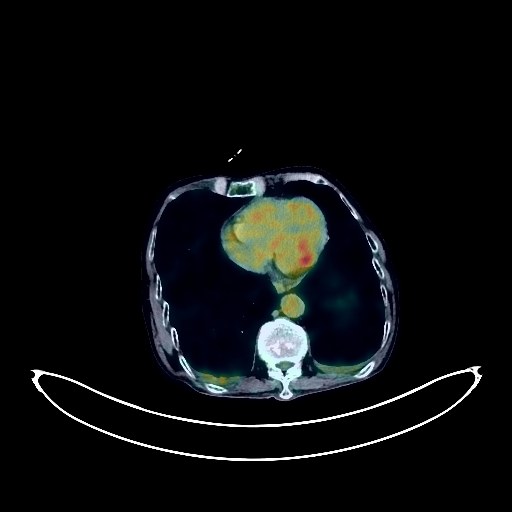
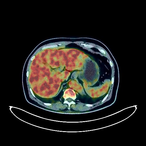
Liver Cancer PET/CT (case 983827-000047 from PETWB-REP)
0 views9 days agoWhole-body 18F-FDG PET/CT scan in a patient with Liver Cancer taken from the PETWB-REP dataset. The following English report (translated from original Chinese) is taken verbatim from the public dataset and has not been modified or otherwise checked for accuracy (see the end for citation). Impression a. A mass in the left lobe of the liver with elevated FDG metabolism, suggestive of malignancy, most likely intrahepatic cholangiocarcinoma. b. Multiple lymph node metastases in the abdominal cavity, retroperitoneum, bilateral inguinal regions, bilateral internal mammary chains, and left supraclavicular fossa. Extensive seeding metastases in the abdominoperineal cavity and right inguinal canal. Abdominal and pelvic effusion. c. Multiple stones in the intrahepatic bile ducts, common bile duct, and gallbladder. Scattered chronic inflammation and fibrosis in both lungs. Small amount of pleural effusion bilaterally. Reactive hyperplasia of hilar and mediastinal lymph nodes bilaterally. Elevated FDG metabolism in some intestinal segments, suggestive of inflammatory or physiological uptake. Kyphosis of the spine, with some old compression fractures of the thoracic and lumbar vertebrae. Spinal degeneration. L4/5 and L5/S1 intervertebral disc bulges. A few ischemic lesions in the deep bilateral cerebral regions, suggestive of age-related brain changes. This case is from PETWB-REP, a curated dataset of whole-body 18F-FDG PET/CT scans and corresponding radiology reports from 490 patients with a broad spectrum of malignancies. The data were retrospectively collected from patients who underwent clinically indicated whole-body 18F-FDG PET/CT scans at the Shanghai Universal Medical Imaging Diagnostic Center between 2021 and 2024. License: Creative Commons Attribution 4.0 International (CC BY 4.0) Citation: Xue, L., Feng, G., Wenbo, Z., Zhang, Y., Li, L., Wang, S., Peng, L., Peng, S., & Gao, X. (2026). PETWB-REP: A Multi-Cancer Whole-Body FDG PET/CT Dataset with Corresponding Radiology Reports [Data set]. Zenodo. https://doi.org/10.5281/zenodo.18670487
Whole BodyPET/CT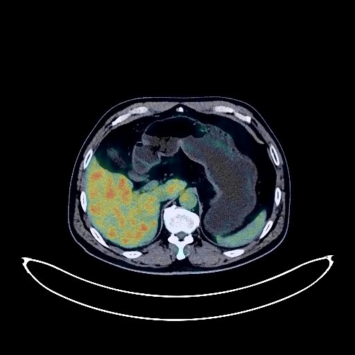
Colon Cancer PET/CT (case 983827-000030 from PETWB-REP)
0 views9 days agoWhole-body 18F-FDG PET/CT scan in a patient with Colon Cancer taken from the PETWB-REP dataset. The following English report (translated from original Chinese) is taken verbatim from the public dataset and has not been modified or otherwise checked for accuracy (see the end for citation). Impression a. Irregular thickening of the sigmoid colon, presenting as a soft tissue mass, with increased FDG metabolism, suggestive of colon cancer. Multiple lymph node metastases are present in the surrounding mesentery, presacral region, left iliac vessels, para-aortic region, and left supraclavicular fossa. b. Multiple liver metastases. Slight thickening of the lower segment of the left ureter, presenting as a cup-shaped stenosis, leading to hydronephrosis at the upper level. Increased FDG uptake in the lesion suggests a space-occupying lesion; specialist examination is recommended. Multiple small chronic inflammatory nodules (solid) in both lungs are highly probable; please use CT comparison to rule out metastasis. A few chronic inflammations and old lesions in both lungs. Small liver cysts. Degenerative changes in the spine. L4/5 and L5/S1 intervertebral disc bulges. No abnormalities were found on cranial scintigraphy. Chronic inflammation of the right maxillary sinus. This case is from PETWB-REP, a curated dataset of whole-body 18F-FDG PET/CT scans and corresponding radiology reports from 490 patients with a broad spectrum of malignancies. The data were retrospectively collected from patients who underwent clinically indicated whole-body 18F-FDG PET/CT scans at the Shanghai Universal Medical Imaging Diagnostic Center between 2021 and 2024. License: Creative Commons Attribution 4.0 International (CC BY 4.0) Citation: Xue, L., Feng, G., Wenbo, Z., Zhang, Y., Li, L., Wang, S., Peng, L., Peng, S., & Gao, X. (2026). PETWB-REP: A Multi-Cancer Whole-Body FDG PET/CT Dataset with Corresponding Radiology Reports [Data set]. Zenodo. https://doi.org/10.5281/zenodo.18670487
Whole BodyPET/CT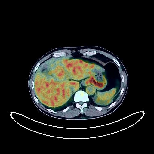
Lung Cancer PET/CT (case 983827-000206 from PETWB-REP)
0 views9 days agoWhole-body 18F-FDG PET/CT scan in a patient with Lung Cancer taken from the PETWB-REP dataset. The following English report (translated from original Chinese) is taken verbatim from the public dataset and has not been modified or otherwise checked for accuracy (see the end for citation). Impression a. Left hilar mass with abnormally elevated FDG metabolism, suggestive of central lung cancer with left lower lobe atelectasis and local involvement of the left upper lobe bronchus. Reactive hyperplasia of bilateral hilar and mediastinal lymph nodes, follow-up recommended. b. Mixed ground-glass opacity nodule in the left upper lobe, no significant abnormalities in FDG metabolism, suggestive of chronic inflammatory nodule or atypical adenomatous hyperplasia, annual HRCT recommended. c. Chronic inflammatory nodule (solid) in the anterior segment of the right upper lobe. Interstitial inflammation in the right lower lobe. Bulla in the left upper lobe. Scattered chronic inflammation and remnants in both lungs. Small amount of pleural effusion on the left side. Partial arteriosclerosis (including coronary arteries). d. Age-related brain changes, deep lacunar infarcts in the brain, MRI recommended. a. A localized, roundish soft tissue density nodule on the inner edge of the right kidney parenchyma, with increased FDG metabolism, suggesting a possible cyst, but renal cell carcinoma cannot be ruled out. Please combine with contrast-enhanced CT or MRI. b. Left renal cyst. Thickening of the bilateral renal septa. Benign prostatic hyperplasia with calcification. Nodular goiter, possibly a left lobe thyroid adenoma with calcification. Ultrasound and thyroid function follow-up are recommended. Increased FDG metabolism in part of the gastric wall, suggesting physiological uptake or chronic inflammatory changes. Degenerative changes in the spine. L2 vertebral islet. L3/4 and L4/5 intervertebral disc bulge. L2/3 and L5/S1 intervertebral disc herniation. L5/S1 intervertebral disc pneumoconiosis and degeneration. L5 lumbarization. Possible old fracture of the left 11th posterior rib. Mild inflammation of the left sphenoid sinus, submucosal cyst of the right maxillary sinus. Reactive hyperplasia of bilateral submandibular lymph nodes. This case is from PETWB-REP, a curated dataset of whole-body 18F-FDG PET/CT scans and corresponding radiology reports from 490 patients with a broad spectrum of malignancies. The data were retrospectively collected from patients who underwent clinically indicated whole-body 18F-FDG PET/CT scans at the Shanghai Universal Medical Imaging Diagnostic Center between 2021 and 2024. License: Creative Commons Attribution 4.0 International (CC BY 4.0) Citation: Xue, L., Feng, G., Wenbo, Z., Zhang, Y., Li, L., Wang, S., Peng, L., Peng, S., & Gao, X. (2026). PETWB-REP: A Multi-Cancer Whole-Body FDG PET/CT Dataset with Corresponding Radiology Reports [Data set]. Zenodo. https://doi.org/10.5281/zenodo.18670487
Whole BodyPET/CT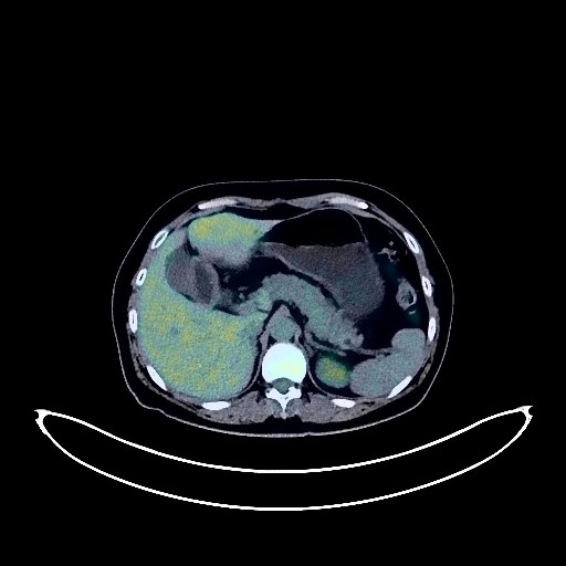
Cervical Cancer PET/CT (case 983827-000266 from PETWB-REP)
0 views9 days agoWhole-body 18F-FDG PET/CT scan in a patient with Cervical Cancer taken from the PETWB-REP dataset. The following English report (translated from original Chinese) is taken verbatim from the public dataset and has not been modified or otherwise checked for accuracy (see the end for citation). Impression Cervical mass with elevated FDG metabolism, consistent with cervical cancer. a. Several ground-glass nodules in both lungs, FDG metabolism normal, suggestive of inflammation or atypical adenomatous hyperplasia; annual HRCT follow-up recommended. b. Chronic inflammatory nodules (solid) in both lungs; CT follow-up recommended. A few post-inflammatory lesions in both lungs. Minor arteriosclerosis in some arteries. Small liver cysts. Gallstones. Post-operative changes following a right adrenal tumor. Chronic inflammatory changes or physiological uptake in the gastric antrum and part of the intestine; endoscopic follow-up recommended. Degenerative changes in the spine; L5/S1 intervertebral disc bulge with posterior calcification. No obvious abnormalities seen on cranial scintigraphy. This case is from PETWB-REP, a curated dataset of whole-body 18F-FDG PET/CT scans and corresponding radiology reports from 490 patients with a broad spectrum of malignancies. The data were retrospectively collected from patients who underwent clinically indicated whole-body 18F-FDG PET/CT scans at the Shanghai Universal Medical Imaging Diagnostic Center between 2021 and 2024. License: Creative Commons Attribution 4.0 International (CC BY 4.0) Citation: Xue, L., Feng, G., Wenbo, Z., Zhang, Y., Li, L., Wang, S., Peng, L., Peng, S., & Gao, X. (2026). PETWB-REP: A Multi-Cancer Whole-Body FDG PET/CT Dataset with Corresponding Radiology Reports [Data set]. Zenodo. https://doi.org/10.5281/zenodo.18670487
Whole BodyPET/CT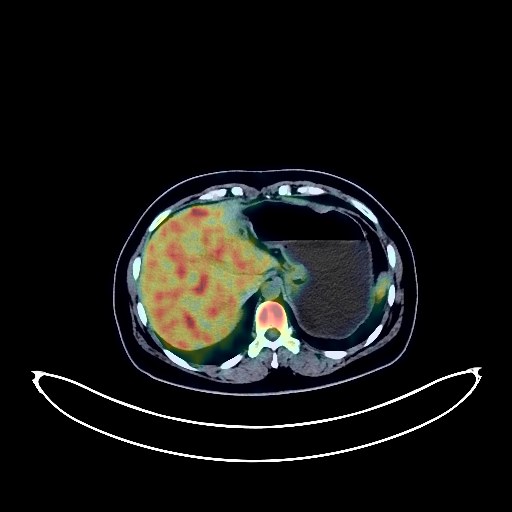
Breast Cancer PET/CT (case 983827-000259 from PETWB-REP)
0 views9 days agoWhole-body 18F-FDG PET/CT scan in a patient with Breast Cancer taken from the PETWB-REP dataset. The following English report (translated from original Chinese) is taken verbatim from the public dataset and has not been modified or otherwise checked for accuracy (see the end for citation). Impression a. Left breast mass with increased FDG metabolism, adjacent skin involvement, suggestive of breast cancer; please correlate with clinicopathology; left axillary lymph node metastasis. b. Right breast hyperplasia; specialist follow-up recommended. Bilateral lung chronic inflammatory micronodules; CT follow-up recommended. A few post-inflammatory lesions in both lungs. Anemia changes. Possibly physiological uptake in the uterine cavity; left ovarian cyst; specialist and ultrasound follow-up recommended. Chronic inflammatory changes or physiological uptake in parts of the stomach wall and intestines; hemorrhoids possible; please correlate with endoscopic follow-up. No obvious abnormalities seen on cranial scintigraphy. Chronic inflammation of the left ethmoid sinus. This case is from PETWB-REP, a curated dataset of whole-body 18F-FDG PET/CT scans and corresponding radiology reports from 490 patients with a broad spectrum of malignancies. The data were retrospectively collected from patients who underwent clinically indicated whole-body 18F-FDG PET/CT scans at the Shanghai Universal Medical Imaging Diagnostic Center between 2021 and 2024. License: Creative Commons Attribution 4.0 International (CC BY 4.0) Citation: Xue, L., Feng, G., Wenbo, Z., Zhang, Y., Li, L., Wang, S., Peng, L., Peng, S., & Gao, X. (2026). PETWB-REP: A Multi-Cancer Whole-Body FDG PET/CT Dataset with Corresponding Radiology Reports [Data set]. Zenodo. https://doi.org/10.5281/zenodo.18670487
Whole BodyPET/CT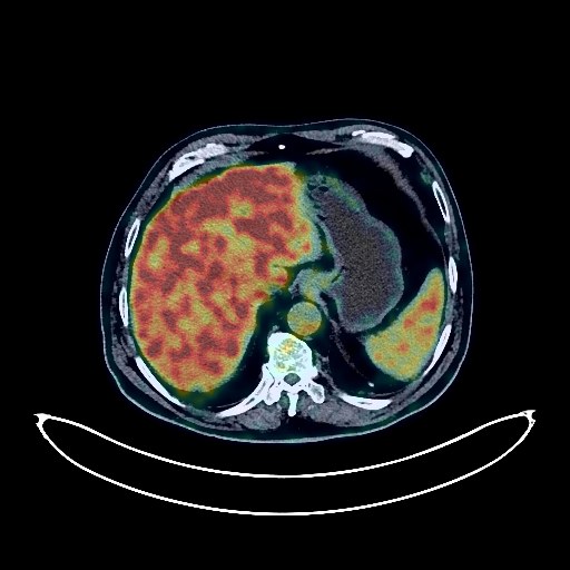
Lung Cancer PET/CT (case 983827-000113 from PETWB-REP)
0 views9 days agoWhole-body 18F-FDG PET/CT scan in a patient with Lung Cancer taken from the PETWB-REP dataset. The following English report (translated from original Chinese) is taken verbatim from the public dataset and has not been modified or otherwise checked for accuracy (see the end for citation). Impression a. A mass located adjacent to the descending aorta in the posterior segment of the left lower lobe, with increased FDG metabolism, consistent with lung cancer. Left hilar lymph node metastasis is possible; please correlate with clinicopathology. b. Mixed ground-glass nodules in the apical and posterior segments of the left upper lobe and the posterior segment of the left lower lobe, with normal FDG uptake, suggest atypical adenomatous hyperplasia or chronic inflammatory nodules; please monitor with CT. c. Multiple solid nodules in the right lower lobe and left lung, with normal FDG uptake, highly suggestive of chronic inflammatory nodules; metastasis needs to be ruled out; please monitor with CT. d. Calcification of some arterial walls (including coronary arteries). Uneven FDG metabolism in the liver parenchyma; contrast-enhanced MRI is recommended to rule out metastasis. Multiple gallstones. Small cyst in the right kidney. Spinal degenerative changes. Postoperative changes following L1 vertebral fracture. A few ischemic lesions in the deep bilateral brain regions; age-related brain damage. MRI is recommended. This case is from PETWB-REP, a curated dataset of whole-body 18F-FDG PET/CT scans and corresponding radiology reports from 490 patients with a broad spectrum of malignancies. The data were retrospectively collected from patients who underwent clinically indicated whole-body 18F-FDG PET/CT scans at the Shanghai Universal Medical Imaging Diagnostic Center between 2021 and 2024. License: Creative Commons Attribution 4.0 International (CC BY 4.0) Citation: Xue, L., Feng, G., Wenbo, Z., Zhang, Y., Li, L., Wang, S., Peng, L., Peng, S., & Gao, X. (2026). PETWB-REP: A Multi-Cancer Whole-Body FDG PET/CT Dataset with Corresponding Radiology Reports [Data set]. Zenodo. https://doi.org/10.5281/zenodo.18670487
Whole BodyPET/CT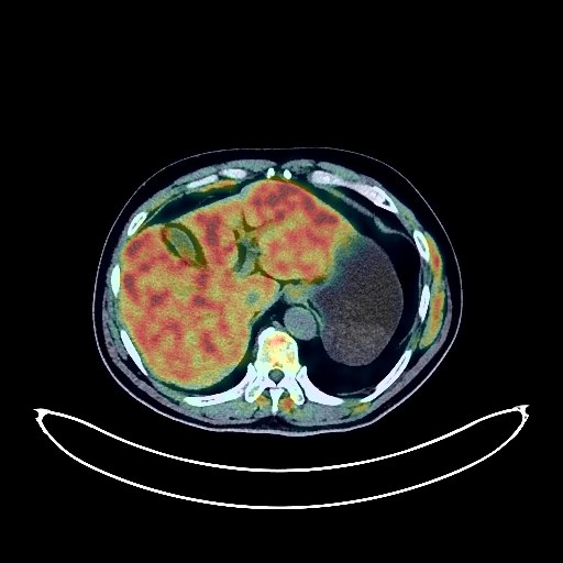
Esophageal Cancer PET/CT (case 983827-000154 from PETWB-REP)
0 views9 days agoWhole-body 18F-FDG PET/CT scan in a patient with Esophageal Cancer taken from the PETWB-REP dataset. The following English report (translated from original Chinese) is taken verbatim from the public dataset and has not been modified or otherwise checked for accuracy (see the end for citation). Impression a. Mass in the lower thoracic esophagus, with increased FDG metabolism, suggestive of esophageal cancer. Multiple lymph node metastases in the mediastinum, lesser curvature of the stomach, and left paramesentery. b. Decreased bone density in the right vertebral arch of T1, with increased FDG metabolism, metastasis to be ruled out; MRI recommended. Chronic inflammatory nodules (solid) in the right middle lobe and anterior basal segment of the right lower lobe. A few chronic inflammations and old lesions in both lungs. Calcification of some arterial walls (including coronary arteries). Liver cyst; splenic calcification. Continuous increased FDG metabolism in the descending colon, sigmoid colon, and rectum, suggestive of inflammatory or physiological uptake; colonoscopy recommended. Prostatic calcification. Degenerative changes in the spine. L4/5 and L5/S1 intervertebral disc bulge. Cranial scintigraphy showed no abnormalities. Mild chronic inflammation of the bilateral maxillary sinuses and bilateral ethmoid sinuses. Increased FDG metabolism in the tongue and left vocal cord, suggesting possible inflammatory uptake; specialist examination is recommended. This case is from PETWB-REP, a curated dataset of whole-body 18F-FDG PET/CT scans and corresponding radiology reports from 490 patients with a broad spectrum of malignancies. The data were retrospectively collected from patients who underwent clinically indicated whole-body 18F-FDG PET/CT scans at the Shanghai Universal Medical Imaging Diagnostic Center between 2021 and 2024. License: Creative Commons Attribution 4.0 International (CC BY 4.0) Citation: Xue, L., Feng, G., Wenbo, Z., Zhang, Y., Li, L., Wang, S., Peng, L., Peng, S., & Gao, X. (2026). PETWB-REP: A Multi-Cancer Whole-Body FDG PET/CT Dataset with Corresponding Radiology Reports [Data set]. Zenodo. https://doi.org/10.5281/zenodo.18670487
Whole BodyPET/CT