Loading...
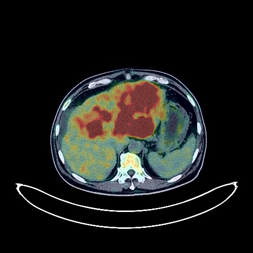
Liver Cancer PET/CT (case 983827-000166 from PETWB-REP)
0 views9 days agoWhole-body 18F-FDG PET/CT scan in a patient with Liver Cancer taken from the PETWB-REP dataset. The following English report (translated from original Chinese) is taken verbatim from the public dataset and has not been modified or otherwise checked for accuracy (see the end for citation). Impression a. Multiple lesions in the liver, increased FDG metabolism, highly suggestive of liver cancer; please correlate with clinicopathology. b. Possible portal vein tumor thrombus; cirrhosis; splenomegaly; portal hypertension. Liver calcifications. Abdominal and pelvic effusions. Flocculent thickening of the greater omentum and mesentery, FDG metabolism normal; please follow up. Several small chronic inflammatory nodules (solid) in both lungs. A small amount of chronic inflammation and old lesions in both lungs. Small amount of pleural effusion on the left side. Anemia. Chronic gastritis; increased FDG metabolism in some intestinal segments, considered inflammatory or physiological uptake. Follow-up gastroscopy and colonoscopy are recommended. Degenerative changes in the spine. L4/5, L5/S1 intervertebral disc bulge. No abnormalities found on cranial scintigraphy. Chronic inflammation of both maxillary sinuses and both ethmoid sinuses. This case is from PETWB-REP, a curated dataset of whole-body 18F-FDG PET/CT scans and corresponding radiology reports from 490 patients with a broad spectrum of malignancies. The data were retrospectively collected from patients who underwent clinically indicated whole-body 18F-FDG PET/CT scans at the Shanghai Universal Medical Imaging Diagnostic Center between 2021 and 2024. License: Creative Commons Attribution 4.0 International (CC BY 4.0) Citation: Xue, L., Feng, G., Wenbo, Z., Zhang, Y., Li, L., Wang, S., Peng, L., Peng, S., & Gao, X. (2026). PETWB-REP: A Multi-Cancer Whole-Body FDG PET/CT Dataset with Corresponding Radiology Reports [Data set]. Zenodo. https://doi.org/10.5281/zenodo.18670487
Whole BodyPET/CT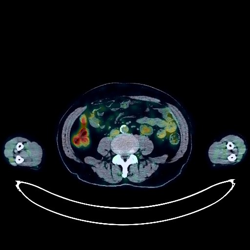
Lymphoma PET/CT (case 983827-000041 from PETWB-REP)
0 views9 days agoWhole-body 18F-FDG PET/CT scan in a patient with Lymphoma taken from the PETWB-REP dataset. The following English report (translated from original Chinese) is taken verbatim from the public dataset and has not been modified or otherwise checked for accuracy (see the end for citation). Impression Post-lymphoma treatment: No enlarged lymph nodes were observed throughout the body; small lymph nodes were visible; FDG metabolism was not increased, roughly similar to the PET/CT results from our center on May 27, 2022, suggesting that tumor activity was largely suppressed after lymphoma treatment. A space-occupying lesion in the intermuscular space of the left groin with increased FDG uptake, slightly larger than before. Considering the medical history, a recurrence of leiomyoma is suspected; please confirm with pathology. a. Several ground-glass nodules in the upper lobes of both lungs, with no abnormal FDG metabolism, suggestive of inflammatory nodules or atypical adenomatous hyperplasia; annual HRCT follow-up is recommended. b. Several small chronic inflammatory nodules (solid) in both lungs. c. A few chronic inflammations and old lesions in both lungs. Calcification of some arterial walls (including coronary arteries). Manifestations of chronic gastritis; gastroscopy follow-up is necessary if needed. Physiological uptake of some intestinal segments. Gallstones. Small liver cysts. Left renal cyst. Prostatic calcification. Spinal degenerative changes. L4/5 disc bulge. Few ischemic lesions in the deep bilateral brain regions, indicative of age-related brain changes. Chronic inflammation of the left sphenoid sinus. Nodular goiter, likely large, similar to the previous findings; ultrasound follow-up is recommended. This case is from PETWB-REP, a curated dataset of whole-body 18F-FDG PET/CT scans and corresponding radiology reports from 490 patients with a broad spectrum of malignancies. The data were retrospectively collected from patients who underwent clinically indicated whole-body 18F-FDG PET/CT scans at the Shanghai Universal Medical Imaging Diagnostic Center between 2021 and 2024. License: Creative Commons Attribution 4.0 International (CC BY 4.0) Citation: Xue, L., Feng, G., Wenbo, Z., Zhang, Y., Li, L., Wang, S., Peng, L., Peng, S., & Gao, X. (2026). PETWB-REP: A Multi-Cancer Whole-Body FDG PET/CT Dataset with Corresponding Radiology Reports [Data set]. Zenodo. https://doi.org/10.5281/zenodo.18670487
Whole BodyPET/CT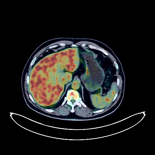
Lung Cancer PET/CT (case 983827-000043 from PETWB-REP)
0 views9 days agoWhole-body 18F-FDG PET/CT scan in a patient with Lung Cancer taken from the PETWB-REP dataset. The following English report (translated from original Chinese) is taken verbatim from the public dataset and has not been modified or otherwise checked for accuracy (see the end for citation). Impression a. A mass in the basal segment of the right lower lobe, with increased FDG metabolism, suggestive of lung cancer; please correlate with clinicopathology. b. Reactive hyperplasia of the hilar, mediastinal, and right supraclavicular lymph nodes is highly probable, with metastasis to be ruled out. Significant changes following fracture of the left 5th and 6th ribs, with metastasis to be ruled out; please correlate with clinical history. Several small, solid, chronic inflammatory nodules in both lungs. A few chronic inflammations and old lesions in both lungs. Calcification of some arterial walls (including coronary arteries). Multiple liver cysts. Intrahepatic calcifications. Small kidney stone in the left kidney. Calcifications in the prostate. Calcifications in the vesicorectal space. Increased FDG uptake in the perineal area; please correlate with clinical examination. Degenerative changes in the spine. Pneumothorax and bulging of the L4/5 and L5/S1 intervertebral discs. Inflammation around the left hip joint. A few ischemic lesions in the deep bilateral brain regions, suggestive of age-related brain changes; follow-up MRI is recommended. This case is from PETWB-REP, a curated dataset of whole-body 18F-FDG PET/CT scans and corresponding radiology reports from 490 patients with a broad spectrum of malignancies. The data were retrospectively collected from patients who underwent clinically indicated whole-body 18F-FDG PET/CT scans at the Shanghai Universal Medical Imaging Diagnostic Center between 2021 and 2024. License: Creative Commons Attribution 4.0 International (CC BY 4.0) Citation: Xue, L., Feng, G., Wenbo, Z., Zhang, Y., Li, L., Wang, S., Peng, L., Peng, S., & Gao, X. (2026). PETWB-REP: A Multi-Cancer Whole-Body FDG PET/CT Dataset with Corresponding Radiology Reports [Data set]. Zenodo. https://doi.org/10.5281/zenodo.18670487
Whole BodyPET/CT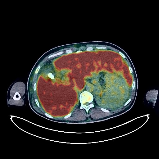
Liver Cancer PET/CT (case 983827-000210 from PETWB-REP)
0 views9 days agoWhole-body 18F-FDG PET/CT scan in a patient with Liver Cancer taken from the PETWB-REP dataset. The following English report (translated from original Chinese) is taken verbatim from the public dataset and has not been modified or otherwise checked for accuracy (see the end for citation). Impression a. Right lobe of liver lesion, FDG uptake slightly reduced compared to background levels, liver cancer suspected based on medical history. Cirrhosis, splenomegaly, fatty liver. b. Portal hypertension with open collateral circulation, ascites/pelvic effusion. a. High-density nodular shadow in the trachea, FDG uptake normal, sputum plug possible, neoplastic lesion to be ruled out, follow-up recommended. b. Ground-glass nodule in the anterior segment of the left upper lobe, FDG uptake normal, chronic inflammatory nodule suspected, follow-up with CT scan recommended. c. Chronic inflammation in the right lower lobe. Scattered post-inflammatory lesions in the remaining lungs. Right pleural thickening. d. Small amount of pericardial effusion. Slightly enlarged cardiac silhouette. Calcification of some arterial walls (including coronary arteries). Low-density thyroid nodule, FDG uptake normal, suggestive of nodular goiter, please repeat ultrasound examination. Reactive hyperplasia of left deep cervical lymph nodes. Cholecystitis, gallstones. Bilateral renal cysts. Prostatic calcification. Bilateral hydrocele. Spinal degenerative changes. L4/5, L5/S1 intervertebral disc bulge, pneumothorax. Subcutaneous edema of abdomen and lower extremities. No obvious abnormalities seen on cranial FDG imaging. This case is from PETWB-REP, a curated dataset of whole-body 18F-FDG PET/CT scans and corresponding radiology reports from 490 patients with a broad spectrum of malignancies. The data were retrospectively collected from patients who underwent clinically indicated whole-body 18F-FDG PET/CT scans at the Shanghai Universal Medical Imaging Diagnostic Center between 2021 and 2024. License: Creative Commons Attribution 4.0 International (CC BY 4.0) Citation: Xue, L., Feng, G., Wenbo, Z., Zhang, Y., Li, L., Wang, S., Peng, L., Peng, S., & Gao, X. (2026). PETWB-REP: A Multi-Cancer Whole-Body FDG PET/CT Dataset with Corresponding Radiology Reports [Data set]. Zenodo. https://doi.org/10.5281/zenodo.18670487
Whole BodyPET/CT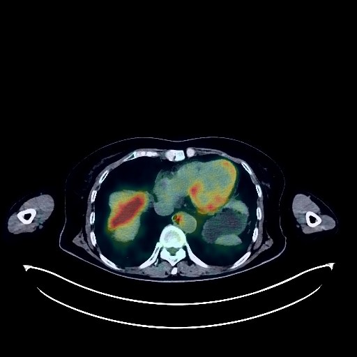
Ovarian Cancer PET/CT (case 983827-000145 from PETWB-REP)
0 views9 days agoWhole-body 18F-FDG PET/CT scan in a patient with Ovarian Cancer taken from the PETWB-REP dataset. The following English report (translated from original Chinese) is taken verbatim from the public dataset and has not been modified or otherwise checked for accuracy (see the end for citation). Impression a. Left adnexal region mass, elevated FDG metabolism, highly suggestive of ovarian cancer. Uterine cavity effusion. b. Extensive peritoneal implantation metastasis in the abdominopelvic cavity. Low-density nodule in the left lobe of the thyroid gland, elevated FDG metabolism, suggestive of adenoma, but thyroid cancer cannot be ruled out; further ultrasound examination recommended. a. Several small, solid, chronic inflammatory nodules in both lungs. A small amount of chronic inflammation and old lesions in both lungs. b. Reactive hyperplasia of hilar and mediastinal lymph nodes in both lungs. Calcification of some arterial walls (including coronary arteries). Liver cyst. Right kidney cyst. Slight thickening of part of the gastric wall, elevated FDG metabolism, suggestive of gastritis; follow-up gastroscopy recommended. Increased FDG uptake in some intestinal segments, possibly due to physiological or inflammatory uptake; endoscopic follow-up is recommended to rule out other possibilities. Degenerative changes in the spine. L4/5 and L5/S1 intervertebral disc bulges. Softening lesions in the right basal ganglia. Mild age-related brain changes. Submucosal cyst of the right maxillary sinus. This case is from PETWB-REP, a curated dataset of whole-body 18F-FDG PET/CT scans and corresponding radiology reports from 490 patients with a broad spectrum of malignancies. The data were retrospectively collected from patients who underwent clinically indicated whole-body 18F-FDG PET/CT scans at the Shanghai Universal Medical Imaging Diagnostic Center between 2021 and 2024. License: Creative Commons Attribution 4.0 International (CC BY 4.0) Citation: Xue, L., Feng, G., Wenbo, Z., Zhang, Y., Li, L., Wang, S., Peng, L., Peng, S., & Gao, X. (2026). PETWB-REP: A Multi-Cancer Whole-Body FDG PET/CT Dataset with Corresponding Radiology Reports [Data set]. Zenodo. https://doi.org/10.5281/zenodo.18670487
Whole BodyPET/CT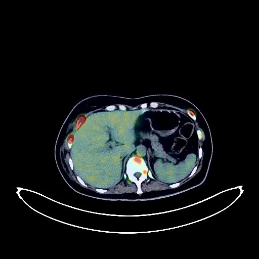
Breast Cancer PET/CT (case 983827-000064 from PETWB-REP)
0 views9 days agoWhole-body 18F-FDG PET/CT scan in a patient with Breast Cancer taken from the PETWB-REP dataset. The following English report (translated from original Chinese) is taken verbatim from the public dataset and has not been modified or otherwise checked for accuracy (see the end for citation). Impression a. Multiple lesions in the right breast with elevated FDG metabolism, suggestive of right breast cancer. b. Multiple lymph node metastases in the right axilla, right internal mammary chain, right interpectoral space, right hilum, and mediastinum. c. Multiple intracranial metastases. Diffuse bone metastases throughout the body. Multiple lung metastases. Liver metastases. Left psoas muscle metastases. A few old lesions in both lungs. Small amount of pleural effusion on the right side. Accessory spleen. Left kidney stone. Small amount of pelvic effusion. Physiological uptake in the uterine cavity. Possible functional cyst in the right adnexa. Mild osteophyte formation in the cervical, thoracic, and lumbar spine. This case is from PETWB-REP, a curated dataset of whole-body 18F-FDG PET/CT scans and corresponding radiology reports from 490 patients with a broad spectrum of malignancies. The data were retrospectively collected from patients who underwent clinically indicated whole-body 18F-FDG PET/CT scans at the Shanghai Universal Medical Imaging Diagnostic Center between 2021 and 2024. License: Creative Commons Attribution 4.0 International (CC BY 4.0) Citation: Xue, L., Feng, G., Wenbo, Z., Zhang, Y., Li, L., Wang, S., Peng, L., Peng, S., & Gao, X. (2026). PETWB-REP: A Multi-Cancer Whole-Body FDG PET/CT Dataset with Corresponding Radiology Reports [Data set]. Zenodo. https://doi.org/10.5281/zenodo.18670487
Whole BodyPET/CT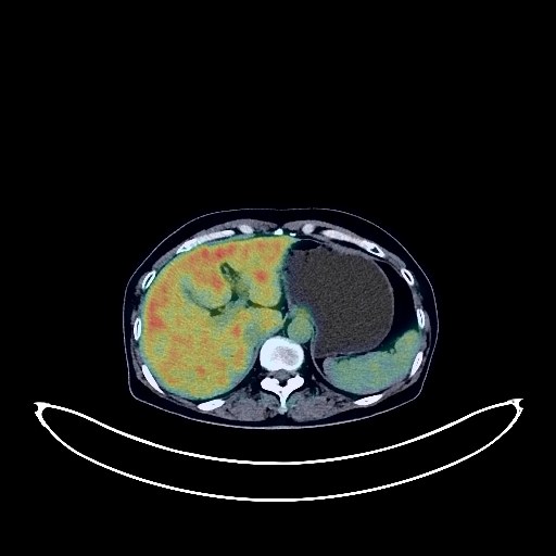
Lung Cancer PET/CT (case 983827-000247 from PETWB-REP)
0 views9 days agoWhole-body 18F-FDG PET/CT scan in a patient with Lung Cancer taken from the PETWB-REP dataset. The following English report (translated from original Chinese) is taken verbatim from the public dataset and has not been modified or otherwise checked for accuracy (see the end for citation). Impression a. Mixed ground-glass nodules in the posterior segment of the right upper lobe, with slightly elevated FDG metabolism, suggestive of lung cancer; please correlate with clinicopathology. b. Chronic inflammatory micronodules (solid) in the right lower lobe; follow-up CT is recommended. A few post-inflammatory lesions in both lungs. Tracheal diverticulum. Partial arteriosclerosis. No definite space-occupying lesion in the left cerebellopontine angle region; FDG metabolism normal; comprehensive analysis with enhanced MRI is recommended. Deep lacunar ischemic lesions in the brain. Chronic inflammatory changes or physiological uptake in some intestinal segments; follow-up endoscopically is recommended. Degenerative changes in the spine; L4/5 intervertebral disc bulge. Left shoulder periarthritis. This case is from PETWB-REP, a curated dataset of whole-body 18F-FDG PET/CT scans and corresponding radiology reports from 490 patients with a broad spectrum of malignancies. The data were retrospectively collected from patients who underwent clinically indicated whole-body 18F-FDG PET/CT scans at the Shanghai Universal Medical Imaging Diagnostic Center between 2021 and 2024. License: Creative Commons Attribution 4.0 International (CC BY 4.0) Citation: Xue, L., Feng, G., Wenbo, Z., Zhang, Y., Li, L., Wang, S., Peng, L., Peng, S., & Gao, X. (2026). PETWB-REP: A Multi-Cancer Whole-Body FDG PET/CT Dataset with Corresponding Radiology Reports [Data set]. Zenodo. https://doi.org/10.5281/zenodo.18670487
Whole BodyPET/CT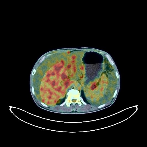
Pancreatic Cancer PET/CT (case 983827-000059 from PETWB-REP)
0 views9 days agoWhole-body 18F-FDG PET/CT scan in a patient with Pancreatic Cancer taken from the PETWB-REP dataset. The following English report (translated from original Chinese) is taken verbatim from the public dataset and has not been modified or otherwise checked for accuracy (see the end for citation). Impression A mass in the body and tail of the pancreas (mainly the tail), with elevated FDG metabolism, strongly suggestive of pancreatic cancer; please correlate with clinical findings. Extensive metastasis to the peritoneum, subcapsular region of the liver, greater omentum, and mesentery. Significant effusion in the abdominal and pelvic cavities. Scattered solid nodules in both lungs, with no elevated FDG metabolism, strongly suggestive of chronic inflammatory nodules; please correlate with CT follow-up. Small liver size, with a slightly low-density lesion in the right posterior lobe, with no elevated FDG metabolism; hemangioma is a possibility; please correlate with contrast-enhanced examination. Chronic cholecystitis with gallstones. Benign prostatic hyperplasia with calcification. Bilateral hydrocele with calcification. Hemorrhoids. Partial vertebral osteophyte formation. L4/5 and L5/S1 intervertebral disc bulge. No obvious abnormalities on cranial scintigraphy. This case is from PETWB-REP, a curated dataset of whole-body 18F-FDG PET/CT scans and corresponding radiology reports from 490 patients with a broad spectrum of malignancies. The data were retrospectively collected from patients who underwent clinically indicated whole-body 18F-FDG PET/CT scans at the Shanghai Universal Medical Imaging Diagnostic Center between 2021 and 2024. License: Creative Commons Attribution 4.0 International (CC BY 4.0) Citation: Xue, L., Feng, G., Wenbo, Z., Zhang, Y., Li, L., Wang, S., Peng, L., Peng, S., & Gao, X. (2026). PETWB-REP: A Multi-Cancer Whole-Body FDG PET/CT Dataset with Corresponding Radiology Reports [Data set]. Zenodo. https://doi.org/10.5281/zenodo.18670487
Whole BodyPET/CT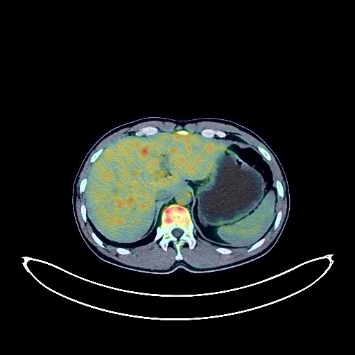
Lung Cancer PET/CT (case 983827-000149 from PETWB-REP)
0 views9 days agoWhole-body 18F-FDG PET/CT scan in a patient with Lung Cancer taken from the PETWB-REP dataset. The following English report (translated from original Chinese) is taken verbatim from the public dataset and has not been modified or otherwise checked for accuracy (see the end for citation). Impression a. A mass near the hilum of the left upper lobe, fused with the left hilar lymph node, with increased FDG metabolism, suggestive of central lung cancer with hilar lymph node metastasis and obstructive inflammation of the left upper lobe. b. A slightly high-density nodule in the left centrum semiovale, surrounded by edema, with increased FDG metabolism, suggestive of brain metastasis. Several small, solid, chronic inflammatory nodules in both lungs. A few chronic inflammations and old lesions in both lungs. Calcifications in the liver. Calcifications in the prostate. Osteophyte formation in the cervical, thoracic, and lumbar vertebrae; L4/5 and L5/S1 intervertebral disc bulges. Small submucosal cysts in both maxillary sinuses. This case is from PETWB-REP, a curated dataset of whole-body 18F-FDG PET/CT scans and corresponding radiology reports from 490 patients with a broad spectrum of malignancies. The data were retrospectively collected from patients who underwent clinically indicated whole-body 18F-FDG PET/CT scans at the Shanghai Universal Medical Imaging Diagnostic Center between 2021 and 2024. License: Creative Commons Attribution 4.0 International (CC BY 4.0) Citation: Xue, L., Feng, G., Wenbo, Z., Zhang, Y., Li, L., Wang, S., Peng, L., Peng, S., & Gao, X. (2026). PETWB-REP: A Multi-Cancer Whole-Body FDG PET/CT Dataset with Corresponding Radiology Reports [Data set]. Zenodo. https://doi.org/10.5281/zenodo.18670487
Whole BodyPET/CT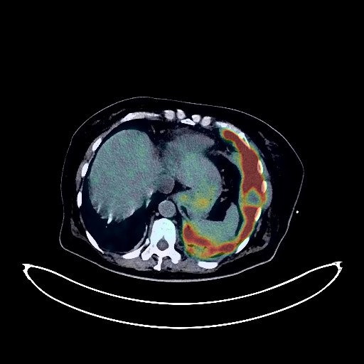
Lung Cancer PET/CT (case 983827-000137 from PETWB-REP)
0 views9 days agoWhole-body 18F-FDG PET/CT scan in a patient with Lung Cancer taken from the PETWB-REP dataset. The following English report (translated from original Chinese) is taken verbatim from the public dataset and has not been modified or otherwise checked for accuracy (see the end for citation). Impression a. Multiple lesions with internal necrosis and hemorrhage in the left pleura and left lung; soft tissue nodules in the right upper lobe; enlarged and swollen lymph nodes in the mediastinum, upper abdomen, and bilateral supraclavicular fossae; significantly increased FDG metabolism in the above lesions; focal FDG metabolism increase in the right iliac spine. All of the above lesions are considered malignant tumors, with the possibility of primary malignant tumors in the left lung or pleura with multiple metastases; metastatic tumors cannot be ruled out. A biopsy is recommended to confirm the diagnosis. b. Postoperative endometrial cancer surgery; no clear signs of tumor recurrence were observed in the surgical area. Changes after placement of the left thoracic drainage tube. Old fibrosis and calcifications in the right upper lobe; a few chronic inflammations in the right middle and lower lobes. Fatty liver; liver cysts; left adrenal hyperplasia; benign cystic lesions near the right external iliac vessels. Poor gastric filling, with physiological uptake by the gastric wall. Post-radiotherapy changes in the lumbosacral spine. Degenerative changes in the spine. Bilateral frozen shoulder. No obvious abnormalities seen on cranial scintigraphy. Minor chronic inflammation of the bilateral maxillary sinuses. Calcifications on both sides of the oropharyngeal wall. The left lobe of the thyroid gland is not clearly visualized; the right lobe shows decreased density with punctate calcifications and increased FDG metabolism. Please refer to the patient's history, ultrasound, and thyroid function tests. Low-density lesions in the muscle layer of the left forearm with increased FDG metabolism, possibly due to drug contamination. Clinical correlation and follow-up are recommended. This case is from PETWB-REP, a curated dataset of whole-body 18F-FDG PET/CT scans and corresponding radiology reports from 490 patients with a broad spectrum of malignancies. The data were retrospectively collected from patients who underwent clinically indicated whole-body 18F-FDG PET/CT scans at the Shanghai Universal Medical Imaging Diagnostic Center between 2021 and 2024. License: Creative Commons Attribution 4.0 International (CC BY 4.0) Citation: Xue, L., Feng, G., Wenbo, Z., Zhang, Y., Li, L., Wang, S., Peng, L., Peng, S., & Gao, X. (2026). PETWB-REP: A Multi-Cancer Whole-Body FDG PET/CT Dataset with Corresponding Radiology Reports [Data set]. Zenodo. https://doi.org/10.5281/zenodo.18670487
Whole BodyPET/CT