Loading...
Renal Cancer PET/CT (case 983827-000263 from PETWB-REP)
0 views9 days agoWhole-body 18F-FDG PET/CT scan in a patient with Renal Cancer taken from the PETWB-REP dataset. The following English report (translated from original Chinese) is taken verbatim from the public dataset and has not been modified or otherwise checked for accuracy (see the end for citation). Impression a. Bone metastases in the right 8th posterior rib, right adnexa of T8, and right iliac bone. b. Right renal mass with increased FDG metabolism, highly suggestive of renal cancer; enhanced MRI is recommended for further examination. Minor chronic inflammation of bilateral maxillary sinuses. Reactive hyperplasia or adenolymphoma of bilateral parotid lymph nodes; ultrasound follow-up is recommended. Scattered chronic inflammatory micronodules (solid) in both lungs; follow-up CT is recommended. Reactive hyperplasia of mediastinal lymph nodes. Calcification of some arterial walls (including coronary arteries). Cardiac enlargement. Small hepatic cysts. Chronic cholecystitis. Left renal calculi. Physiological changes or inflammatory lesions in the gastric antrum; gastroscopy follow-up is recommended. Bilateral pars interarticularis fracture of L5, anterior slippage of L4 and L5 vertebral bodies. Spinal osteophyte formation. L1-S1 intervertebral disc bulge. A few ischemic lesions deep in the brain, age-related brain changes. Nodular goiter; ultrasound follow-up is recommended. This case is from PETWB-REP, a curated dataset of whole-body 18F-FDG PET/CT scans and corresponding radiology reports from 490 patients with a broad spectrum of malignancies. The data were retrospectively collected from patients who underwent clinically indicated whole-body 18F-FDG PET/CT scans at the Shanghai Universal Medical Imaging Diagnostic Center between 2021 and 2024. License: Creative Commons Attribution 4.0 International (CC BY 4.0) Citation: Xue, L., Feng, G., Wenbo, Z., Zhang, Y., Li, L., Wang, S., Peng, L., Peng, S., & Gao, X. (2026). PETWB-REP: A Multi-Cancer Whole-Body FDG PET/CT Dataset with Corresponding Radiology Reports [Data set]. Zenodo. https://doi.org/10.5281/zenodo.18670487
Whole BodyPET/CT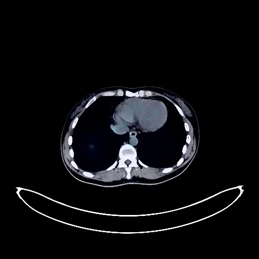
Ovarian Cancer PET/CT (case 983827-000063 from PETWB-REP)
0 views9 days agoWhole-body 18F-FDG PET/CT scan in a patient with Ovarian Cancer taken from the PETWB-REP dataset. The following English report (translated from original Chinese) is taken verbatim from the public dataset and has not been modified or otherwise checked for accuracy (see the end for citation). Impression a. Bilateral adnexal cystic-solid lesions with increased FDG metabolism in the solid component, suggestive of ovarian cancer. b. Multiple lymph node metastases in the bilateral pelvic walls, bilateral groins, bilateral diaphragmatic crura, right anterior diaphragmatic group, retroperitoneum, and left supraclavicular fossa. Metastasis to the right hilar and mediastinal lymph nodes is highly probable. c. Increased density in the mesentery and greater omentum of the abdominoperineal cavity with mild FDG uptake, suggesting implantation metastasis is highly probable; further clinical correlation is needed. Small amount of pelvic effusion. a. Ground-glass nodules in the apical segment of the right upper lobe, the lateral segment of the middle lobe, and the posterior basal segment of the lower lobe, suggestive of chronic inflammation or atypical adenomatous hyperplasia; annual HRCT follow-up is recommended. b. Multiple inflammatory nodules in the remaining two lungs. A few old lesions in the left lower lobe. Calcification in the left medial lobe of the liver. Uterine fibroids. Spinal osteophyte formation. No obvious abnormalities were observed on cranial imaging. Reactive hyperplasia of bilateral deep cervical spaces, submandibular and submental lymph nodes. This case is from PETWB-REP, a curated dataset of whole-body 18F-FDG PET/CT scans and corresponding radiology reports from 490 patients with a broad spectrum of malignancies. The data were retrospectively collected from patients who underwent clinically indicated whole-body 18F-FDG PET/CT scans at the Shanghai Universal Medical Imaging Diagnostic Center between 2021 and 2024. License: Creative Commons Attribution 4.0 International (CC BY 4.0) Citation: Xue, L., Feng, G., Wenbo, Z., Zhang, Y., Li, L., Wang, S., Peng, L., Peng, S., & Gao, X. (2026). PETWB-REP: A Multi-Cancer Whole-Body FDG PET/CT Dataset with Corresponding Radiology Reports [Data set]. Zenodo. https://doi.org/10.5281/zenodo.18670487
Whole BodyPET/CT
Cervical Cancer PET/CT (case 983827-000029 from PETWB-REP)
0 views9 days agoWhole-body 18F-FDG PET/CT scan in a patient with Cervical Cancer taken from the PETWB-REP dataset. The following English report (translated from original Chinese) is taken verbatim from the public dataset and has not been modified or otherwise checked for accuracy (see the end for citation). Impression a. Cervical mass with elevated FDG metabolism, consistent with cervical cancer; bilateral iliac lymph node metastasis to be ruled out, follow-up recommended; b. Intrauterine device insertion, multiple uterine fibroids, some with possible degeneration, left adnexal cyst, please combine with specialist examination. Chronic inflammatory nodules in both lungs, CT follow-up recommended. A few post-inflammatory lesions in both lungs. Reactive hyperplasia of right hilar and mediastinal lymph nodes. Anemia. Bilateral breast hyperplasia, please follow up with specialist. Liver cyst. Chronic inflammatory changes or physiological uptake in part of the gastric wall and ascending colon, please combine with endoscopy follow-up. Mild vertebral osteophyte, L4/5 and L5/S1 intervertebral disc bulge. No obvious abnormalities seen on cranial scintigraphy. This case is from PETWB-REP, a curated dataset of whole-body 18F-FDG PET/CT scans and corresponding radiology reports from 490 patients with a broad spectrum of malignancies. The data were retrospectively collected from patients who underwent clinically indicated whole-body 18F-FDG PET/CT scans at the Shanghai Universal Medical Imaging Diagnostic Center between 2021 and 2024. License: Creative Commons Attribution 4.0 International (CC BY 4.0) Citation: Xue, L., Feng, G., Wenbo, Z., Zhang, Y., Li, L., Wang, S., Peng, L., Peng, S., & Gao, X. (2026). PETWB-REP: A Multi-Cancer Whole-Body FDG PET/CT Dataset with Corresponding Radiology Reports [Data set]. Zenodo. https://doi.org/10.5281/zenodo.18670487
Whole BodyPET/CT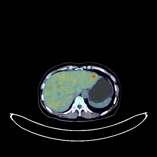
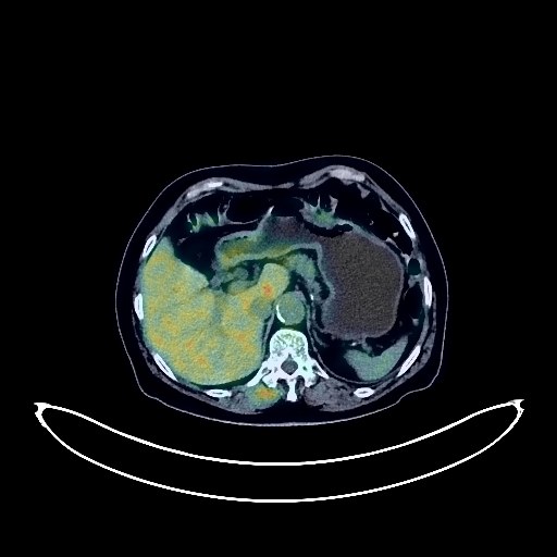
Liver Cancer PET/CT (case 983827-000069 from PETWB-REP)
0 views9 days agoWhole-body 18F-FDG PET/CT scan in a patient with Liver Cancer taken from the PETWB-REP dataset. The following English report (translated from original Chinese) is taken verbatim from the public dataset and has not been modified or otherwise checked for accuracy (see the end for citation). Impression a. Multiple space-occupying lesions in the liver with increased FDG metabolism, suggestive of malignancy, possibly multiple metastases or hepatocellular carcinoma with intrahepatic metastasis. The boundary with the right portal vein branch is unclear; please combine clinical findings with enhanced MRI for comprehensive analysis. b. Right anterior diaphragmatic lymph node metastasis and left pelvic wall metastasis. c. Possible partial peritoneal tumor infiltration, pelvic effusion. d. Focal increased FDG uptake in the right 4th posterior rib, metastasis to be ruled out; follow-up is recommended. Multiple metastases in both lungs. Pulmonary fibrosis. Bilateral breast hyperplasia. Slightly thickened gastric antrum wall with increased FDG metabolism, and increased FDG metabolism in some intestinal segments, suggestive of chronic inflammation; please combine endoscopic follow-up and rule out other possibilities. Gallstones. Left renal cyst, complex cyst. Partial vertebral osteophyte formation. L4/5 and L5/S1 intervertebral disc bulge. No obvious abnormalities seen on cranial scintigraphy. Chronic inflammation of bilateral ethmoid and maxillary sinuses. The thyroid gland has uneven density, and no abnormalities were found in FDG metabolism. Nodular goiter is suspected. Ultrasound follow-up is recommended. This case is from PETWB-REP, a curated dataset of whole-body 18F-FDG PET/CT scans and corresponding radiology reports from 490 patients with a broad spectrum of malignancies. The data were retrospectively collected from patients who underwent clinically indicated whole-body 18F-FDG PET/CT scans at the Shanghai Universal Medical Imaging Diagnostic Center between 2021 and 2024. License: Creative Commons Attribution 4.0 International (CC BY 4.0) Citation: Xue, L., Feng, G., Wenbo, Z., Zhang, Y., Li, L., Wang, S., Peng, L., Peng, S., & Gao, X. (2026). PETWB-REP: A Multi-Cancer Whole-Body FDG PET/CT Dataset with Corresponding Radiology Reports [Data set]. Zenodo. https://doi.org/10.5281/zenodo.18670487
Whole BodyPET/CT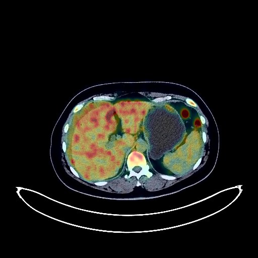
Ovarian Cancer PET/CT (case 983827-000117 from PETWB-REP)
0 views9 days agoWhole-body 18F-FDG PET/CT scan in a patient with Ovarian Cancer taken from the PETWB-REP dataset. The following English report (translated from original Chinese) is taken verbatim from the public dataset and has not been modified or otherwise checked for accuracy (see the end for citation). Impression a. Bilateral adnexal regions are full, structures are not clearly visible, FDG metabolism is increased, suggesting a high probability of malignancy, either primary or metastatic. Please combine with enhanced MRI for comprehensive analysis. b. Extensive implantation metastases in the abdominopelvic cavity. Liver metastasis is possible, and capsular metastasis involving the liver cannot be ruled out. Portocaval space and retroperitoneal lymph node metastases. Abdominal and pelvic effusions. c. Cystic low-density lesion on the left side of the uterus, with local nodular consolidation and increased FDG metabolism, suggesting possible cystic degeneration of uterine fibroids, with local malignancy to be ruled out. Please combine with enhanced MRI for comprehensive analysis. d. Splenic angioma is highly probable, ultrasound follow-up is recommended. Bilateral adrenal hyperplasia. Right middle cranial fossa floor mass, mildly increased FDG metabolism, suggesting a high probability of meningioma with calcification, with chondrosarcoma to be ruled out. Further enhanced MRI examination is recommended. Chronic inflammatory nodules (solid) in the posterior segment of the right upper lobe. A few chronic inflammations and old lesions in both lungs. Some arterial wall calcifications. Degenerative changes in the spine. L4/5 and L5/S1 intervertebral disc bulges. This case is from PETWB-REP, a curated dataset of whole-body 18F-FDG PET/CT scans and corresponding radiology reports from 490 patients with a broad spectrum of malignancies. The data were retrospectively collected from patients who underwent clinically indicated whole-body 18F-FDG PET/CT scans at the Shanghai Universal Medical Imaging Diagnostic Center between 2021 and 2024. License: Creative Commons Attribution 4.0 International (CC BY 4.0) Citation: Xue, L., Feng, G., Wenbo, Z., Zhang, Y., Li, L., Wang, S., Peng, L., Peng, S., & Gao, X. (2026). PETWB-REP: A Multi-Cancer Whole-Body FDG PET/CT Dataset with Corresponding Radiology Reports [Data set]. Zenodo. https://doi.org/10.5281/zenodo.18670487
Whole BodyPET/CT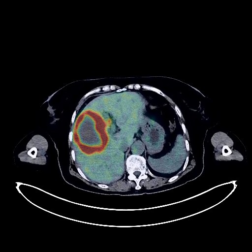
Cholangiocarcinoma PET/CT (case 983827-000216 from PETWB-REP)
0 views9 days agoWhole-body 18F-FDG PET/CT scan in a patient with Cholangiocarcinoma taken from the PETWB-REP dataset. The following English report (translated from original Chinese) is taken verbatim from the public dataset and has not been modified or otherwise checked for accuracy (see the end for citation). Impression a. A mass in the junction of the left and right lobes of the liver, with unevenly increased FDG metabolism, suggestive of a malignant tumor, most likely cholangiocarcinoma invading the gallbladder and hepatic flexure of the colon; please refer to pathology reports. Slight dilation of intrahepatic bile ducts and common bile duct. b. Metastasis to the hilar lymph nodes and root of the mesentery. c. Bilateral lung metastases. Mild interstitial changes in both lungs with a few post-inflammatory remnants. Signs of anemia. Calcification of some arterial walls. Main pancreatic duct visible. Senile uterus. Mildly increased FDG metabolism in some gastric walls, suggestive of physiological uptake or chronic inflammatory changes. Osteoporosis. Spinal degenerative changes. L3/4 and L4/5 intervertebral disc bulge. A few lacunar ischemic foci in the deep bilateral brain regions, with mild age-related brain changes. Sclerotic mastoid process on the left side. This case is from PETWB-REP, a curated dataset of whole-body 18F-FDG PET/CT scans and corresponding radiology reports from 490 patients with a broad spectrum of malignancies. The data were retrospectively collected from patients who underwent clinically indicated whole-body 18F-FDG PET/CT scans at the Shanghai Universal Medical Imaging Diagnostic Center between 2021 and 2024. License: Creative Commons Attribution 4.0 International (CC BY 4.0) Citation: Xue, L., Feng, G., Wenbo, Z., Zhang, Y., Li, L., Wang, S., Peng, L., Peng, S., & Gao, X. (2026). PETWB-REP: A Multi-Cancer Whole-Body FDG PET/CT Dataset with Corresponding Radiology Reports [Data set]. Zenodo. https://doi.org/10.5281/zenodo.18670487
Whole BodyPET/CT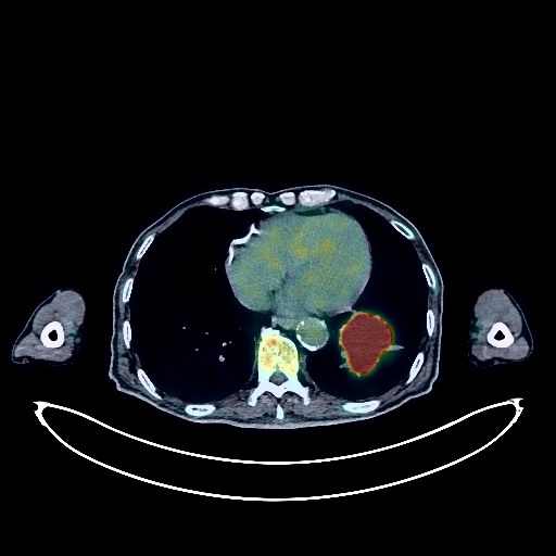
Lung Cancer PET/CT (case 983827-000147 from PETWB-REP)
0 views9 days agoWhole-body 18F-FDG PET/CT scan in a patient with Lung Cancer taken from the PETWB-REP dataset. The following English report (translated from original Chinese) is taken verbatim from the public dataset and has not been modified or otherwise checked for accuracy (see the end for citation). Impression a. Mass in the lower lobe of the left lung, with increased FDG metabolism, suggestive of lung cancer; please confirm with pathology. b. High probability of metastasis to the left hilar and left subcarinal lymph nodes. c. Scattered chronic inflammation and sequelae in both lungs. Emphysema in both lungs. Post-coronary artery stenting. Calcification of the aortic arch and abdominal aortic wall. Scattered punctate calcifications in the spleen. Right kidney stone. Multiple cysts in the left kidney. Continuous increased FDG metabolism in parts of the colon and rectum, likely due to inflammatory or physiological uptake; colonoscopy follow-up is recommended. Partial vertebral osteophyte formation. L5/S1 intervertebral disc bulge with pneumothorax. A few ischemic lesions deep in the brain. Age-related brain changes suggest an MRI scan. This case is from PETWB-REP, a curated dataset of whole-body 18F-FDG PET/CT scans and corresponding radiology reports from 490 patients with a broad spectrum of malignancies. The data were retrospectively collected from patients who underwent clinically indicated whole-body 18F-FDG PET/CT scans at the Shanghai Universal Medical Imaging Diagnostic Center between 2021 and 2024. License: Creative Commons Attribution 4.0 International (CC BY 4.0) Citation: Xue, L., Feng, G., Wenbo, Z., Zhang, Y., Li, L., Wang, S., Peng, L., Peng, S., & Gao, X. (2026). PETWB-REP: A Multi-Cancer Whole-Body FDG PET/CT Dataset with Corresponding Radiology Reports [Data set]. Zenodo. https://doi.org/10.5281/zenodo.18670487
Whole BodyPET/CT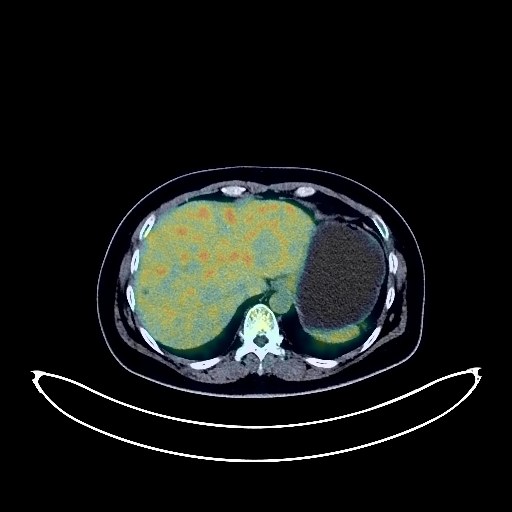
Ovarian Cancer PET/CT (case 983827-000135 from PETWB-REP)
0 views9 days agoWhole-body 18F-FDG PET/CT scan in a patient with Ovarian Cancer taken from the PETWB-REP dataset. The following English report (translated from original Chinese) is taken verbatim from the public dataset and has not been modified or otherwise checked for accuracy (see the end for citation). Impression a. Multiple space-occupying lesions in the bilateral adnexa and rectouterine pouch, multiple enlarged and swollen lymph nodes in the abdominopelvic region (as described above), increased FDG metabolism. The above suggests malignancy, with ovarian cancer and multiple metastases being highly probable. Metastatic tumors should be ruled out. Some lesions have unclear boundaries with the uterus and adjacent intestines; please consult pathology. b. Uterine fibroids with partial degeneration. a. Chronic inflammatory ground-glass nodules or atypical adenomatous hyperplasia in the apical segment of the right upper lobe; chronic inflammatory solid nodules in the medial segment of the right middle lobe and the oblique fissure of the left lung. Please repeat HRCT. b. A few post-inflammatory remnants in the right middle lobe; calcifications and small air-filled cysts in the right upper lobe. c. Bilateral breast proliferative changes; reactive hyperplasia of small axillary lymph nodes bilaterally. Thyroid gland is full and uneven in density with increased FDG metabolism, suggesting inflammatory uptake; please follow up with ultrasound. Small cysts and calcifications in the right lobe of the liver. Post-cholecystectomy changes. Manifestations of chronic gastritis. Degenerative changes in the spine, L3/4 intervertebral disc bulge. No obvious abnormalities seen on cranial scintigraphy. Minor chronic inflammation of the left ethmoid sinus. Physiological uptake in the nasopharynx. This case is from PETWB-REP, a curated dataset of whole-body 18F-FDG PET/CT scans and corresponding radiology reports from 490 patients with a broad spectrum of malignancies. The data were retrospectively collected from patients who underwent clinically indicated whole-body 18F-FDG PET/CT scans at the Shanghai Universal Medical Imaging Diagnostic Center between 2021 and 2024. License: Creative Commons Attribution 4.0 International (CC BY 4.0) Citation: Xue, L., Feng, G., Wenbo, Z., Zhang, Y., Li, L., Wang, S., Peng, L., Peng, S., & Gao, X. (2026). PETWB-REP: A Multi-Cancer Whole-Body FDG PET/CT Dataset with Corresponding Radiology Reports [Data set]. Zenodo. https://doi.org/10.5281/zenodo.18670487
Whole BodyPET/CT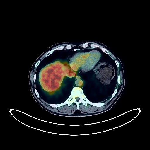
Lung Cancer PET/CT (case 983827-000212 from PETWB-REP)
0 views9 days agoWhole-body 18F-FDG PET/CT scan in a patient with Lung Cancer taken from the PETWB-REP dataset. The following English report (translated from original Chinese) is taken verbatim from the public dataset and has not been modified or otherwise checked for accuracy (see the end for citation). Impression a. A space-occupying lesion in the left upper lobe of the lung, adjacent to the mediastinum, with increased FDG metabolism, consistent with lung cancer with surrounding obstructive inflammation. b. Multiple chronic inflammatory micronodules in both lungs, mild bronchial dilatation in the right lower and middle lobes, and a few post-inflammatory remnants in both lungs. Slight pleural thickening bilaterally. Reactive hyperplasia of mediastinal lymph nodes. Small renal calculus in the left kidney. Multiple reactive hyperplasia of bilateral inguinal lymph nodes. Small amount of hydrocele in the left testis. Multiple diverticula in the ascending colon. Scoliosis with osteophyte formation. L3/4, L4/5, and L5/S1 intervertebral disc bulges. Old fractures of the left 1st-10th ribs. Age-related brain changes; MRI recommended. Minor inflammation of the right maxillary sinus. This case is from PETWB-REP, a curated dataset of whole-body 18F-FDG PET/CT scans and corresponding radiology reports from 490 patients with a broad spectrum of malignancies. The data were retrospectively collected from patients who underwent clinically indicated whole-body 18F-FDG PET/CT scans at the Shanghai Universal Medical Imaging Diagnostic Center between 2021 and 2024. License: Creative Commons Attribution 4.0 International (CC BY 4.0) Citation: Xue, L., Feng, G., Wenbo, Z., Zhang, Y., Li, L., Wang, S., Peng, L., Peng, S., & Gao, X. (2026). PETWB-REP: A Multi-Cancer Whole-Body FDG PET/CT Dataset with Corresponding Radiology Reports [Data set]. Zenodo. https://doi.org/10.5281/zenodo.18670487
Whole BodyPET/CT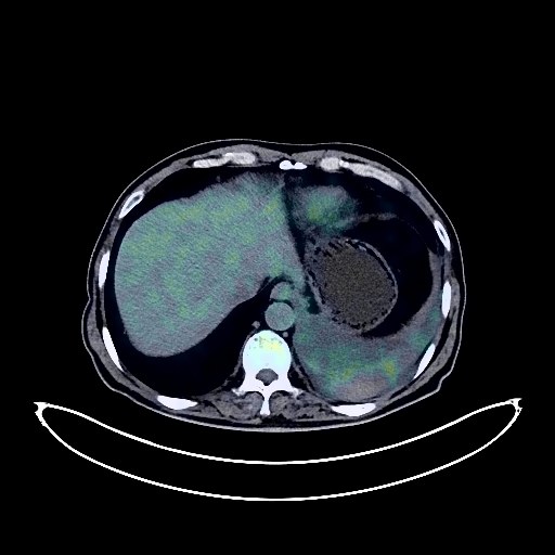
Lung Cancer PET/CT (case 983827-000198 from PETWB-REP)
0 views9 days agoWhole-body 18F-FDG PET/CT scan in a patient with Lung Cancer taken from the PETWB-REP dataset. The following English report (translated from original Chinese) is taken verbatim from the public dataset and has not been modified or otherwise checked for accuracy (see the end for citation). Impression a. A mass near the hilum of the left lower lobe, with elevated FDG metabolism, consistent with central lung cancer with peripheral obstructive inflammation. Left pleural effusion. Multiple lymph node metastases in the left hilum and mediastinum. Possible metastases to the left internal mammary chain and left anterior diaphragmatic lymph nodes; follow-up is recommended. ? b. A mixed ground-glass nodule in the anterior segment of the right upper lobe, with normal FDG metabolism, suggestive of lung cancer; please correlate with clinicopathology. ? c. Multiple pure ground-glass nodules in the apical-posterior segment of the left upper lobe, the anterior segment of the right upper lobe, and the anterior-basal segment of the lower lobe, with normal FDG metabolism; suggestive of atypical adenomatous hyperplasia or inflammatory nodules; annual HRCT follow-up is recommended. ? d. Multiple chronic inflammatory micronodules (solid) in both lungs. ? e. High probability of left adrenal hyperplasia; follow-up CT is recommended to rule out metastasis. Focal increased FDG uptake in the anal area, highly suggestive of hemorrhoids; digital rectal examination is recommended to rule out tumors. Small cyst in the left inner lobe of the liver. Small cyst in the left kidney. Subcutaneous calcification in the left mid-abdomen. Calcification in the prostate. Small amount of hydrocele in both testes. Reactive hyperplasia of multiple lymph nodes in the hepatogastric space, portal vena cava, retroperitoneum, and bilateral inguinal regions. Scoliosis with bone hyperplasia. L3/4 and L4/5 intervertebral disc bulge. Age-related brain changes; contrast-enhanced MRI is recommended. This case is from PETWB-REP, a curated dataset of whole-body 18F-FDG PET/CT scans and corresponding radiology reports from 490 patients with a broad spectrum of malignancies. The data were retrospectively collected from patients who underwent clinically indicated whole-body 18F-FDG PET/CT scans at the Shanghai Universal Medical Imaging Diagnostic Center between 2021 and 2024. License: Creative Commons Attribution 4.0 International (CC BY 4.0) Citation: Xue, L., Feng, G., Wenbo, Z., Zhang, Y., Li, L., Wang, S., Peng, L., Peng, S., & Gao, X. (2026). PETWB-REP: A Multi-Cancer Whole-Body FDG PET/CT Dataset with Corresponding Radiology Reports [Data set]. Zenodo. https://doi.org/10.5281/zenodo.18670487
Whole BodyPET/CT