Loading...
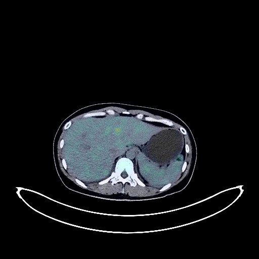
Liver Cancer PET/CT (case 983827-000157 from PETWB-REP)
0 views9 days agoWhole-body 18F-FDG PET/CT scan in a patient with Liver Cancer taken from the PETWB-REP dataset. The following English report (translated from original Chinese) is taken verbatim from the public dataset and has not been modified or otherwise checked for accuracy (see the end for citation). Impression a. Diffuse intrahepatic lesion with increased FDG metabolism, suggestive of malignancy; please refer to pathology. b. Multiple lymph node metastases in the prediaphragmatic group, abdominal cavity, and retroperitoneum. Small amount of pelvic effusion. c. Chronic cholecystitis. Gallstones. a. Ground-glass nodules in the upper lobes of both lungs and the middle lobe of the right lung, with normal FDG metabolism, suggestive of atypical adenomatous hyperplasia. Early lung cancer cannot be ruled out in the apical-posterior segment nodule of the left upper lobe. HRCT follow-up is recommended in 3 months. b. Several small chronic inflammatory nodules (solid) in both lungs, excluding some metastatic tumors; close observation is recommended. A small amount of chronic inflammation and old lesions in both lungs. c. Small amount of pleural effusion bilaterally, partial atelectasis in the lower lobes of both lungs. Slight pericardial thickening. Anemia. Bilateral breast proliferative changes. Physiological uptake in the uterine cavity. Physiological ovarian cyst in the right adnexal region. No abnormalities found on cranial scintigraphy. This case is from PETWB-REP, a curated dataset of whole-body 18F-FDG PET/CT scans and corresponding radiology reports from 490 patients with a broad spectrum of malignancies. The data were retrospectively collected from patients who underwent clinically indicated whole-body 18F-FDG PET/CT scans at the Shanghai Universal Medical Imaging Diagnostic Center between 2021 and 2024. License: Creative Commons Attribution 4.0 International (CC BY 4.0) Citation: Xue, L., Feng, G., Wenbo, Z., Zhang, Y., Li, L., Wang, S., Peng, L., Peng, S., & Gao, X. (2026). PETWB-REP: A Multi-Cancer Whole-Body FDG PET/CT Dataset with Corresponding Radiology Reports [Data set]. Zenodo. https://doi.org/10.5281/zenodo.18670487
Whole BodyPET/CT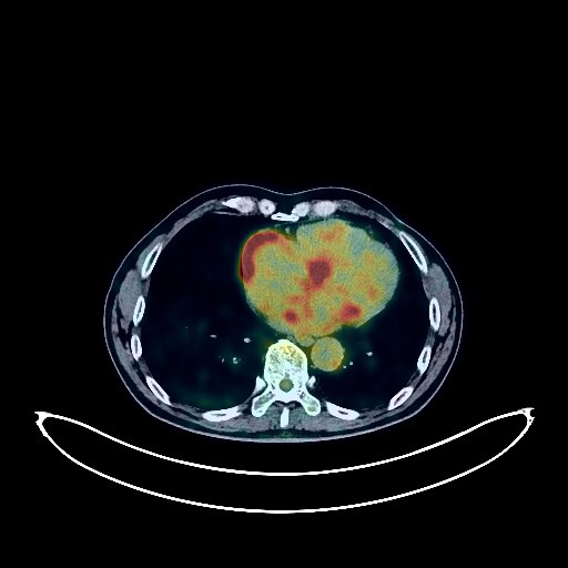
Lung Cancer PET/CT (case 983827-000163 from PETWB-REP)
0 views9 days agoWhole-body 18F-FDG PET/CT scan in a patient with Lung Cancer taken from the PETWB-REP dataset. The following English report (translated from original Chinese) is taken verbatim from the public dataset and has not been modified or otherwise checked for accuracy (see the end for citation). Impression a. A mass in the left upper lobe of the lung, adjacent to the mediastinum, with increased FDG metabolism, consistent with lung cancer with surrounding inflammation. The boundary with the left subclavian artery and aortic arch is unclear. b. A lesion in the apical segment of the right upper lobe, with slightly increased FDG metabolism, suggesting a high probability of inflammatory changes. Regular CT follow-up is recommended to rule out metastasis. Chronic inflammatory lymph nodes in the left hilum and mediastinum are highly likely, with possible metastasis. Follow-up is advised. c. Several small, solid, chronic inflammatory nodules in both lungs. A few chronic inflammations and old lesions in both lungs. Emphysema and multiple bullae in both lungs. Enlarged cardiac silhouette, with partial calcification of arterial walls. A slightly low-density nodule in the upper segment of the right posterior lobe of the liver, with decreased FDG metabolism, suggesting a small cyst or hemangioma. Comprehensive analysis with enhanced MRI is recommended. Liver cyst. Rough gallbladder wall. Bilateral renal cysts. Benign prostatic hyperplasia with calcifications. Mildly increased FDG metabolism in part of the gastric wall, considered physiological uptake or chronic inflammatory changes; please follow up with endoscopy. Spinal degenerative changes. L4/5 and L5/S1 intervertebral disc herniation. Subcutaneous calcification in the left buttock. Slight thickening of the bilateral nasopharyngeal walls with increased FDG metabolism, considered inflammatory changes. No obvious abnormalities seen on cranial scintigraphy. Sinusitis with a submucosal cyst in the left maxillary sinus. This case is from PETWB-REP, a curated dataset of whole-body 18F-FDG PET/CT scans and corresponding radiology reports from 490 patients with a broad spectrum of malignancies. The data were retrospectively collected from patients who underwent clinically indicated whole-body 18F-FDG PET/CT scans at the Shanghai Universal Medical Imaging Diagnostic Center between 2021 and 2024. License: Creative Commons Attribution 4.0 International (CC BY 4.0) Citation: Xue, L., Feng, G., Wenbo, Z., Zhang, Y., Li, L., Wang, S., Peng, L., Peng, S., & Gao, X. (2026). PETWB-REP: A Multi-Cancer Whole-Body FDG PET/CT Dataset with Corresponding Radiology Reports [Data set]. Zenodo. https://doi.org/10.5281/zenodo.18670487
Whole BodyPET/CT
Liver Cancer PET/CT (case 983827-000061 from PETWB-REP)
0 views9 days agoWhole-body 18F-FDG PET/CT scan in a patient with Liver Cancer taken from the PETWB-REP dataset. The following English report (translated from original Chinese) is taken verbatim from the public dataset and has not been modified or otherwise checked for accuracy (see the end for citation). Impression a. Mass in the left lobe of the liver, with elevated FDG metabolism, suggestive of malignancy, possibly primary or metastatic. b. Extensive implantation and metastasis in the abdominopelvic cavity. Abdominal and pelvic effusion. c. Bilateral pleural effusion. Metastasis to the anterior diaphragmatic lymph nodes. d. Bilateral adnexal structures are poorly visualized; clinical findings and enhanced MRI are recommended to rule out ovarian tumors. Liver cirrhosis. Liver cysts. Multiple small chronic inflammatory nodules (solid) in both lungs. A few chronic inflammatory lesions and old lesions in both lungs. Degenerative changes in the spine. L4/5 and L5/S1 intervertebral disc bulges. Bilateral nasal polyps. Chronic inflammation of the left maxillary sinus. A few ischemic lesions in the deep bilateral brain regions, age-related brain changes. Nodular goiter. This case is from PETWB-REP, a curated dataset of whole-body 18F-FDG PET/CT scans and corresponding radiology reports from 490 patients with a broad spectrum of malignancies. The data were retrospectively collected from patients who underwent clinically indicated whole-body 18F-FDG PET/CT scans at the Shanghai Universal Medical Imaging Diagnostic Center between 2021 and 2024. License: Creative Commons Attribution 4.0 International (CC BY 4.0) Citation: Xue, L., Feng, G., Wenbo, Z., Zhang, Y., Li, L., Wang, S., Peng, L., Peng, S., & Gao, X. (2026). PETWB-REP: A Multi-Cancer Whole-Body FDG PET/CT Dataset with Corresponding Radiology Reports [Data set]. Zenodo. https://doi.org/10.5281/zenodo.18670487
Whole BodyPET/CT
Esophageal Cancer PET/CT (case 983827-000119 from PETWB-REP)
0 views9 days agoWhole-body 18F-FDG PET/CT scan in a patient with Esophageal Cancer taken from the PETWB-REP dataset. The following English report (translated from original Chinese) is taken verbatim from the public dataset and has not been modified or otherwise checked for accuracy (see the end for citation). Impression a. Postoperative changes after esophageal cancer surgery; thickening of soft tissue in the hypopharynx and cervical esophagus, increased FDG metabolism, suggestive of malignancy; please refer to pathology reports. b. Right deep cervical lymph node metastasis. Reactive hyperplasia of left deep cervical lymph nodes is highly probable. Left renal mass with increased FDG metabolism, highly probable of renal cell carcinoma; please provide a comprehensive analysis with contrast-enhanced MRI. Chronic inflammatory micronodules in the left lung. Chronic inflammation and sequelae in both lungs, emphysema. Reactive hyperplasia of small mediastinal lymph nodes. Partial arteriosclerosis (including coronary arteries). Liver cysts. Liver calcifications. Gallstones, chronic cholecystitis. Left adrenal hyperplasia. Increased FDG metabolism in parts of the intestine, suggestive of physiological uptake or chronic inflammation. Scoliosis with degeneration. L4/5 disc bulge, L4/5 and L5/S1 disc pneumatosis and degeneration. Multiple old rib fractures on the right side. Bilateral deep lacunar infarcts, age-related brain. Chronic inflammation of the bilateral maxillary and ethmoid sinuses. This case is from PETWB-REP, a curated dataset of whole-body 18F-FDG PET/CT scans and corresponding radiology reports from 490 patients with a broad spectrum of malignancies. The data were retrospectively collected from patients who underwent clinically indicated whole-body 18F-FDG PET/CT scans at the Shanghai Universal Medical Imaging Diagnostic Center between 2021 and 2024. License: Creative Commons Attribution 4.0 International (CC BY 4.0) Citation: Xue, L., Feng, G., Wenbo, Z., Zhang, Y., Li, L., Wang, S., Peng, L., Peng, S., & Gao, X. (2026). PETWB-REP: A Multi-Cancer Whole-Body FDG PET/CT Dataset with Corresponding Radiology Reports [Data set]. Zenodo. https://doi.org/10.5281/zenodo.18670487
Whole BodyPET/CT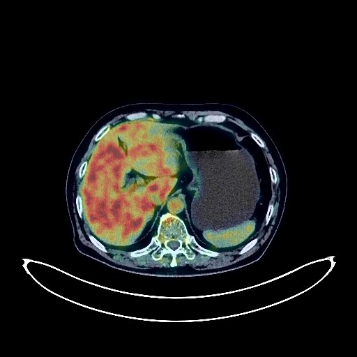
Lung Cancer PET/CT (case 983827-000103 from PETWB-REP)
0 views9 days agoWhole-body 18F-FDG PET/CT scan in a patient with Lung Cancer taken from the PETWB-REP dataset. The following English report (translated from original Chinese) is taken verbatim from the public dataset and has not been modified or otherwise checked for accuracy (see the end for citation). Impression a. Mixed-density ground-glass nodule in the posterior segment of the left lower lobe, with mildly increased FDG uptake, suggestive of peripheral lung cancer. b. Ground-glass nodule in the anterior-interior basal segment of the left lower lobe, with normal FDG uptake, suggestive of atypical adenomatous hyperplasia, early lung cancer to be ruled out. c. Several small chronic inflammatory nodules (solid) in both lungs. A few chronic inflammations and old lesions in both lungs. Emphysema with bullae in both lungs. Reactive hyperplasia of hilar and mediastinal lymph nodes in both lungs. Calcification of some arterial walls (including coronary arteries). Sellar region mass, increased FDG uptake, suggestive of pituitary adenoma, metastasis to be ruled out; enhanced MRI is recommended for further examination. Multiple liver cysts. Bilateral renal cysts. Prostatic calcifications. Chronic antral gastritis. Increased continuous FDG metabolism in parts of the colon and rectum, suggestive of inflammatory or physiological uptake. Duodenal diverticulum. Osteoporosis. Degenerative changes in the spine. L4/5 and L5/S1 intervertebral disc bulge. Inflammatory uptake in the L3/4 interspinous region. A few ischemic foci in the deep bilateral brain regions, age-related brain changes. This case is from PETWB-REP, a curated dataset of whole-body 18F-FDG PET/CT scans and corresponding radiology reports from 490 patients with a broad spectrum of malignancies. The data were retrospectively collected from patients who underwent clinically indicated whole-body 18F-FDG PET/CT scans at the Shanghai Universal Medical Imaging Diagnostic Center between 2021 and 2024. License: Creative Commons Attribution 4.0 International (CC BY 4.0) Citation: Xue, L., Feng, G., Wenbo, Z., Zhang, Y., Li, L., Wang, S., Peng, L., Peng, S., & Gao, X. (2026). PETWB-REP: A Multi-Cancer Whole-Body FDG PET/CT Dataset with Corresponding Radiology Reports [Data set]. Zenodo. https://doi.org/10.5281/zenodo.18670487
Whole BodyPET/CT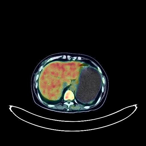
Cervical Cancer PET/CT (case 983827-000151 from PETWB-REP)
0 views9 days agoWhole-body 18F-FDG PET/CT scan in a patient with Cervical Cancer taken from the PETWB-REP dataset. The following English report (translated from original Chinese) is taken verbatim from the public dataset and has not been modified or otherwise checked for accuracy (see the end for citation). Impression a. Irregular lesions in the cervix and adjacent uterine body with increased FDG metabolism, consistent with cervical cancer; multiple lymph node metastases in the bilateral iliac vessels and retroperitoneum; possible reactive hyperplasia of bilateral inguinal lymph nodes. b. Full uterus with irregular margins; no abnormalities in FDG metabolism; please correlate with clinical findings. Pelvic effusion. Chronic inflammatory nodule in the lower lobe of the left lung; CT follow-up is recommended. Reactive hyperplasia of bilateral axillary lymph nodes. Anemia. Small cyst in the left lobe of the liver; possible hemangioma in the right lobe. Chronic cholecystitis. Chronic inflammatory changes or physiological uptake in parts of the intestine; please correlate with endoscopic follow-up. Mild vertebral osteophyte formation; L4/5 and L5/S1 intervertebral disc bulge. Uneven thyroid density and elevated FDG metabolism suggest possible inflammation. Please combine thyroid function tests and ultrasound examination. No obvious abnormalities were found on cranial scintigraphy. Reactive hyperplasia of bilateral cervical lymph nodes. This case is from PETWB-REP, a curated dataset of whole-body 18F-FDG PET/CT scans and corresponding radiology reports from 490 patients with a broad spectrum of malignancies. The data were retrospectively collected from patients who underwent clinically indicated whole-body 18F-FDG PET/CT scans at the Shanghai Universal Medical Imaging Diagnostic Center between 2021 and 2024. License: Creative Commons Attribution 4.0 International (CC BY 4.0) Citation: Xue, L., Feng, G., Wenbo, Z., Zhang, Y., Li, L., Wang, S., Peng, L., Peng, S., & Gao, X. (2026). PETWB-REP: A Multi-Cancer Whole-Body FDG PET/CT Dataset with Corresponding Radiology Reports [Data set]. Zenodo. https://doi.org/10.5281/zenodo.18670487
Whole BodyPET/CT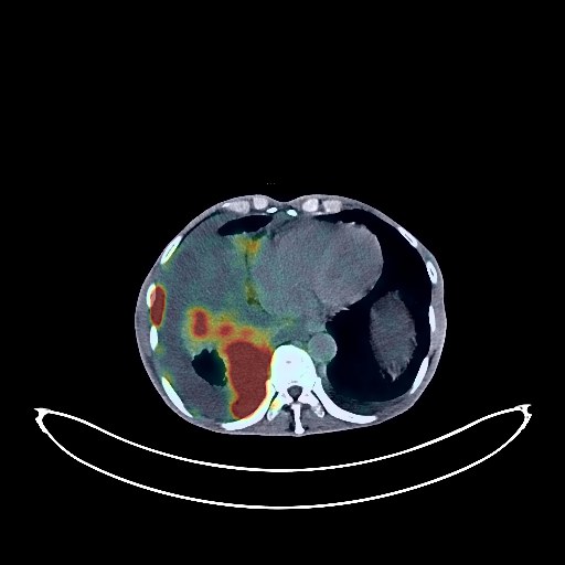
Lung Cancer PET/CT (case 983827-000126 from PETWB-REP)
0 views9 days agoWhole-body 18F-FDG PET/CT scan in a patient with Lung Cancer taken from the PETWB-REP dataset. The following English report (translated from original Chinese) is taken verbatim from the public dataset and has not been modified or otherwise checked for accuracy (see the end for citation). Impression a. Right hilar mass with elevated FDG metabolism, suggestive of central lung cancer with obstructive changes. b. Multiple lymph node metastases in the right hilum, mediastinum, and right supraclavicular fossa. c. Extensive right pleural metastasis. Located pleural effusion on the right side, small amount of pleural effusion on the left side. d. Peritoneal seeding metastasis. Multiple bone metastases throughout the body. Multiple muscle metastases throughout the body. Large amounts of abdominal and pelvic effusions. e. Possible bilateral adrenal metastases. Bilateral emphysema with bullae. Scattered chronic inflammation in the remaining lungs. Calcification of some arterial walls. Anemia. Multiple liver cysts. Degenerative changes in the spine. L4/5 and L5/S1 intervertebral disc bulges. A few ischemic lesions in the deep bilateral brain regions, indicative of age-related brain disorders. This case is from PETWB-REP, a curated dataset of whole-body 18F-FDG PET/CT scans and corresponding radiology reports from 490 patients with a broad spectrum of malignancies. The data were retrospectively collected from patients who underwent clinically indicated whole-body 18F-FDG PET/CT scans at the Shanghai Universal Medical Imaging Diagnostic Center between 2021 and 2024. License: Creative Commons Attribution 4.0 International (CC BY 4.0) Citation: Xue, L., Feng, G., Wenbo, Z., Zhang, Y., Li, L., Wang, S., Peng, L., Peng, S., & Gao, X. (2026). PETWB-REP: A Multi-Cancer Whole-Body FDG PET/CT Dataset with Corresponding Radiology Reports [Data set]. Zenodo. https://doi.org/10.5281/zenodo.18670487
Whole BodyPET/CT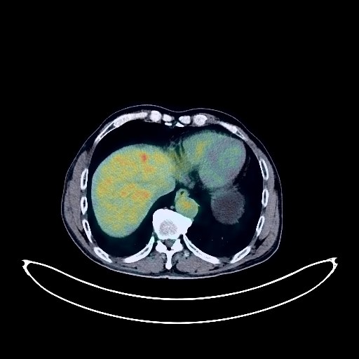
Nasopharyngeal Cancer PET/CT (case 983827-000100 from PETWB-REP)
0 views9 days agoWhole-body 18F-FDG PET/CT scan in a patient with Nasopharyngeal Cancer taken from the PETWB-REP dataset. The following English report (translated from original Chinese) is taken verbatim from the public dataset and has not been modified or otherwise checked for accuracy (see the end for citation). Impression A mass in the nasopharynx with elevated FDG metabolism, consistent with nasopharyngeal carcinoma and invasion of the adjacent skull base; multiple lymph node metastases in the right retropharyngeal space and bilateral deep cervical spaces. Enlarged left thyroid lobe with heterogeneous density and elevated FDG metabolism; thyroid cancer to be ruled out; further ultrasound examination recommended. Chronic inflammatory micronodules in both lungs. A few post-inflammatory lesions in both lungs. Minor arteriosclerosis in some arteries. Gallstones. Right renal cyst. Residual contrast agent in the urinary tract. Benign prostatic hyperplasia with calcification. Chronic inflammatory changes in the cardia and antrum of the stomach; please follow up with endoscopy. Mild posterior slippage of the L1 vertebral body. Degenerative changes in the spine; L4/5 disc herniation. Age-related brain; deep lacunar infarcts; please combine with MRI examination. This case is from PETWB-REP, a curated dataset of whole-body 18F-FDG PET/CT scans and corresponding radiology reports from 490 patients with a broad spectrum of malignancies. The data were retrospectively collected from patients who underwent clinically indicated whole-body 18F-FDG PET/CT scans at the Shanghai Universal Medical Imaging Diagnostic Center between 2021 and 2024. License: Creative Commons Attribution 4.0 International (CC BY 4.0) Citation: Xue, L., Feng, G., Wenbo, Z., Zhang, Y., Li, L., Wang, S., Peng, L., Peng, S., & Gao, X. (2026). PETWB-REP: A Multi-Cancer Whole-Body FDG PET/CT Dataset with Corresponding Radiology Reports [Data set]. Zenodo. https://doi.org/10.5281/zenodo.18670487
Whole BodyPET/CT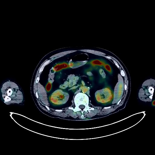
Lung Cancer PET/CT (case 983827-000062 from PETWB-REP)
1 views9 days agoWhole-body 18F-FDG PET/CT scan in a patient with Lung Cancer taken from the PETWB-REP dataset. The following English report (translated from original Chinese) is taken verbatim from the public dataset and has not been modified or otherwise checked for accuracy (see the end for citation). Impression a. Irregular nodules in the anterior segment of the right upper lobe, with increased FDG metabolism, suggestive of lung cancer; please correlate with clinicopathology. b. Right hilar and subcarinal lymph node metastasis to be ruled out; bilateral axillary lymph node reactive hyperplasia is highly probable; close follow-up is recommended. c. Multiple solid nodules in both lungs with normal FDG metabolism, highly probable as chronic inflammatory nodules; regular CT scans are recommended to rule out other complications. Interstitial inflammation in the lower lobes of both lungs. Calcification of some arterial walls (including coronary arteries). Liver calcifications. Chronic cholecystitis, cholestasis or sludge-like stones in the gallbladder. Benign prostatic hyperplasia. Continuous increased FDG metabolism in the ascending colon, descending colon, sigmoid colon, and rectum, suggestive of inflammatory lesions; colonoscopy is recommended. Hemorrhoidal changes. Degenerative changes in the spine. Lumbar instability. L3/5 disc bulge, L2/3 and L4/5 disc pneumatosis and degeneration. Increased FDG metabolism around the left elbow, both knees, both ankles, and small joints of both hands and feet, suggesting inflammatory changes. A mass in the sellar region is suspected, possibly a pituitary tumor. There are a few ischemic lesions in the deep bilateral brain regions, indicative of age-related brain conditions. Further enhanced MRI is recommended. This case is from PETWB-REP, a curated dataset of whole-body 18F-FDG PET/CT scans and corresponding radiology reports from 490 patients with a broad spectrum of malignancies. The data were retrospectively collected from patients who underwent clinically indicated whole-body 18F-FDG PET/CT scans at the Shanghai Universal Medical Imaging Diagnostic Center between 2021 and 2024. License: Creative Commons Attribution 4.0 International (CC BY 4.0) Citation: Xue, L., Feng, G., Wenbo, Z., Zhang, Y., Li, L., Wang, S., Peng, L., Peng, S., & Gao, X. (2026). PETWB-REP: A Multi-Cancer Whole-Body FDG PET/CT Dataset with Corresponding Radiology Reports [Data set]. Zenodo. https://doi.org/10.5281/zenodo.18670487
Whole BodyPET/CT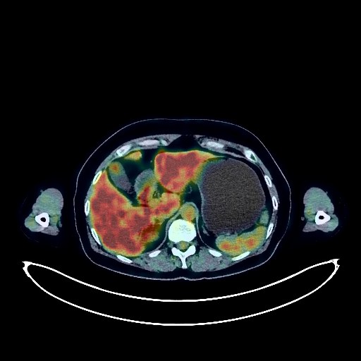
Cervical Cancer PET/CT (case 983827-000218 from PETWB-REP)
0 views9 days agoWhole-body 18F-FDG PET/CT scan in a patient with Cervical Cancer taken from the PETWB-REP dataset. The following English report (translated from original Chinese) is taken verbatim from the public dataset and has not been modified or otherwise checked for accuracy (see the end for citation). Impression Uterine atrophy; no abnormal FDG metabolic foci seen in the cervix; previously enlarged lymph nodes in the left pelvic wall and retroperitoneum were not clearly visible this time. Considering the above, tumor activity has been largely suppressed after treatment; please consult a specialist and undergo enhanced MRI. Bilateral inguinal lymph node reactive hyperplasia. Bilateral knee joint effusion; no obvious space-occupying lesion seen in the left knee joint; please consult a specialist for follow-up. No significant abnormalities in intracranial FDG metabolism. Bilateral ethmoid sinusitis, right maxillary sinusitis, slight nasal septum deviation, bilateral turbinate hypertrophy. Chronic inflammation of the oropharynx. Bilateral chronic inflammatory small lymph nodes in the neck. Chronic miliary nodules in the posterior basal segment of the right lower lobe; chronic inflammation and remnants in both lungs. Bilateral pleural thickening. Bilateral breast proliferative changes; please follow up with a clinical specialist. Calcification in the right anterior lobe of the liver. Thickening of the gastric antrum wall with increased FDG metabolism; increased FDG metabolism in some intestinal segments, suggesting a high probability of chronic inflammatory changes; follow-up gastroscopy and colonoscopy are recommended. Nodular soft tissue density shadow on the right side of the anus with increased FDG metabolism, suggesting a high probability of inflammatory lesions; specialist examination is recommended. Degenerative changes in the spine. Tensile take-up in some muscles of both upper and lower limbs. This case is from PETWB-REP, a curated dataset of whole-body 18F-FDG PET/CT scans and corresponding radiology reports from 490 patients with a broad spectrum of malignancies. The data were retrospectively collected from patients who underwent clinically indicated whole-body 18F-FDG PET/CT scans at the Shanghai Universal Medical Imaging Diagnostic Center between 2021 and 2024. License: Creative Commons Attribution 4.0 International (CC BY 4.0) Citation: Xue, L., Feng, G., Wenbo, Z., Zhang, Y., Li, L., Wang, S., Peng, L., Peng, S., & Gao, X. (2026). PETWB-REP: A Multi-Cancer Whole-Body FDG PET/CT Dataset with Corresponding Radiology Reports [Data set]. Zenodo. https://doi.org/10.5281/zenodo.18670487
Whole BodyPET/CT