Loading...
Gastric Cancer PET/CT (case 983827-000068 from PETWB-REP)
0 views9 days agoWhole-body 18F-FDG PET/CT scan in a patient with Gastric Cancer taken from the PETWB-REP dataset. The following English report (translated from original Chinese) is taken verbatim from the public dataset and has not been modified or otherwise checked for accuracy (see the end for citation). Impression a. Irregular thickening of the gastric wall at the gastric angle, with increased FDG metabolism, consistent with gastric cancer. b. Several small lymph nodes in the hepatogastric space and beside the abdominal aorta, with normal FDG metabolism, suggesting likely reactive hyperplasia; follow-up is recommended to rule out other complications. A mass in the sellar region, with increased FDG metabolism, suggesting a possible pituitary adenoma; metastasis needs to be ruled out. Further enhanced MRI is recommended. Several small, solid, chronic inflammatory nodules in both lungs. Chronic inflammation and old lesions in the lower lobes of both lungs. Small cysts in the liver. Degenerative changes in the spine. L4/5 intervertebral disc bulge. Right basal ganglia softening lesion, a few ischemic lesions in the deep bilateral brain, senile encephalopathy. Chronic inflammation of the left maxillary sinus and left ethmoid sinus. This case is from PETWB-REP, a curated dataset of whole-body 18F-FDG PET/CT scans and corresponding radiology reports from 490 patients with a broad spectrum of malignancies. The data were retrospectively collected from patients who underwent clinically indicated whole-body 18F-FDG PET/CT scans at the Shanghai Universal Medical Imaging Diagnostic Center between 2021 and 2024. License: Creative Commons Attribution 4.0 International (CC BY 4.0) Citation: Xue, L., Feng, G., Wenbo, Z., Zhang, Y., Li, L., Wang, S., Peng, L., Peng, S., & Gao, X. (2026). PETWB-REP: A Multi-Cancer Whole-Body FDG PET/CT Dataset with Corresponding Radiology Reports [Data set]. Zenodo. https://doi.org/10.5281/zenodo.18670487
Whole BodyPET/CT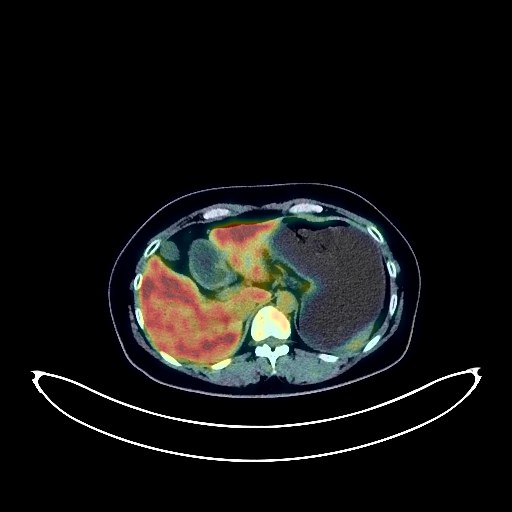
Cervical Cancer PET/CT (case 983827-000093 from PETWB-REP)
0 views9 days agoWhole-body 18F-FDG PET/CT scan in a patient with Cervical Cancer taken from the PETWB-REP dataset. The following English report (translated from original Chinese) is taken verbatim from the public dataset and has not been modified or otherwise checked for accuracy (see the end for citation). Impression Cervical mass with elevated FDG metabolism, highly suggestive of cervical cancer; please correlate with clinicopathology. Possible metastasis to the left iliac lymph nodes. Chronic inflammatory micronodules in the left lung. A few post-inflammatory lesions in both lungs. Small cyst in the right kidney. Chronic gastritis; please follow up with endoscopy. Mild vertebral osteophyte formation; slight bulging of the L4/5 and L5/S1 intervertebral discs. No obvious abnormalities on cranial scintigraphy. A few chronic inflammations in both ethmoid sinuses. Physiological uptake in the glottic region. This case is from PETWB-REP, a curated dataset of whole-body 18F-FDG PET/CT scans and corresponding radiology reports from 490 patients with a broad spectrum of malignancies. The data were retrospectively collected from patients who underwent clinically indicated whole-body 18F-FDG PET/CT scans at the Shanghai Universal Medical Imaging Diagnostic Center between 2021 and 2024. License: Creative Commons Attribution 4.0 International (CC BY 4.0) Citation: Xue, L., Feng, G., Wenbo, Z., Zhang, Y., Li, L., Wang, S., Peng, L., Peng, S., & Gao, X. (2026). PETWB-REP: A Multi-Cancer Whole-Body FDG PET/CT Dataset with Corresponding Radiology Reports [Data set]. Zenodo. https://doi.org/10.5281/zenodo.18670487
Whole BodyPET/CT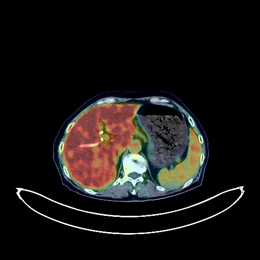
Gallbladder Cancer PET/CT (case 983827-000200 from PETWB-REP)
0 views9 days agoWhole-body 18F-FDG PET/CT scan in a patient with Gallbladder Cancer taken from the PETWB-REP dataset. The following English report (translated from original Chinese) is taken verbatim from the public dataset and has not been modified or otherwise checked for accuracy (see the end for citation). Impression Gallbladder mass with elevated FDG metabolism, highly suggestive of gallbladder cancer; please correlate with clinicopathology. Possible metastasis to lymph nodes in the hepatic hilum, hilar space, and retroperitoneum. Indwelling bile duct drainage tube. Chronic inflammatory micronodules in the right lung. A few post-inflammatory lesions in both lungs. Bilateral pleural thickening. Anemic changes, partial arterial wall calcification (including coronary arteries). Accessory spleen. Small renal cysts bilaterally. Residual contrast agent in the cystostomy chamber. Chronic inflammatory changes or physiological uptake in some intestinal segments; please correlate with endoscopic follow-up. Osteoporosis, degenerative changes in the spine, anterior slippage of the L4 vertebral body. L3/4, L4/5, and L5/S1 intervertebral disc bulges. Bilateral subcutaneous calcifications in the buttocks. Age-related brain lesions, deep lacunar infarcts. Submucosal cyst of the right maxillary sinus. This case is from PETWB-REP, a curated dataset of whole-body 18F-FDG PET/CT scans and corresponding radiology reports from 490 patients with a broad spectrum of malignancies. The data were retrospectively collected from patients who underwent clinically indicated whole-body 18F-FDG PET/CT scans at the Shanghai Universal Medical Imaging Diagnostic Center between 2021 and 2024. License: Creative Commons Attribution 4.0 International (CC BY 4.0) Citation: Xue, L., Feng, G., Wenbo, Z., Zhang, Y., Li, L., Wang, S., Peng, L., Peng, S., & Gao, X. (2026). PETWB-REP: A Multi-Cancer Whole-Body FDG PET/CT Dataset with Corresponding Radiology Reports [Data set]. Zenodo. https://doi.org/10.5281/zenodo.18670487
Whole BodyPET/CT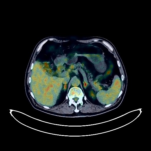
Colon Cancer PET/CT (case 983827-000204 from PETWB-REP)
0 views9 days agoWhole-body 18F-FDG PET/CT scan in a patient with Colon Cancer taken from the PETWB-REP dataset. The following English report (translated from original Chinese) is taken verbatim from the public dataset and has not been modified or otherwise checked for accuracy (see the end for citation). Impression a. Masses in the transverse colon and sigmoid colon with increased FDG metabolism, suggestive of colon cancer with surrounding mesenteric infiltration. Continuous increased FDG metabolism in the remaining colon and rectum suggests possible inflammatory uptake. b. Metastasis to lymph nodes around the lesion, in the local mesenteric region, and bilaterally to the iliac vessels. Anemia. a. Soft tissue mass in the anterior medial basal segment of the left lower lobe with increased FDG metabolism, highly suggestive of lung cancer; please confirm with pathology. b. Scattered solid nodules in both lungs, with normal FDG uptake, suggestive of chronic inflammatory nodules; please follow up with CT to rule out metastasis. Low-density nodule in the left lobe of the thyroid gland, with normal FDG uptake, suggestive of adenoma; local malignancy to be ruled out; please follow up with ultrasound and fine-needle aspiration. Mild fatty liver. Post-cholecystectomy. Bilateral renal cysts. Spinal degenerative changes. L4/5 and L5/S1 intervertebral disc bulges. Multiple old fractures of the bilateral pubic bones. Reactive hyperplasia of bilateral deep cervical and submandibular lymph nodes. No obvious abnormalities were found on cranial FDG imaging. Bilateral maxillary sinusitis. This case is from PETWB-REP, a curated dataset of whole-body 18F-FDG PET/CT scans and corresponding radiology reports from 490 patients with a broad spectrum of malignancies. The data were retrospectively collected from patients who underwent clinically indicated whole-body 18F-FDG PET/CT scans at the Shanghai Universal Medical Imaging Diagnostic Center between 2021 and 2024. License: Creative Commons Attribution 4.0 International (CC BY 4.0) Citation: Xue, L., Feng, G., Wenbo, Z., Zhang, Y., Li, L., Wang, S., Peng, L., Peng, S., & Gao, X. (2026). PETWB-REP: A Multi-Cancer Whole-Body FDG PET/CT Dataset with Corresponding Radiology Reports [Data set]. Zenodo. https://doi.org/10.5281/zenodo.18670487
Whole BodyPET/CT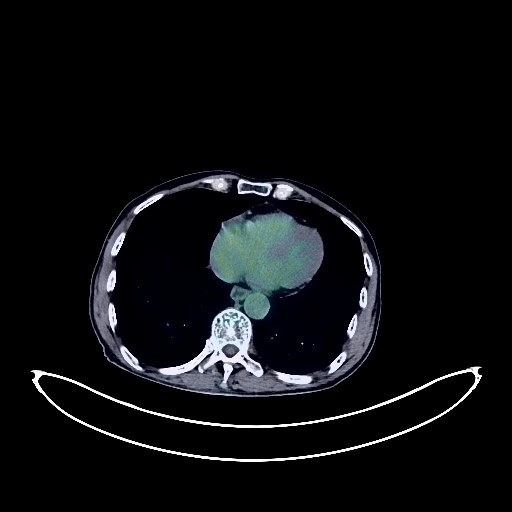
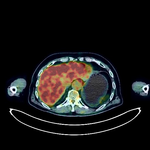
Liver Cancer PET/CT (case 983827-000165 from PETWB-REP)
0 views9 days agoWhole-body 18F-FDG PET/CT scan in a patient with Liver Cancer taken from the PETWB-REP dataset. The following English report (translated from original Chinese) is taken verbatim from the public dataset and has not been modified or otherwise checked for accuracy (see the end for citation). Impression a. A large, slightly low-density mass in the lower segment of the right lobe of the liver, with increased FDG metabolism. Combined with enhanced MRI from our center, this is considered a malignant tumor, most likely hepatocellular carcinoma, with tumor thrombus formation in the main portal vein and its left and right branches. b. Possible metastasis to the hilar lymph nodes; reactive hyperplasia of the hepatogastric space and retroperitoneal lymph nodes, to be ruled out as mixed metastasis. Please follow up. c. A slightly low-density nodule in the lower segment of the left lateral lobe of the liver, with decreased FDG metabolism. Combined with enhanced MRI from our center, this is considered a hemangioma. A cyst in the left medial lobe of the liver. Calcification in the diaphragmatic dome of the right lobe of the liver. a. Patchy areas of increased density near the hilum in the right middle lobe of the lung with increased FDG metabolism; multiple nodules in both lungs, some with increased FDG metabolism. Inflammatory lesions are considered possible. A space-occupying lesion in the right middle lobe of the lung is to be ruled out, and metastasis of some nodules is to be ruled out. Please closely monitor with clinical findings and CT scans. b. Chronic inflammatory lymph nodes in the bilateral hilar and mediastinal regions, pending metastasis; follow-up is required. Partial thickening and calcification of the bilateral pleura. c. A few post-inflammatory lesions in the remaining lungs, emphysema in both lungs. Calcification of some arterial walls (including coronary arteries). Slightly distended gallbladder. Mild atrophy of the left kidney, multiple cysts in the left kidney (some complex). Small vascular leiomyolipomas in the upper pole of the right kidney. Benign prostatic hyperplasia with calcification. Increased FDG metabolism in some gastric walls, likely due to chronic inflammation. Osteoporosis. Scoliosis with degenerative changes. L3/4 and L4/5 intervertebral disc bulging and calcification, L4/5 intervertebral disc pneumoconiosis and degeneration. Bilateral deep lacunar infarcts, age-related brain changes. Chronic inflammation of the right maxillary sinus. Reactive hyperplasia of the bilateral deep cervical spaces and submandibular lymph nodes. This case is from PETWB-REP, a curated dataset of whole-body 18F-FDG PET/CT scans and corresponding radiology reports from 490 patients with a broad spectrum of malignancies. The data were retrospectively collected from patients who underwent clinically indicated whole-body 18F-FDG PET/CT scans at the Shanghai Universal Medical Imaging Diagnostic Center between 2021 and 2024. License: Creative Commons Attribution 4.0 International (CC BY 4.0) Citation: Xue, L., Feng, G., Wenbo, Z., Zhang, Y., Li, L., Wang, S., Peng, L., Peng, S., & Gao, X. (2026). PETWB-REP: A Multi-Cancer Whole-Body FDG PET/CT Dataset with Corresponding Radiology Reports [Data set]. Zenodo. https://doi.org/10.5281/zenodo.18670487
Whole BodyPET/CT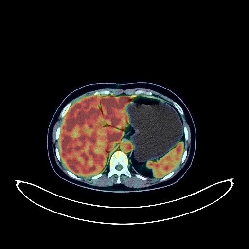
Lung Cancer PET/CT (case 983827-000032 from PETWB-REP)
0 views9 days agoWhole-body 18F-FDG PET/CT scan in a patient with Lung Cancer taken from the PETWB-REP dataset. The following English report (translated from original Chinese) is taken verbatim from the public dataset and has not been modified or otherwise checked for accuracy (see the end for citation). Impression a. Postoperative changes in the right upper lobe of lung cancer, no signs of tumor recurrence in the surgical area; b. Chronic inflammatory nodules in both lungs, follow-up CT recommended. Scattered post-inflammatory lesions in both lungs. Slight local thickening of the right pleura. Reactive hyperplasia of bilateral axillary lymph nodes. Bilateral adrenal hyperplasia, possible small renal cysts, please follow up; local protrusion on the medial border of the spleen, possibly normal variation, please use ultrasound to rule out space-occupying lesions. Reactive hyperplasia of lymph nodes in the hepatogastric space, interhilar cavity, retroperitoneum, bilateral pelvic walls, and bilateral inguinal regions. Chronic inflammatory changes in the antrum of the stomach and duodenal bulb, hemorrhoidal changes, please use endoscopy for follow-up. Mild vertebral osteophyte formation. Changes following fracture of the right 4th and 5th ribs. Benign bone disease of the auricular surface of the right iliac bone, please follow up. Uneven thyroid density, increased FDG metabolism, please use ultrasound and laboratory tests. No obvious abnormalities were found on cranial scintigraphy. Reactive hyperplasia of bilateral cervical lymph nodes was observed. This case is from PETWB-REP, a curated dataset of whole-body 18F-FDG PET/CT scans and corresponding radiology reports from 490 patients with a broad spectrum of malignancies. The data were retrospectively collected from patients who underwent clinically indicated whole-body 18F-FDG PET/CT scans at the Shanghai Universal Medical Imaging Diagnostic Center between 2021 and 2024. License: Creative Commons Attribution 4.0 International (CC BY 4.0) Citation: Xue, L., Feng, G., Wenbo, Z., Zhang, Y., Li, L., Wang, S., Peng, L., Peng, S., & Gao, X. (2026). PETWB-REP: A Multi-Cancer Whole-Body FDG PET/CT Dataset with Corresponding Radiology Reports [Data set]. Zenodo. https://doi.org/10.5281/zenodo.18670487
Whole BodyPET/CT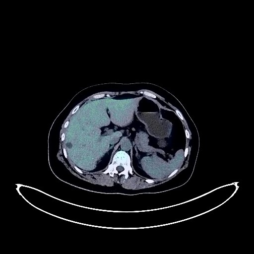
Cervical Cancer PET/CT (case 983827-000140 from PETWB-REP)
0 views9 days agoWhole-body 18F-FDG PET/CT scan in a patient with Cervical Cancer taken from the PETWB-REP dataset. The following English report (translated from original Chinese) is taken verbatim from the public dataset and has not been modified or otherwise checked for accuracy (see the end for citation). Impression a. Cervical mass with elevated FDG metabolism, consistent with cervical cancer. b. Nabothian cyst of the cervix. Reactive hyperplasia of small lymph nodes beside the bilateral iliac vessels. Left adnexal cyst or small amount of pelvic effusion. c. Possibly large bony island on the right iliac bone; follow-up is recommended to rule out other possibilities. Chronic inflammatory nodules in both lungs. A few post-inflammatory lesions in both lungs. Anemia changes, partial arteriosclerosis. Bilateral breast hyperplasia, possibly fibroadenoma of the right breast; please confirm with ultrasound examination. Right lobe cyst of the liver. Accessory spleen. Chronic inflammatory changes in the cardia and antrum of the stomach; please confirm with endoscopic follow-up. Degenerative changes in the spine, with L4/5 and L5/S1 intervertebral disc bulges. No obvious abnormalities were found on cranial scintigraphy. Physiological uptake of the glottic area. This case is from PETWB-REP, a curated dataset of whole-body 18F-FDG PET/CT scans and corresponding radiology reports from 490 patients with a broad spectrum of malignancies. The data were retrospectively collected from patients who underwent clinically indicated whole-body 18F-FDG PET/CT scans at the Shanghai Universal Medical Imaging Diagnostic Center between 2021 and 2024. License: Creative Commons Attribution 4.0 International (CC BY 4.0) Citation: Xue, L., Feng, G., Wenbo, Z., Zhang, Y., Li, L., Wang, S., Peng, L., Peng, S., & Gao, X. (2026). PETWB-REP: A Multi-Cancer Whole-Body FDG PET/CT Dataset with Corresponding Radiology Reports [Data set]. Zenodo. https://doi.org/10.5281/zenodo.18670487
Whole BodyPET/CT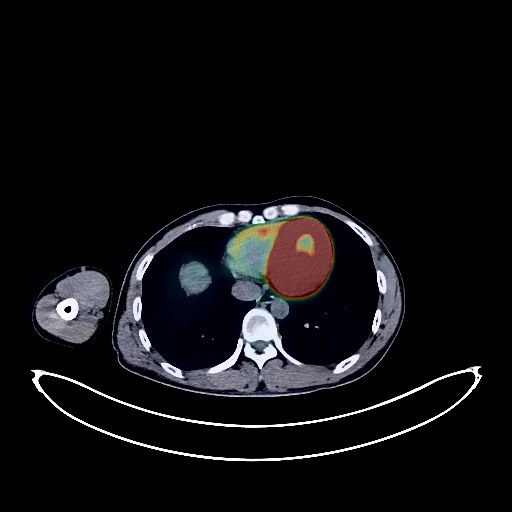
Glioma PET/CT (case 983827-000124 from PETWB-REP)
0 views9 days agoWhole-body 18F-FDG PET/CT scan in a patient with Glioma taken from the PETWB-REP dataset. The following English report (translated from original Chinese) is taken verbatim from the public dataset and has not been modified or otherwise checked for accuracy (see the end for citation). Impression A mass in the left pons, midbrain, and thalamus, with increased FDG uptake, strongly suggestive of a neoplastic lesion, possibly a glioma or germ cell tumor; lymphoma is a possibility. Please combine this with functional MRI for comprehensive analysis. Several small, solid, chronic inflammatory nodules in both lungs. A few chronic inflammations and old lesions in both lungs. Para-septal emphysema in the right lung. Calcification in the upper lobe of the left lung. Slight thickening of the walls of part of the gastric body and antrum, with mildly increased FDG uptake, suggestive of chronic gastritis; increased FDG uptake in part of the intestinal tract, suggestive of inflammatory or physiological uptake. Follow-up with gastroscopy and colonoscopy is recommended. Postoperative changes in the lower rectum. Left inguinal hernia. Mild vertebral osteophyte formation. Tonic uptake of the right longus capitis and right psoas major muscles. This case is from PETWB-REP, a curated dataset of whole-body 18F-FDG PET/CT scans and corresponding radiology reports from 490 patients with a broad spectrum of malignancies. The data were retrospectively collected from patients who underwent clinically indicated whole-body 18F-FDG PET/CT scans at the Shanghai Universal Medical Imaging Diagnostic Center between 2021 and 2024. License: Creative Commons Attribution 4.0 International (CC BY 4.0) Citation: Xue, L., Feng, G., Wenbo, Z., Zhang, Y., Li, L., Wang, S., Peng, L., Peng, S., & Gao, X. (2026). PETWB-REP: A Multi-Cancer Whole-Body FDG PET/CT Dataset with Corresponding Radiology Reports [Data set]. Zenodo. https://doi.org/10.5281/zenodo.18670487
Whole BodyPET/CT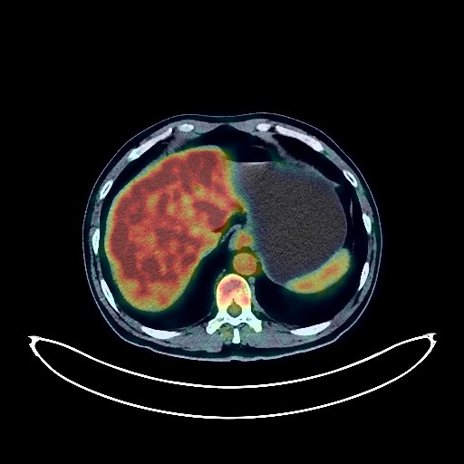
Lung Cancer PET/CT (case 983827-000207 from PETWB-REP)
0 views9 days agoWhole-body 18F-FDG PET/CT scan in a patient with Lung Cancer taken from the PETWB-REP dataset. The following English report (translated from original Chinese) is taken verbatim from the public dataset and has not been modified or otherwise checked for accuracy (see the end for citation). Impression a. A mass adjacent to the oblique fissure in the lower lobe of the left lung, with increased FDG metabolism, suggestive of lung cancer; please correlate with clinicopathology. b. A ground-glass nodule in the lateral basal segment of the lower lobe of the left lung, with normal FDG metabolism, suggestive of inflammatory nodule or atypical adenomatous hyperplasia; annual HRCT follow-up is recommended. c. A few chronic inflammations and old lesions in both lungs. Liver cysts; liver calcifications. Mild fatty liver. Pancreatic fatty infiltration. Possible cholestasis; ultrasound examination recommended. Right renal cyst. Continuous increased FDG metabolism in part of the colon and rectum, suggestive of inflammatory or physiological uptake; colonoscopy recommended. Prostatic calcification; follow-up PSA and ultrasound examination recommended. Degenerative changes in the spine. L4/5 and L5/S1 disc bulges. A few ischemic lesions in the deep bilateral brain regions; MRI follow-up is recommended. Chronic inflammation of the bilateral maxillary and ethmoid sinuses. This case is from PETWB-REP, a curated dataset of whole-body 18F-FDG PET/CT scans and corresponding radiology reports from 490 patients with a broad spectrum of malignancies. The data were retrospectively collected from patients who underwent clinically indicated whole-body 18F-FDG PET/CT scans at the Shanghai Universal Medical Imaging Diagnostic Center between 2021 and 2024. License: Creative Commons Attribution 4.0 International (CC BY 4.0) Citation: Xue, L., Feng, G., Wenbo, Z., Zhang, Y., Li, L., Wang, S., Peng, L., Peng, S., & Gao, X. (2026). PETWB-REP: A Multi-Cancer Whole-Body FDG PET/CT Dataset with Corresponding Radiology Reports [Data set]. Zenodo. https://doi.org/10.5281/zenodo.18670487
Whole BodyPET/CT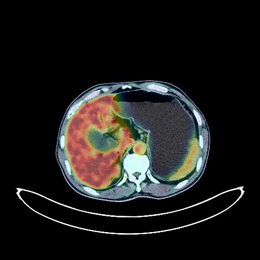
Pancreatic Cancer PET/CT (case 983827-000074 from PETWB-REP)
0 views9 days agoWhole-body 18F-FDG PET/CT scan in a patient with Pancreatic Cancer taken from the PETWB-REP dataset. The following English report (translated from original Chinese) is taken verbatim from the public dataset and has not been modified or otherwise checked for accuracy (see the end for citation). Impression A mass in the head of the pancreas with elevated FDG metabolism is suggestive of a malignant tumor, most likely pancreatic cancer with obstructive inflammation; small peripancreatic lymph nodes show no abnormalities in FDG metabolism, follow-up is recommended. Chronic inflammatory nodules in both lungs. A few post-inflammatory lesions in both lungs. Calcification of some arterial walls (including coronary arteries). Multiple liver cysts. Gallbladder effusion. Residual contrast agent in the urinary tract. Small cysts in both kidneys. Benign prostatic hyperplasia with calcification. Calcification foci in the tunica vaginalis of both testes. Physiological uptake or chronic inflammatory changes in the gastric antrum; follow-up with endoscopy is recommended. Degenerative changes in the spine, mild anterior slippage of the L4 vertebral body, L4/5 and L5/S1 intervertebral disc bulges. Subcutaneous lipoma in the left posterior neck. Left shoulder periarthritis. No obvious abnormalities were found on cranial scintigraphy. Chronic inflammation of the left maxillary sinus. This case is from PETWB-REP, a curated dataset of whole-body 18F-FDG PET/CT scans and corresponding radiology reports from 490 patients with a broad spectrum of malignancies. The data were retrospectively collected from patients who underwent clinically indicated whole-body 18F-FDG PET/CT scans at the Shanghai Universal Medical Imaging Diagnostic Center between 2021 and 2024. License: Creative Commons Attribution 4.0 International (CC BY 4.0) Citation: Xue, L., Feng, G., Wenbo, Z., Zhang, Y., Li, L., Wang, S., Peng, L., Peng, S., & Gao, X. (2026). PETWB-REP: A Multi-Cancer Whole-Body FDG PET/CT Dataset with Corresponding Radiology Reports [Data set]. Zenodo. https://doi.org/10.5281/zenodo.18670487
Whole BodyPET/CT