Loading...
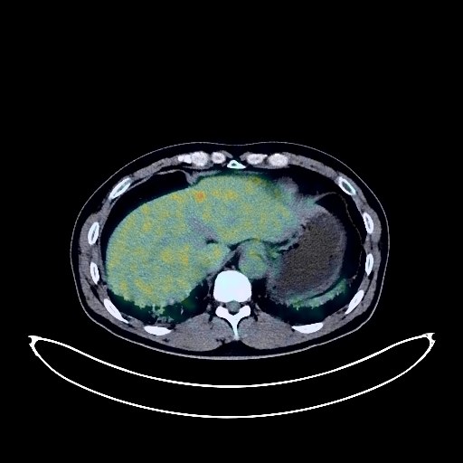
Lung Cancer PET/CT (case 983827-000102 from PETWB-REP)
0 views9 days agoWhole-body 18F-FDG PET/CT scan in a patient with Lung Cancer taken from the PETWB-REP dataset. The following English report (translated from original Chinese) is taken verbatim from the public dataset and has not been modified or otherwise checked for accuracy (see the end for citation). Impression a. Masses in the right lower lobe and right hilum with increased FDG metabolism, suggestive of lung cancer with obstructive changes; multiple lymph node metastases in the right hilum and mediastinum. b. Bilateral emphysema, bilateral interstitial pneumonia. Bilateral pleural thickening. Calcification of some arterial walls (including coronary arteries). Left adrenal hyperplasia. Bilateral calcifications of the tunica vaginalis. Chronic inflammatory changes in the gastric antrum; please follow up with endoscopy. Degenerative changes in the spine, L4/5 and L5/S1 intervertebral disc bulges. No obvious abnormalities seen on cranial scintigraphy. This case is from PETWB-REP, a curated dataset of whole-body 18F-FDG PET/CT scans and corresponding radiology reports from 490 patients with a broad spectrum of malignancies. The data were retrospectively collected from patients who underwent clinically indicated whole-body 18F-FDG PET/CT scans at the Shanghai Universal Medical Imaging Diagnostic Center between 2021 and 2024. License: Creative Commons Attribution 4.0 International (CC BY 4.0) Citation: Xue, L., Feng, G., Wenbo, Z., Zhang, Y., Li, L., Wang, S., Peng, L., Peng, S., & Gao, X. (2026). PETWB-REP: A Multi-Cancer Whole-Body FDG PET/CT Dataset with Corresponding Radiology Reports [Data set]. Zenodo. https://doi.org/10.5281/zenodo.18670487
Whole BodyPET/CT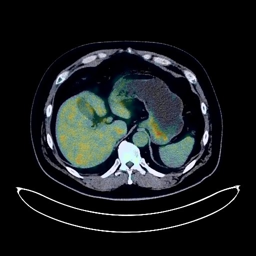
Lung Cancer PET/CT (case 983827-000018 from PETWB-REP)
0 views9 days agoWhole-body 18F-FDG PET/CT scan in a patient with Lung Cancer taken from the PETWB-REP dataset. The following English report (translated from original Chinese) is taken verbatim from the public dataset and has not been modified or otherwise checked for accuracy (see the end for citation). Impression a. A mass in the lateral basal segment of the right lower lobe with mildly increased FDG uptake, suggestive of cystic lung cancer. b. Several miliary chronic inflammatory nodules in the remaining right lung. Slight thickening of the pleura bilaterally. Slight thickening of the gastric fundus wall with mildly increased FDG uptake, suggestive of chronic gastritis; increased FDG uptake in some intestinal segments, suggestive of inflammatory or physiological uptake. Follow-up gastroscopy and colonoscopy are recommended. Liver cyst. Spleen density is uneven, FDG uptake is normal; follow-up ultrasound is recommended. Degenerative changes in the spine. L4/5 and L5/S1 intervertebral disc bulges. Cranial scintigraphy is normal. Bilateral chronic ethmoid sinusitis. Bilateral chronic inflammation of the palatine tonsils and oropharynx. This case is from PETWB-REP, a curated dataset of whole-body 18F-FDG PET/CT scans and corresponding radiology reports from 490 patients with a broad spectrum of malignancies. The data were retrospectively collected from patients who underwent clinically indicated whole-body 18F-FDG PET/CT scans at the Shanghai Universal Medical Imaging Diagnostic Center between 2021 and 2024. License: Creative Commons Attribution 4.0 International (CC BY 4.0) Citation: Xue, L., Feng, G., Wenbo, Z., Zhang, Y., Li, L., Wang, S., Peng, L., Peng, S., & Gao, X. (2026). PETWB-REP: A Multi-Cancer Whole-Body FDG PET/CT Dataset with Corresponding Radiology Reports [Data set]. Zenodo. https://doi.org/10.5281/zenodo.18670487
Whole BodyPET/CT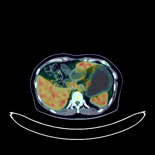
Ovarian Cancer PET/CT (case 983827-000269 from PETWB-REP)
0 views9 days agoWhole-body 18F-FDG PET/CT scan in a patient with Ovarian Cancer taken from the PETWB-REP dataset. The following English report (translated from original Chinese) is taken verbatim from the public dataset and has not been modified or otherwise checked for accuracy (see the end for citation). Impression a. Thickening of the sigmoid colon wall with increased FDG metabolism, consistent with malignant tumor (lesion activity still present). Comparison with previous images and follow-up examination are recommended. b. Multilocular cystic-solid lesions in the abdominopelvic cavity with increased FDG metabolism in the solid component, suggestive of malignant tumor with activity; ovarian cancer is the primary consideration, metastatic tumor to be ruled out. Abdominal and pelvic peritoneal seeding metastasis with activity. Small amount of pelvic effusion. c. Retroperitoneal para-aortic, mesenteric, and bilateral anterior diaphragmatic lymph nodes show increased FDG metabolism in some areas, suggesting post-treatment changes from metastatic tumors or reactive lymph node hyperplasia. Comparison with previous images and follow-up examination are recommended. d. Liver cyst; no abnormal FDG metabolic foci seen in the liver parenchyma. Follow-up with enhanced MRI is recommended. a. Multiple solid nodules in both lungs, FDG metabolism normal, suggest inflammatory nodules or post-treatment changes after metastatic tumor treatment. Comparison with old images and follow-up examination are recommended. b. Pure ground-glass nodules in both lungs, FDG metabolism normal, suggest chronic inflammatory nodules or atypical adenomatous hyperplasia. Annual HRCT follow-up is recommended. Bilateral pulmonary fibrosis. Reactive hyperplasia of hilar and mediastinal lymph nodes. Increased fibrous glands in the upper outer quadrant of the right breast compared to the left, with punctate calcifications. FDG metabolism normal, suggest proliferative changes. Ultrasound follow-up is recommended to rule out other possibilities. Uneven thyroid density, FDG metabolism normal. Ultrasound follow-up is recommended. Reactive hyperplasia of cervical lymph nodes. Spinal osteophyte formation. L5 and S1 vertebral endplate inflammation. L4/5 and L5/S1 disc herniation with pneumoconiosis and degeneration. Left temporal pole arachnoid cyst; FDG scintigraphy of the brain showed no obvious abnormalities. This case is from PETWB-REP, a curated dataset of whole-body 18F-FDG PET/CT scans and corresponding radiology reports from 490 patients with a broad spectrum of malignancies. The data were retrospectively collected from patients who underwent clinically indicated whole-body 18F-FDG PET/CT scans at the Shanghai Universal Medical Imaging Diagnostic Center between 2021 and 2024. License: Creative Commons Attribution 4.0 International (CC BY 4.0) Citation: Xue, L., Feng, G., Wenbo, Z., Zhang, Y., Li, L., Wang, S., Peng, L., Peng, S., & Gao, X. (2026). PETWB-REP: A Multi-Cancer Whole-Body FDG PET/CT Dataset with Corresponding Radiology Reports [Data set]. Zenodo. https://doi.org/10.5281/zenodo.18670487
Whole BodyPET/CT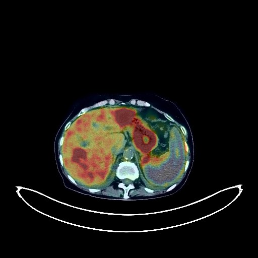
Pancreatic Cancer PET/CT (case 983827-000193 from PETWB-REP)
0 views9 days agoWhole-body 18F-FDG PET/CT scan in a patient with Pancreatic Cancer taken from the PETWB-REP dataset. The following English report (translated from original Chinese) is taken verbatim from the public dataset and has not been modified or otherwise checked for accuracy (see the end for citation). Impression a. A mass in the uncinate process of the pancreas with increased FDG metabolism. Combined with enhanced MRI from our center, pancreatic cancer is highly probable, involving adjacent blood vessels and accompanied by low-level biliary obstruction. Clinical correlation is required. b. Multiple liver metastases. Tumor thrombi in the splenic vascular zone with large-area splenic infarction are highly probable. Multiple bone metastases throughout the body. c. Multiple lymph nodes are visible in the bilateral cardiophrenic angles, posterior diaphragmatic crura, hepatic hilum, and around the pancreatic head. Some have increased FDG metabolism, suggesting possible metastases. Follow-up is recommended. d. Blurred mesenteric fat space in the abdominal cavity, with normal FDG metabolism, suggests possible inflammatory changes. Follow-up is recommended. Small amount of pelvic effusion. Interstitial lung changes with multiple inflammations in both lungs. Post-treatment CT re-examination is recommended to rule out hidden lesions. Small amount of bilateral pleural effusion. Anemia. Slight pericardial thickening. Calcification of some arterial walls (including coronary arteries). Gallbladder diverticulum, chronic cholecystitis. Please combine with ultrasound examination. Bilateral renal cysts. Bilateral adrenal hyperplasia is highly probable. Slight thickening of the walls of part of the gastric body and antrum, increased FDG uptake, suggestive of chronic gastritis; continuous increased FDG metabolism in the colon and rectum, suggestive of inflammatory or physiological uptake. Follow-up gastroscopy and colonoscopy are recommended for the above. Degenerative changes in the spine. L5/S1 intervertebral disc pneumoconiosis. L4/5, L5/S1 intervertebral disc bulge. A few ischemic foci in the deep bilateral brain; senile encephalopathy. This case is from PETWB-REP, a curated dataset of whole-body 18F-FDG PET/CT scans and corresponding radiology reports from 490 patients with a broad spectrum of malignancies. The data were retrospectively collected from patients who underwent clinically indicated whole-body 18F-FDG PET/CT scans at the Shanghai Universal Medical Imaging Diagnostic Center between 2021 and 2024. License: Creative Commons Attribution 4.0 International (CC BY 4.0) Citation: Xue, L., Feng, G., Wenbo, Z., Zhang, Y., Li, L., Wang, S., Peng, L., Peng, S., & Gao, X. (2026). PETWB-REP: A Multi-Cancer Whole-Body FDG PET/CT Dataset with Corresponding Radiology Reports [Data set]. Zenodo. https://doi.org/10.5281/zenodo.18670487
Whole BodyPET/CT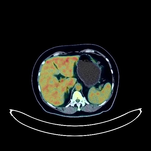
Ovarian Cancer PET/CT (case 983827-000167 from PETWB-REP)
0 views9 days agoWhole-body 18F-FDG PET/CT scan in a patient with Ovarian Cancer taken from the PETWB-REP dataset. The following English report (translated from original Chinese) is taken verbatim from the public dataset and has not been modified or otherwise checked for accuracy (see the end for citation). Impression a. Post-ovarian cancer surgery and chemotherapy changes; new soft tissue nodules with increased FDG metabolism in the right pelvic region, highly suggestive of metastasis; further analysis with contrast-enhanced MRI is recommended. b. Reactive hyperplasia of lymph nodes around the abdominal aorta and in both inguinal regions, similar to previous findings. New bladder lesion with increased FDG metabolism, highly suggestive of bladder cancer; metastasis to be ruled out; further cystoscopy is recommended. Chronic inflammatory nodules in both lungs, similar to previous findings; follow-up with CT is recommended. Multiple bullae in both lungs. A few old lesions in both lungs. Mild anemia. Bilateral breast hyperplasia. Liver cyst. Left renal angiomyolipoma, similar to previous findings. Chronic inflammatory changes in the gastric antrum and part of the intestine; endoscopic follow-up is recommended. Cervical spondylosis. L4/5 disc bulge. No abnormalities found on cranial scintigraphy. Inflammatory lymph nodes in the bilateral deep cervical spaces and submandibular region. This case is from PETWB-REP, a curated dataset of whole-body 18F-FDG PET/CT scans and corresponding radiology reports from 490 patients with a broad spectrum of malignancies. The data were retrospectively collected from patients who underwent clinically indicated whole-body 18F-FDG PET/CT scans at the Shanghai Universal Medical Imaging Diagnostic Center between 2021 and 2024. License: Creative Commons Attribution 4.0 International (CC BY 4.0) Citation: Xue, L., Feng, G., Wenbo, Z., Zhang, Y., Li, L., Wang, S., Peng, L., Peng, S., & Gao, X. (2026). PETWB-REP: A Multi-Cancer Whole-Body FDG PET/CT Dataset with Corresponding Radiology Reports [Data set]. Zenodo. https://doi.org/10.5281/zenodo.18670487
Whole BodyPET/CT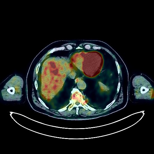
Lung Cancer PET/CT (case 983827-000183 from PETWB-REP)
0 views9 days agoWhole-body 18F-FDG PET/CT scan in a patient with Lung Cancer taken from the PETWB-REP dataset. The following English report (translated from original Chinese) is taken verbatim from the public dataset and has not been modified or otherwise checked for accuracy (see the end for citation). Impression a. Right middle lobe lung mass, increased FDG metabolism, suggestive of peripheral lung cancer; please correlate with clinicopathology. b. Scattered chronic inflammation and old lesions in both lungs. Bilateral emphysema. Highly likely reactive hyperplasia of hilar and mediastinal lymph nodes; follow-up examination recommended. Calcification of some arterial walls (including coronary arteries). Chronic cholecystitis; gallstones. Right renal cyst. Right renal calculus. Left adrenal medullary lipoma; CT follow-up recommended. Slight thickening of the walls of part of the gastric body and antrum, mildly increased FDG uptake, suggestive of chronic gastritis; increased FDG metabolism in part of the intestines, suggestive of inflammatory or physiological uptake. Hemorrhoids. Follow-up gastroscopy and colonoscopy are recommended for all of the above. Enlargement of the left inguinal canal; please correlate with clinical findings. Reactive hyperplasia of bilateral inguinal lymph nodes. Degenerative changes in the spine. L4/5 and L5/S1 intervertebral disc bulges. A few ischemic lesions in the deep bilateral brain regions; age-related brain abnormalities. A left parotid gland lymphoma is highly probable; ultrasound follow-up is recommended. This case is from PETWB-REP, a curated dataset of whole-body 18F-FDG PET/CT scans and corresponding radiology reports from 490 patients with a broad spectrum of malignancies. The data were retrospectively collected from patients who underwent clinically indicated whole-body 18F-FDG PET/CT scans at the Shanghai Universal Medical Imaging Diagnostic Center between 2021 and 2024. License: Creative Commons Attribution 4.0 International (CC BY 4.0) Citation: Xue, L., Feng, G., Wenbo, Z., Zhang, Y., Li, L., Wang, S., Peng, L., Peng, S., & Gao, X. (2026). PETWB-REP: A Multi-Cancer Whole-Body FDG PET/CT Dataset with Corresponding Radiology Reports [Data set]. Zenodo. https://doi.org/10.5281/zenodo.18670487
Whole BodyPET/CT
Lung Cancer PET/CT (case 983827-000192 from PETWB-REP)
0 views9 days agoWhole-body 18F-FDG PET/CT scan in a patient with Lung Cancer taken from the PETWB-REP dataset. The following English report (translated from original Chinese) is taken verbatim from the public dataset and has not been modified or otherwise checked for accuracy (see the end for citation). Impression a. A mass in the apical segment of the right upper lobe, with increased FDG metabolism in the solid component, strongly suggestive of lung cancer; please correlate with clinicopathology. b. Bilateral emphysema with bullae, a few post-inflammatory lesions in both lungs. Reactive hyperplasia of mediastinal lymph nodes. Partial calcification of the aorta and coronary artery walls. Possible left adrenal hyperplasia, adenoma to be ruled out; MRI follow-up recommended. Prostatic calcification. Small amount of hydrocele in both testes. Scoliosis with degenerative changes. L3/4 and L4/5 intervertebral disc bulge. Right femoral head herniation. Small softening lesion in the right frontal lobe, ischemic lesion in the left basal ganglia, white matter degeneration, age-related brain changes; MRI follow-up recommended. This case is from PETWB-REP, a curated dataset of whole-body 18F-FDG PET/CT scans and corresponding radiology reports from 490 patients with a broad spectrum of malignancies. The data were retrospectively collected from patients who underwent clinically indicated whole-body 18F-FDG PET/CT scans at the Shanghai Universal Medical Imaging Diagnostic Center between 2021 and 2024. License: Creative Commons Attribution 4.0 International (CC BY 4.0) Citation: Xue, L., Feng, G., Wenbo, Z., Zhang, Y., Li, L., Wang, S., Peng, L., Peng, S., & Gao, X. (2026). PETWB-REP: A Multi-Cancer Whole-Body FDG PET/CT Dataset with Corresponding Radiology Reports [Data set]. Zenodo. https://doi.org/10.5281/zenodo.18670487
Whole BodyPET/CT
Renal Cancer PET/CT (case 983827-000260 from PETWB-REP)
0 views9 days agoWhole-body 18F-FDG PET/CT scan in a patient with Renal Cancer taken from the PETWB-REP dataset. The following English report (translated from original Chinese) is taken verbatim from the public dataset and has not been modified or otherwise checked for accuracy (see the end for citation). Impression a. Post-right renal cell carcinoma surgery, right renal mass with increased FDG metabolism, suggestive of tumor recurrence; reactive hyperplasia of retroperitoneal lymph nodes. b. Osteophyte formation and fracture at the left anterior margin of the L2 vertebral body, increased FDG uptake, suggestive of post-fracture changes due to osteophyte formation; follow-up is recommended. Elderly brain, softening lesion in the left basal ganglia, deep lacunar ischemic lesion in the brain; follow-up with MRI is recommended. Nodular goiter; ultrasound follow-up is recommended. Chronic inflammatory nodules in both lungs; CT follow-up is recommended. A few post-inflammatory lesions in both lungs. Calcification of some arterial walls (including coronary arteries). Cyst in the left lobe of the liver. Hyperplasia of the left adrenal gland, calcification of the right adrenal gland. Chronic inflammatory changes in some gastric walls; follow-up with endoscopy is recommended. Osteoporosis, degenerative changes in the spine, mild anterior slippage of the L4 vertebral body, L4/5 and L5/S1 intervertebral disc bulge. Bilateral gluteal and thigh muscle atrophy. Left shoulder periarthritis. This case is from PETWB-REP, a curated dataset of whole-body 18F-FDG PET/CT scans and corresponding radiology reports from 490 patients with a broad spectrum of malignancies. The data were retrospectively collected from patients who underwent clinically indicated whole-body 18F-FDG PET/CT scans at the Shanghai Universal Medical Imaging Diagnostic Center between 2021 and 2024. License: Creative Commons Attribution 4.0 International (CC BY 4.0) Citation: Xue, L., Feng, G., Wenbo, Z., Zhang, Y., Li, L., Wang, S., Peng, L., Peng, S., & Gao, X. (2026). PETWB-REP: A Multi-Cancer Whole-Body FDG PET/CT Dataset with Corresponding Radiology Reports [Data set]. Zenodo. https://doi.org/10.5281/zenodo.18670487
Whole BodyPET/CT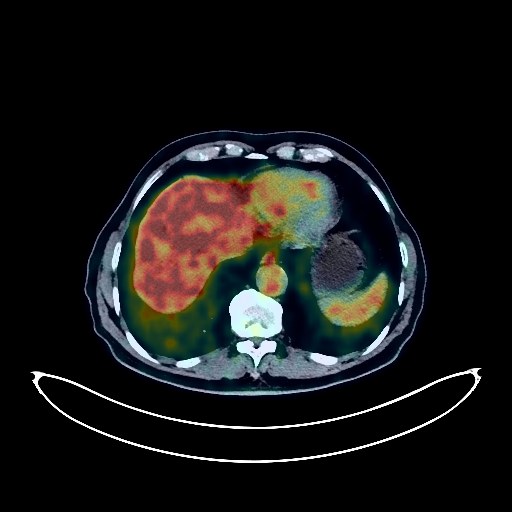
Cholangiocarcinoma PET/CT (case 983827-000007 from PETWB-REP)
0 views9 days agoWhole-body 18F-FDG PET/CT scan in a patient with Cholangiocarcinoma taken from the PETWB-REP dataset. The following English report (translated from original Chinese) is taken verbatim from the public dataset and has not been modified or otherwise checked for accuracy (see the end for citation). Impression Mass in the hepatic hilum, with elevated FDG metabolism, suggestive of hilar cholangiocarcinoma. Metastasis to the parapancreatic lymph nodes is highly probable; further clinical correlation is needed. Bilateral emphysema with bullae. Ground-glass nodule in the posterior segment of the right lower lobe, FDG metabolism normal, suggestive of inflammatory nodule; atypical adenomatous hyperplasia to be ruled out. Annual HRCT follow-up recommended. Scattered chronic inflammation and remnants in both lungs. Partial arterial wall calcification. Hepatic cyst. Gallbladder mural stones. Pancreatic tail calcification. Bilateral renal cysts, with a complex cyst in the left kidney. Benign prostatic hyperplasia. Bilateral testicular tunica vaginalis calcification. Partial vertebral osteophyte formation. L4/5 and L5/S1 intervertebral disc bulge. L4/5 intervertebral disc pneumothorax. A few ischemic lesions deep in the brain. Age-related brain changes. Reactive hyperplasia of bilateral deep cervical and submandibular lymph nodes. This case is from PETWB-REP, a curated dataset of whole-body 18F-FDG PET/CT scans and corresponding radiology reports from 490 patients with a broad spectrum of malignancies. The data were retrospectively collected from patients who underwent clinically indicated whole-body 18F-FDG PET/CT scans at the Shanghai Universal Medical Imaging Diagnostic Center between 2021 and 2024. License: Creative Commons Attribution 4.0 International (CC BY 4.0) Citation: Xue, L., Feng, G., Wenbo, Z., Zhang, Y., Li, L., Wang, S., Peng, L., Peng, S., & Gao, X. (2026). PETWB-REP: A Multi-Cancer Whole-Body FDG PET/CT Dataset with Corresponding Radiology Reports [Data set]. Zenodo. https://doi.org/10.5281/zenodo.18670487
Whole BodyPET/CT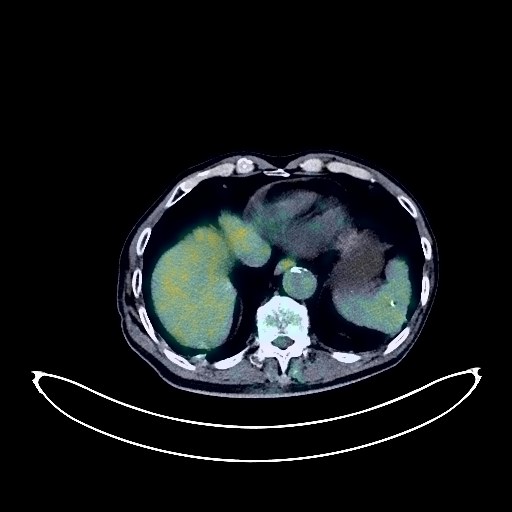
Lymphoma PET/CT (case 983827-000091 from PETWB-REP)
0 views9 days agoWhole-body 18F-FDG PET/CT scan in a patient with Lymphoma taken from the PETWB-REP dataset. The following English report (translated from original Chinese) is taken verbatim from the public dataset and has not been modified or otherwise checked for accuracy (see the end for citation). Impression Newly observed multiple enlarged lymph nodes bilaterally in the supraclavicular fossa, left hilum, and mediastinum, accompanied by increased FDG metabolism, suggestive of a neoplastic lesion (most likely lymphoma). Other lymphocytic proliferative diseases need to be ruled out; please correlate with clinicopathology. Small patchy slightly low-density lesion adjacent to the posterior horn of the right lateral ventricle, and punctate low-density lesions in the deep brain regions bilaterally, neither showing significant mass effect. FDG metabolism was not significantly abnormal, suggesting possible ischemic lesions. Please combine clinical findings with enhanced MRI for comprehensive judgment. Age-related brain changes. a. Multiple bullae in the upper lobes of both lungs and the lower lobe of the right lung. Among them, one bulla in the posterior segment of the right lower lobe adjacent to the spine has shrunk compared to the previous examination, with thickened and roughened walls. FDG metabolism was not abnormal, suggesting a high probability of concurrent infection. Please follow up with CT. b. Old lesions in the posterior segment of the right lower lobe, fibrotic lesions in both lungs, and newly observed scattered subpleural inflammation in the right middle lobe and the upper and lower lobes of the left lung. CT follow-up is recommended after anti-infective treatment. Bilateral pleural thickening. Pericardial thickening with a small amount of effusion. Anemia. Liver calcifications. Splenic calcifications. Multiple arteriosclerosis throughout the body. Possible chronic inflammation of the lower esophagus, part of the stomach wall, and intestines; please combine with endoscopic examination. Hemorrhoids. Mildly elevated FDG metabolism in the prostatic parenchyma, suggesting possible chronic prostatitis; please combine with PSA follow-up. Osteoporosis, degenerative changes in the cervical, thoracic, and lumbar spine. Multiple intervertebral disc bulges with pneumoconiosis. Left sacroiliitis. Postoperative changes at T12-L2 vertebral bodies. Uneven density in the left and right lobes of the thyroid gland, with mild FDG uptake, highly suggestive of chronic thyroiditis; please combine with ultrasound follow-up. Chronic inflammation of the left sphenoid sinus. Right inferior alveolar ulceritis. This case is from PETWB-REP, a curated dataset of whole-body 18F-FDG PET/CT scans and corresponding radiology reports from 490 patients with a broad spectrum of malignancies. The data were retrospectively collected from patients who underwent clinically indicated whole-body 18F-FDG PET/CT scans at the Shanghai Universal Medical Imaging Diagnostic Center between 2021 and 2024. License: Creative Commons Attribution 4.0 International (CC BY 4.0) Citation: Xue, L., Feng, G., Wenbo, Z., Zhang, Y., Li, L., Wang, S., Peng, L., Peng, S., & Gao, X. (2026). PETWB-REP: A Multi-Cancer Whole-Body FDG PET/CT Dataset with Corresponding Radiology Reports [Data set]. Zenodo. https://doi.org/10.5281/zenodo.18670487
Whole BodyPET/CT