Loading...
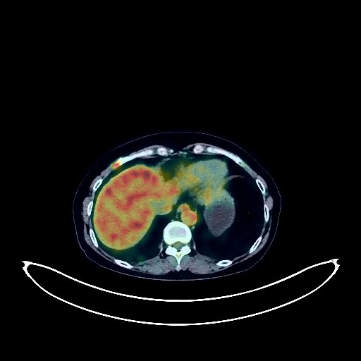
Renal Cancer PET/CT (case 983827-000094 from PETWB-REP)
0 views9 days agoWhole-body 18F-FDG PET/CT scan in a patient with Renal Cancer taken from the PETWB-REP dataset. The following English report (translated from original Chinese) is taken verbatim from the public dataset and has not been modified or otherwise checked for accuracy (see the end for citation). Impression a. Mass on the upper pole of the left kidney, with increased FDG metabolism, strongly suggesting renal cell carcinoma; please combine with enhanced MRI for comprehensive analysis. Multiple renal cysts and stones in both kidneys. b. Absence of the spleen with metallic density shadow on the stomach wall, suggesting postoperative changes and spleen regeneration. Gallstones and cholestasis. c. Trend towards cirrhosis, no abnormal FDG metabolism seen in the liver; please combine with enhanced MRI for comprehensive analysis. d. Increased FDG metabolism in the right 6th anterior rib, suggesting possible post-traumatic changes; metastasis to be ruled out; please consider clinical history and follow up. Increased FDG metabolism on the right nasopharyngeal wall, suggesting possible inflammation; specialist examination recommended to rule out tumors. Reactive hyperplasia of bilateral deep cervical spaces, submandibular, and submental lymph nodes. Chronic inflammatory micronodules in the lower lobe of the left lung. A few fibrotic lesions in the middle lobe of the right lung. Reactive hyperplasia of right axillary lymph nodes. Spinal osteophyte formation, sacral canal cyst at the S1 vertebral level. No obvious abnormalities seen on cranial imaging. There is a high probability of reactive hyperplasia of the subcutaneous lymph nodes in the right upper arm. Follow-up examination is recommended to rule out other possibilities. This case is from PETWB-REP, a curated dataset of whole-body 18F-FDG PET/CT scans and corresponding radiology reports from 490 patients with a broad spectrum of malignancies. The data were retrospectively collected from patients who underwent clinically indicated whole-body 18F-FDG PET/CT scans at the Shanghai Universal Medical Imaging Diagnostic Center between 2021 and 2024. License: Creative Commons Attribution 4.0 International (CC BY 4.0) Citation: Xue, L., Feng, G., Wenbo, Z., Zhang, Y., Li, L., Wang, S., Peng, L., Peng, S., & Gao, X. (2026). PETWB-REP: A Multi-Cancer Whole-Body FDG PET/CT Dataset with Corresponding Radiology Reports [Data set]. Zenodo. https://doi.org/10.5281/zenodo.18670487
Whole BodyPET/CT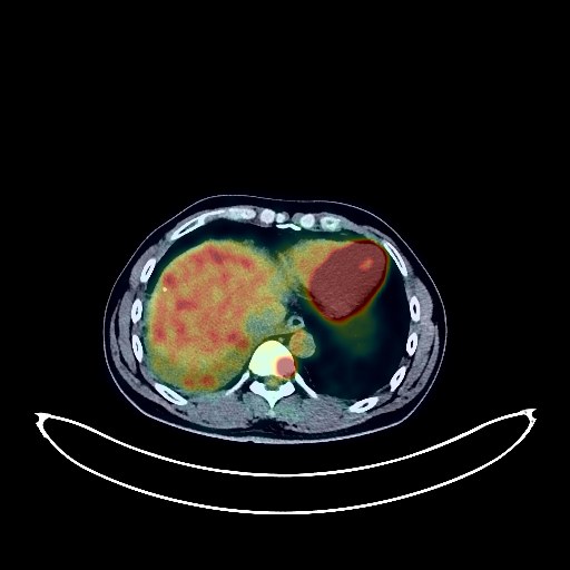
Lung Cancer PET/CT (case 983827-000172 from PETWB-REP)
0 views9 days agoWhole-body 18F-FDG PET/CT scan in a patient with Lung Cancer taken from the PETWB-REP dataset. The following English report (translated from original Chinese) is taken verbatim from the public dataset and has not been modified or otherwise checked for accuracy (see the end for citation). Impression a. Multiple lesions in the right lung and subpleural region, with elevated FDG metabolism, suggestive of lung cancer with metastases. The nodule in the lateral basal segment of the right lower lobe is most likely the primary lesion; please correlate with clinicopathology. b. Multiple small nodules in both lungs, suggestive of metastases. Multiple lymph node metastases in the right hilum and mediastinum. Right pleural metastasis. T9 vertebral body bone metastasis. c. Moderate pleural effusion on the right side. Right cerebellar calcification. Cervical, thoracic, and lumbar vertebral osteophytes. L4/5 and L5/S1 intervertebral disc bulges. This case is from PETWB-REP, a curated dataset of whole-body 18F-FDG PET/CT scans and corresponding radiology reports from 490 patients with a broad spectrum of malignancies. The data were retrospectively collected from patients who underwent clinically indicated whole-body 18F-FDG PET/CT scans at the Shanghai Universal Medical Imaging Diagnostic Center between 2021 and 2024. License: Creative Commons Attribution 4.0 International (CC BY 4.0) Citation: Xue, L., Feng, G., Wenbo, Z., Zhang, Y., Li, L., Wang, S., Peng, L., Peng, S., & Gao, X. (2026). PETWB-REP: A Multi-Cancer Whole-Body FDG PET/CT Dataset with Corresponding Radiology Reports [Data set]. Zenodo. https://doi.org/10.5281/zenodo.18670487
Whole BodyPET/CT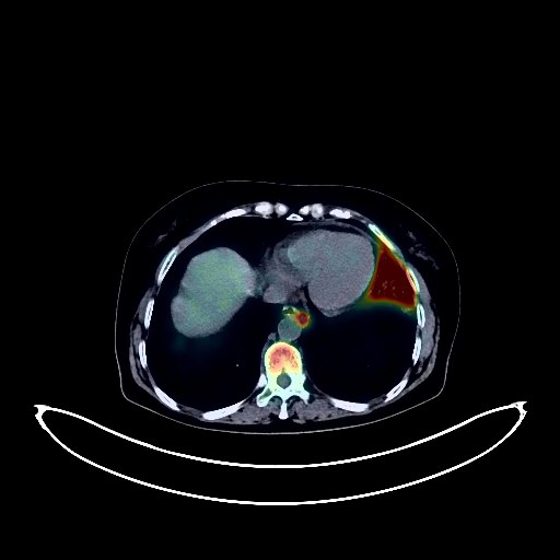
Lung Cancer PET/CT (case 983827-000265 from PETWB-REP)
0 views9 days agoWhole-body 18F-FDG PET/CT scan in a patient with Lung Cancer taken from the PETWB-REP dataset. The following English report (translated from original Chinese) is taken verbatim from the public dataset and has not been modified or otherwise checked for accuracy (see the end for citation). Impression a. A space-occupying lesion with cavity formation in the left upper lobe, with unevenly increased FDG metabolism, consistent with lung cancer, accompanied by obstructive inflammation in the left upper lobe; partial lymph node metastasis in both hilar and mediastinal regions. b. Chronic inflammatory nodules in both lungs; follow-up CT scan recommended. Scattered post-inflammatory lesions in both lungs. Pericardial thickening with a small amount of effusion, mild anemia, and partial calcification of arterial walls (including coronary arteries). Bilateral breast hyperplasia, with calcification in the right breast. Mild fatty liver, small cyst in the left lobe of the liver. Increased FDG metabolism in the spleen, suggestive of reactive hyperplasia. Left adrenal hyperplasia. Possibly due to physiological uptake in the uterine cavity; follow-up ultrasound is recommended to rule out other possibilities. Partial chronic inflammatory changes in the gastric wall; follow-up endoscopically recommended. Osteoporosis, degenerative changes in the spine, multiple intervertebral disc bulges with pneumoconiosis. Age-related brain abnormalities, deep lacunar infarcts. This case is from PETWB-REP, a curated dataset of whole-body 18F-FDG PET/CT scans and corresponding radiology reports from 490 patients with a broad spectrum of malignancies. The data were retrospectively collected from patients who underwent clinically indicated whole-body 18F-FDG PET/CT scans at the Shanghai Universal Medical Imaging Diagnostic Center between 2021 and 2024. License: Creative Commons Attribution 4.0 International (CC BY 4.0) Citation: Xue, L., Feng, G., Wenbo, Z., Zhang, Y., Li, L., Wang, S., Peng, L., Peng, S., & Gao, X. (2026). PETWB-REP: A Multi-Cancer Whole-Body FDG PET/CT Dataset with Corresponding Radiology Reports [Data set]. Zenodo. https://doi.org/10.5281/zenodo.18670487
Whole BodyPET/CT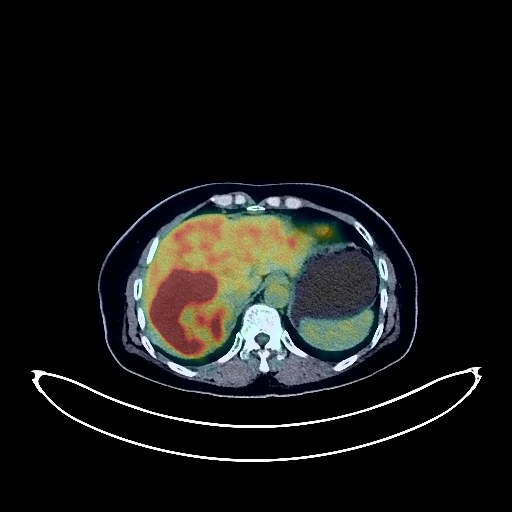
Liver Cancer PET/CT (case 983827-000186 from PETWB-REP)
0 views9 days agoWhole-body 18F-FDG PET/CT scan in a patient with Liver Cancer taken from the PETWB-REP dataset. The following English report (translated from original Chinese) is taken verbatim from the public dataset and has not been modified or otherwise checked for accuracy (see the end for citation). Impression A mass in the right lobe of the liver with increased FDG metabolism is suggestive of malignancy, possibly hepatocellular carcinoma. Reactive hyperplasia of the abdominal and retroperitoneal lymph nodes is highly likely; follow-up is recommended to rule out mixed metastases. Slight thickening of the walls of part of the gastric body and antrum, with mildly increased FDG uptake, suggests possible chronic gastritis; follow-up with gastroscopy is recommended. Chronic inflammatory micronodules (solid) in the lateral basal segment of the left lower lobe. A few chronic inflammations and old lesions in both lungs. Increased FDG metabolism in part of the intestinal tract, considered physiological uptake. Degenerative changes in the spine. L4/5 and L5/S1 intervertebral disc bulges. Right-sided frozen shoulder. Ischemic lesions in the right basal ganglia region. This case is from PETWB-REP, a curated dataset of whole-body 18F-FDG PET/CT scans and corresponding radiology reports from 490 patients with a broad spectrum of malignancies. The data were retrospectively collected from patients who underwent clinically indicated whole-body 18F-FDG PET/CT scans at the Shanghai Universal Medical Imaging Diagnostic Center between 2021 and 2024. License: Creative Commons Attribution 4.0 International (CC BY 4.0) Citation: Xue, L., Feng, G., Wenbo, Z., Zhang, Y., Li, L., Wang, S., Peng, L., Peng, S., & Gao, X. (2026). PETWB-REP: A Multi-Cancer Whole-Body FDG PET/CT Dataset with Corresponding Radiology Reports [Data set]. Zenodo. https://doi.org/10.5281/zenodo.18670487
Whole BodyPET/CT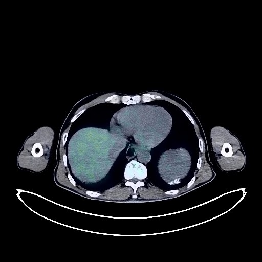
Gastric Cancer PET/CT (case 983827-000175 from PETWB-REP)
0 views9 days agoWhole-body 18F-FDG PET/CT scan in a patient with Gastric Cancer taken from the PETWB-REP dataset. The following English report (translated from original Chinese) is taken verbatim from the public dataset and has not been modified or otherwise checked for accuracy (see the end for citation). Impression a. Post-gastric cancer surgery and chemotherapy, no obvious signs of tumor recurrence were observed locally. b. Mass in the porta hepatis, mildly increased FDG metabolism, cholangiocarcinoma or metastasis to be ruled out; enhanced MRI is recommended for further examination. c. Intrahepatic bile duct dilation, stone in the left lobe bile duct. a. Rectal wall thickening and edema, increased FDG metabolism, inflammation is suspected; colonoscopy follow-up is recommended. b. Increased FDG uptake in strips of the colon and small intestine, considered inflammatory or physiological uptake. Left renal cyst. Calcification at the spleen margin. Right testicular hydrocele with calcification. Multiple chronic inflammatory micronodules in the apical-posterior segment of the left upper lobe and the medial segment of the right middle lobe; an air-filled cavity in the posterior basal segment of the right lower lobe; chronic inflammation and remnants in both lungs. Partial calcification of the aorta and coronary artery walls. Low-density nodule in the lower pole of the left thyroid lobe, with increased FDG metabolism, suggestive of nodular thyroiditis; ultrasound follow-up recommended. Scoliosis with degenerative changes. L3/4 and L4/5 intervertebral disc bulge. Small bone islands in the right femoral head and L4 vertebral body. Bilateral basal ganglia ischemic lesions, age-related brain changes. Minor inflammation of the left maxillary sinus. This case is from PETWB-REP, a curated dataset of whole-body 18F-FDG PET/CT scans and corresponding radiology reports from 490 patients with a broad spectrum of malignancies. The data were retrospectively collected from patients who underwent clinically indicated whole-body 18F-FDG PET/CT scans at the Shanghai Universal Medical Imaging Diagnostic Center between 2021 and 2024. License: Creative Commons Attribution 4.0 International (CC BY 4.0) Citation: Xue, L., Feng, G., Wenbo, Z., Zhang, Y., Li, L., Wang, S., Peng, L., Peng, S., & Gao, X. (2026). PETWB-REP: A Multi-Cancer Whole-Body FDG PET/CT Dataset with Corresponding Radiology Reports [Data set]. Zenodo. https://doi.org/10.5281/zenodo.18670487
Whole BodyPET/CT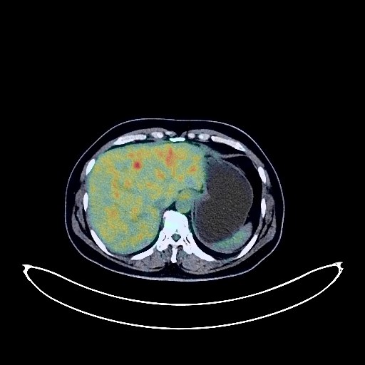
Cervical Cancer PET/CT (case 983827-000036 from PETWB-REP)
0 views9 days agoWhole-body 18F-FDG PET/CT scan in a patient with Cervical Cancer taken from the PETWB-REP dataset. The following English report (translated from original Chinese) is taken verbatim from the public dataset and has not been modified or otherwise checked for accuracy (see the end for citation). Impression a. Cervical mass with elevated FDG metabolism, consistent with cervical cancer. b. Possible uterine fibroids, uterine cavity effusion, bilateral thickened fallopian tubes with effusion. Small amount of pelvic effusion. Chronic inflammatory nodules in both lungs; CT follow-up is recommended. A few post-inflammatory lesions in both lungs. Small cyst in the left lobe of the liver. Chronic cholecystitis, gallstones. Chronic inflammatory changes or physiological uptake in the gastric antrum and part of the intestine; please follow up with endoscopy. Degenerative changes in the spine; L3/4 intervertebral disc bulge with posterior margin calcification. No obvious abnormalities seen on cranial scintigraphy. A few chronic inflammations in both ethmoid sinuses. This case is from PETWB-REP, a curated dataset of whole-body 18F-FDG PET/CT scans and corresponding radiology reports from 490 patients with a broad spectrum of malignancies. The data were retrospectively collected from patients who underwent clinically indicated whole-body 18F-FDG PET/CT scans at the Shanghai Universal Medical Imaging Diagnostic Center between 2021 and 2024. License: Creative Commons Attribution 4.0 International (CC BY 4.0) Citation: Xue, L., Feng, G., Wenbo, Z., Zhang, Y., Li, L., Wang, S., Peng, L., Peng, S., & Gao, X. (2026). PETWB-REP: A Multi-Cancer Whole-Body FDG PET/CT Dataset with Corresponding Radiology Reports [Data set]. Zenodo. https://doi.org/10.5281/zenodo.18670487
Whole BodyPET/CT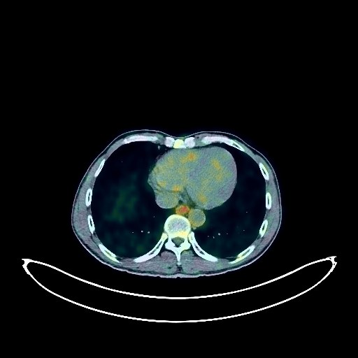
Gastric Cancer PET/CT (case 983827-000195 from PETWB-REP)
0 views9 days agoWhole-body 18F-FDG PET/CT scan in a patient with Gastric Cancer taken from the PETWB-REP dataset. The following English report (translated from original Chinese) is taken verbatim from the public dataset and has not been modified or otherwise checked for accuracy (see the end for citation). Impression Post-gastric cancer treatment: Slight thickening of the gastric wall on the greater curvature of the residual stomach with increased FDG metabolism, consistent with gastric cancer findings based on pathology. Reactive hyperplasia of retroperitoneal and mesenteric lymph nodes. Small amount of pelvic effusion. Increased FDG metabolism at the intestinal anastomosis site in the right upper quadrant; segmental FDG metabolism increases in the remaining intestinal segments, without obvious space-occupying lesions. Physiological uptake or chronic inflammation is considered; please follow up with endoscopy. Ground-glass nodules in the paravertebral and lateral basal segments of the right lower lobe; solid miliary nodules in both upper lobes. FDG metabolism is normal; chronic inflammatory nodules are considered; please have an annual HRCT scan. A few fibrotic lesions in both lungs; calcification in the right upper lobe. Paraseptal emphysema in both upper lobes. Partial arteriosclerosis (including coronary arteries). Localized thickening of the gallbladder wall, FDG metabolism normal, polyp suspected, further ultrasound examination recommended to rule out other possibilities. Partial cervical, thoracic and lumbar vertebrae osteophyte formation. L2/3, L3/4, L4/5 and L5/S1 intervertebral disc bulge. No obvious abnormalities seen on cranial scintigraphy. Bilateral maxillary sinusitis. Bilateral cervical lymph node reactive hyperplasia is highly probable. This case is from PETWB-REP, a curated dataset of whole-body 18F-FDG PET/CT scans and corresponding radiology reports from 490 patients with a broad spectrum of malignancies. The data were retrospectively collected from patients who underwent clinically indicated whole-body 18F-FDG PET/CT scans at the Shanghai Universal Medical Imaging Diagnostic Center between 2021 and 2024. License: Creative Commons Attribution 4.0 International (CC BY 4.0) Citation: Xue, L., Feng, G., Wenbo, Z., Zhang, Y., Li, L., Wang, S., Peng, L., Peng, S., & Gao, X. (2026). PETWB-REP: A Multi-Cancer Whole-Body FDG PET/CT Dataset with Corresponding Radiology Reports [Data set]. Zenodo. https://doi.org/10.5281/zenodo.18670487
Whole BodyPET/CT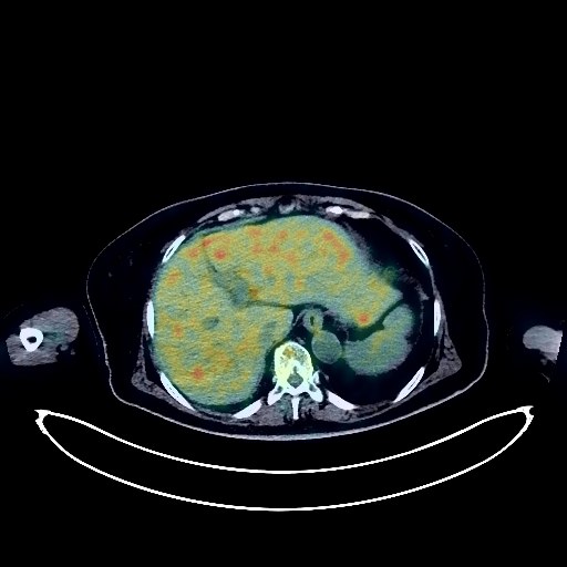
Pancreatic Cancer PET/CT (case 983827-000131 from PETWB-REP)
0 views9 days agoWhole-body 18F-FDG PET/CT scan in a patient with Pancreatic Cancer taken from the PETWB-REP dataset. The following English report (translated from original Chinese) is taken verbatim from the public dataset and has not been modified or otherwise checked for accuracy (see the end for citation). Impression a. Mass in the head and neck of the pancreas, with increased FDG metabolism, suggestive of malignancy, possibly pancreatic cancer or intraductal papillary tumor of the pancreas. Please correlate with clinicopathology. b. Lymph node metastasis to the left anterior aorta. Peritoneal seeding metastasis. Possible metastasis to the second hepatic hilum. Please correlate with contrast-enhanced MRI. Small amount of pelvic effusion. c. Localized low-density nodule in the pancreatic body, with normal FDG metabolism, suggestive of a cyst. Localized thickening and dense shadow in the gastric fundus wall, with increased FDG metabolism, tumor cannot be ruled out. Gastroscopy is recommended. Multiple low-density lesions in the liver, with normal FDG uptake, suggestive of hemangioma or cyst. Several ground-glass nodules in both lungs, suggestive of inflammatory nodules or atypical adenomatous hyperplasia. Annual HRCT follow-up is recommended. Scattered chronic inflammation and old lesions in both lungs. Cardiac shadow is full. Calcification of some arterial walls (including coronary arteries). Continuous increased FDG metabolism in the colon and rectum, suggestive of inflammation or physiological uptake. Focal increased FDG metabolism in the tongue, likely physiological; clinical correlation is recommended. Degenerative changes in the spine. L4/5 and L5/S1 intervertebral disc bulges. Benign osteopathy of the right temporomandibular joint. A few ischemic lesions in the deep bilateral brain, suggestive of age-related encephalopathy. This case is from PETWB-REP, a curated dataset of whole-body 18F-FDG PET/CT scans and corresponding radiology reports from 490 patients with a broad spectrum of malignancies. The data were retrospectively collected from patients who underwent clinically indicated whole-body 18F-FDG PET/CT scans at the Shanghai Universal Medical Imaging Diagnostic Center between 2021 and 2024. License: Creative Commons Attribution 4.0 International (CC BY 4.0) Citation: Xue, L., Feng, G., Wenbo, Z., Zhang, Y., Li, L., Wang, S., Peng, L., Peng, S., & Gao, X. (2026). PETWB-REP: A Multi-Cancer Whole-Body FDG PET/CT Dataset with Corresponding Radiology Reports [Data set]. Zenodo. https://doi.org/10.5281/zenodo.18670487
Whole BodyPET/CT
Colon Cancer PET/CT (case 983827-000220 from PETWB-REP)
0 views9 days agoWhole-body 18F-FDG PET/CT scan in a patient with Colon Cancer taken from the PETWB-REP dataset. The following English report (translated from original Chinese) is taken verbatim from the public dataset and has not been modified or otherwise checked for accuracy (see the end for citation). Impression a. Post-colon cancer treatment: No abnormal FDG metabolic foci were observed in the surgical area. b. Post-duodenal lesion treatment: The mesentery around the surgical area was slightly thickened, and FDG metabolism was increased, suggesting possible post-operative changes. Tumor recurrence needs to be ruled out; close observation is recommended. Chronic miliary nodules in the lateral basal segment of the left lower lobe, chronic inflammation and sequelae in both lungs. Reactive hyperplasia of the hilar and mediastinal lymph nodes in both lungs. Partial arteriosclerosis (including coronary arteries). Several calcifications in the right breast. Mild fatty liver, hepatic hemangioma is highly probable; ultrasound follow-up is recommended. Gallstones. Slightly low-density lesions in the spleen, with FDG showing background uptake, suggesting a high probability of hemangioma; ultrasound follow-up is recommended. Scoliosis with degenerative changes. T12/L1 disc herniation with pneumothorax and degeneration. Bilateral deep lacunar infarcts. Thyroid gland density is uneven; FDG metabolism is normal. Ultrasound follow-up is recommended. Bilateral cervical lymph node reactive hyperplasia. This case is from PETWB-REP, a curated dataset of whole-body 18F-FDG PET/CT scans and corresponding radiology reports from 490 patients with a broad spectrum of malignancies. The data were retrospectively collected from patients who underwent clinically indicated whole-body 18F-FDG PET/CT scans at the Shanghai Universal Medical Imaging Diagnostic Center between 2021 and 2024. License: Creative Commons Attribution 4.0 International (CC BY 4.0) Citation: Xue, L., Feng, G., Wenbo, Z., Zhang, Y., Li, L., Wang, S., Peng, L., Peng, S., & Gao, X. (2026). PETWB-REP: A Multi-Cancer Whole-Body FDG PET/CT Dataset with Corresponding Radiology Reports [Data set]. Zenodo. https://doi.org/10.5281/zenodo.18670487
Whole BodyPET/CT
Lung Cancer PET/CT (case 983827-000225 from PETWB-REP)
0 views9 days agoWhole-body 18F-FDG PET/CT scan in a patient with Lung Cancer taken from the PETWB-REP dataset. The following English report (translated from original Chinese) is taken verbatim from the public dataset and has not been modified or otherwise checked for accuracy (see the end for citation). Impression a. Ground-glass nodule in the apical-posterior segment of the left upper lobe, FDG metabolism normal, likely early-stage lung cancer; please correlate with clinicopathology. b. Ground-glass nodule in the superior lingular segment of the left upper lobe, FDG metabolism normal, suggest inflammation or atypical adenomatous hyperplasia; annual HRCT follow-up recommended. c. Chronic inflammatory micronodule (solid) in the left lung. Mild anemia changes. Patchy FDG elevation in the spleen, slightly decreased density on same-slice CT, space-occupying lesion to be ruled out; further enhanced MRI recommended. Focal FDG elevation in the left lobe of the thyroid gland, suggestive of adenomatous nodule, malignancy to be ruled out; further ultrasound examination recommended. Bilateral breast hyperplasia. Reactive hyperplasia of bilateral axillary lymph nodes. Physiological uptake in the uterine cavity; cystic lesion in the left adnexa; follow-up ultrasound is recommended. Small amount of pelvic effusion. Possible ischemic lesion in the left frontal lobe; follow-up MRI is recommended to rule out other possibilities. This case is from PETWB-REP, a curated dataset of whole-body 18F-FDG PET/CT scans and corresponding radiology reports from 490 patients with a broad spectrum of malignancies. The data were retrospectively collected from patients who underwent clinically indicated whole-body 18F-FDG PET/CT scans at the Shanghai Universal Medical Imaging Diagnostic Center between 2021 and 2024. License: Creative Commons Attribution 4.0 International (CC BY 4.0) Citation: Xue, L., Feng, G., Wenbo, Z., Zhang, Y., Li, L., Wang, S., Peng, L., Peng, S., & Gao, X. (2026). PETWB-REP: A Multi-Cancer Whole-Body FDG PET/CT Dataset with Corresponding Radiology Reports [Data set]. Zenodo. https://doi.org/10.5281/zenodo.18670487
Whole BodyPET/CT