Loading...
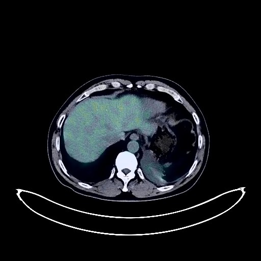
Lung Cancer PET/CT (case 983827-000141 from PETWB-REP)
0 views9 days agoWhole-body 18F-FDG PET/CT scan in a patient with Lung Cancer taken from the PETWB-REP dataset. The following English report (translated from original Chinese) is taken verbatim from the public dataset and has not been modified or otherwise checked for accuracy (see the end for citation). Impression a. A mass in the lower lobe of the left lung with increased FDG metabolism, highly suggestive of lung cancer with surrounding obstructive changes; please correlate with clinicopathology. Multiple lymph node metastases in the bilateral hilar, mediastinal, posterior pancreatic body, and perivena cava areas. Small amount of pleural effusion on the left side. b. Multiple chronic inflammatory micronodules in both lungs are highly probable; follow-up CT is recommended. Bilateral pleural thickening with right-sided calcification. Increased FDG metabolism in the nasopharynx, suggesting possible inflammation; specialist follow-up is recommended. Inflammation of the left maxillary sinus. Small cyst in the left lateral lobe of the liver. Small kidney stone and cyst in the left kidney. Calcification in the prostate. Small amount of hydrocele in both testes. Slightly thickened gastric antral wall with increased FDG uptake, highly suggestive of gastritis; follow-up gastroscopy is recommended. Increased FDG metabolism in the distal rectum, suggesting hemorrhoids or inflammatory uptake. Spinal degenerative changes. L2/3, L3/4, L4/5, L5/S1 intervertebral disc bulge. Bilateral frozen shoulder. Small softening lesion in the left basal ganglia region, age-related brain changes; MRI is recommended. This case is from PETWB-REP, a curated dataset of whole-body 18F-FDG PET/CT scans and corresponding radiology reports from 490 patients with a broad spectrum of malignancies. The data were retrospectively collected from patients who underwent clinically indicated whole-body 18F-FDG PET/CT scans at the Shanghai Universal Medical Imaging Diagnostic Center between 2021 and 2024. License: Creative Commons Attribution 4.0 International (CC BY 4.0) Citation: Xue, L., Feng, G., Wenbo, Z., Zhang, Y., Li, L., Wang, S., Peng, L., Peng, S., & Gao, X. (2026). PETWB-REP: A Multi-Cancer Whole-Body FDG PET/CT Dataset with Corresponding Radiology Reports [Data set]. Zenodo. https://doi.org/10.5281/zenodo.18670487
Whole BodyPET/CT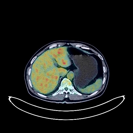
Renal Cancer PET/CT (case 983827-000179 from PETWB-REP)
0 views9 days agoWhole-body 18F-FDG PET/CT scan in a patient with Renal Cancer taken from the PETWB-REP dataset. The following English report (translated from original Chinese) is taken verbatim from the public dataset and has not been modified or otherwise checked for accuracy (see the end for citation). Impression a. Mass on the upper pole of the left kidney, with increased FDG metabolism, suggestive of renal cell carcinoma. b. Multiple lymph node metastases in the left renal hilum and para-aortic region. Extensive metastases in both lungs. Metastasis to the diaphragmatic roof of the liver. High probability of left adrenal metastasis. Minor chronic inflammation and old lesions in both lungs. Slight pleural thickening bilaterally. Reactive hyperplasia of mediastinal lymph nodes. Calcification of some arterial walls (including coronary arteries). Benign prostatic hyperplasia with calcification, mildly increased FDG metabolism, suggestive of inflammatory or physiological uptake; follow-up PSA and ultrasound examination recommended. Nuchal ligament calcification. Osteophyte formation in the cervical, thoracic, and lumbar vertebrae; L4/5 and L5/S1 intervertebral disc bulges. Minor ischemic lesions in the deep bilateral brain; senile encephalopathy. Chronic inflammation of the bilateral maxillary sinuses. Inflammation of some periodontal tissues. This case is from PETWB-REP, a curated dataset of whole-body 18F-FDG PET/CT scans and corresponding radiology reports from 490 patients with a broad spectrum of malignancies. The data were retrospectively collected from patients who underwent clinically indicated whole-body 18F-FDG PET/CT scans at the Shanghai Universal Medical Imaging Diagnostic Center between 2021 and 2024. License: Creative Commons Attribution 4.0 International (CC BY 4.0) Citation: Xue, L., Feng, G., Wenbo, Z., Zhang, Y., Li, L., Wang, S., Peng, L., Peng, S., & Gao, X. (2026). PETWB-REP: A Multi-Cancer Whole-Body FDG PET/CT Dataset with Corresponding Radiology Reports [Data set]. Zenodo. https://doi.org/10.5281/zenodo.18670487
Whole BodyPET/CT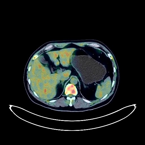
Lung Cancer PET/CT (case 983827-000240 from PETWB-REP)
0 views9 days agoWhole-body 18F-FDG PET/CT scan in a patient with Lung Cancer taken from the PETWB-REP dataset. The following English report (translated from original Chinese) is taken verbatim from the public dataset and has not been modified or otherwise checked for accuracy (see the end for citation). Impression a. A soft tissue mass in the posterior segment of the left upper lobe with unevenly increased FDG metabolism, suggestive of lung cancer. Please confirm with pathology. b. Metastasis to the left hilar and mediastinal lymph nodes, and the right supraclavicular fossa. c. Mild interstitial changes in both lungs with a few chronic inflammations and sequelae, emphysema in the upper lobes of both lungs. Mild thickening of the pleura on both sides. Calcification of some arterial walls (including coronary arteries). d. Left adrenal hyperplasia is highly probable; please follow up with ultrasound to rule out metastasis. Multiple lesions in the right cerebellum and left frontoparietal lobe after Gamma Knife treatment: a. Multiple mixed slightly high-density masses in the right cerebellar hemisphere and left frontoparietal lobe, with decreased FDG metabolism, suggestive of post-treatment changes accompanied by peritumoral edema. It is recommended to have a follow-up enhanced MRI at intervals to rule out residual tumor activity. ? b. Lacunar infarcts in both basal ganglia regions; please follow up with MRI. A few chronic inflammations and submucosal cysts in the right maxillary sinus. ? c. Inflammatory or physiological uptake of both nasopharyngeal walls. Reactive hyperplasia of both deep cervical spaces and submandibular lymph nodes. No clear signs of fatty liver, liver cysts. Splenic artery aneurysm is highly likely; CTA follow-up is necessary if needed. Small cyst in the left kidney. Prostatic calcifications. Increased FDG metabolism in parts of the esophagus, stomach wall, and intestines, suggesting chronic inflammatory changes or physiological uptake; please combine with endoscopic examination. Spinal degenerative changes. Mild bulging of L4/5 and L5/S1 intervertebral discs. Physiological uptake in multiple muscles throughout the body; inflammatory uptake near the right shoulder joint. This case is from PETWB-REP, a curated dataset of whole-body 18F-FDG PET/CT scans and corresponding radiology reports from 490 patients with a broad spectrum of malignancies. The data were retrospectively collected from patients who underwent clinically indicated whole-body 18F-FDG PET/CT scans at the Shanghai Universal Medical Imaging Diagnostic Center between 2021 and 2024. License: Creative Commons Attribution 4.0 International (CC BY 4.0) Citation: Xue, L., Feng, G., Wenbo, Z., Zhang, Y., Li, L., Wang, S., Peng, L., Peng, S., & Gao, X. (2026). PETWB-REP: A Multi-Cancer Whole-Body FDG PET/CT Dataset with Corresponding Radiology Reports [Data set]. Zenodo. https://doi.org/10.5281/zenodo.18670487
Whole BodyPET/CT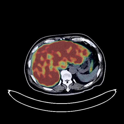
Lung Cancer PET/CT (case 983827-000251 from PETWB-REP)
0 views9 days agoWhole-body 18F-FDG PET/CT scan in a patient with Lung Cancer taken from the PETWB-REP dataset. The following English report (translated from original Chinese) is taken verbatim from the public dataset and has not been modified or otherwise checked for accuracy (see the end for citation). Impression a. A mass near the hilum in the apical segment of the right upper lobe, with elevated FDG metabolism, strongly suggestive of central lung cancer; please corroborate clinicopathology. Multiple lymph node metastases in bilateral hilar regions, mediastinum, prediaphragmatic group, hepatogastric space, hepatic hilum, peripancreatic region, bilateral supraclavicular fossa, and right deep cervical space. Nodular metastases in the apical segment of the right upper lobe and the posterior segment of the left lower lobe. Multiple bone metastases throughout the body. b. Mesenteric proliferation in the left and right paracolic gutter, with elevated FDG metabolism, suggesting possible metastasis. Significant effusion in the abdomen and pelvis. c. Diffuse lesions in the liver, with unevenly elevated FDG metabolism, suggestive of malignancy, with a higher probability of metastasis than the primary tumor. Liver cysts. Chronic bronchitis and emphysematous changes in both lungs. Scattered small chronic inflammatory nodules (solid) in the remaining lungs; please follow up with CT scans. A few post-inflammatory remnants and calcifications in both lungs. Mild thickening of the pleura bilaterally. Partial arteriosclerosis (including coronary arteries). Chronic cholecystitis. Bilateral renal cysts, with the right kidney showing a complex cyst. Benign prostatic hyperplasia. Bilateral hydrocele. Increased FDG metabolism in parts of the colon and rectum, considered physiological uptake or chronic inflammatory changes; please follow up with endoscopy. Degenerative changes in the spine. L3/4, L4/5, and L5/S1 intervertebral disc bulging with pneumoconiosis and degeneration. Age-related brain changes, deep lacunar infarcts in the brain; MRI is recommended. Minor inflammation of the right maxillary sinus. Right sclerotic mastoid process. This case is from PETWB-REP, a curated dataset of whole-body 18F-FDG PET/CT scans and corresponding radiology reports from 490 patients with a broad spectrum of malignancies. The data were retrospectively collected from patients who underwent clinically indicated whole-body 18F-FDG PET/CT scans at the Shanghai Universal Medical Imaging Diagnostic Center between 2021 and 2024. License: Creative Commons Attribution 4.0 International (CC BY 4.0) Citation: Xue, L., Feng, G., Wenbo, Z., Zhang, Y., Li, L., Wang, S., Peng, L., Peng, S., & Gao, X. (2026). PETWB-REP: A Multi-Cancer Whole-Body FDG PET/CT Dataset with Corresponding Radiology Reports [Data set]. Zenodo. https://doi.org/10.5281/zenodo.18670487
Whole BodyPET/CT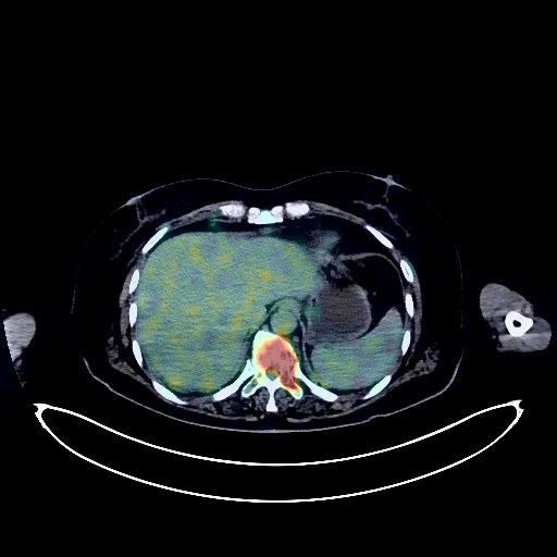
Rectal Cancer PET/CT (case 983827-000014 from PETWB-REP)
0 views9 days agoWhole-body 18F-FDG PET/CT scan in a patient with Rectal Cancer taken from the PETWB-REP dataset. The following English report (translated from original Chinese) is taken verbatim from the public dataset and has not been modified or otherwise checked for accuracy (see the end for citation). Impression a. Post-rectal cancer surgery, slight thickening of the anastomotic wall, no abnormalities in FDG metabolism, suggesting post-operative changes are highly likely; colonoscopy follow-up is recommended. b. Multiple enlarged lymph nodes throughout the body, multiple areas of decreased bone density, all with increased FDG metabolism, suggest malignancy, with a high probability of metastasis; further investigation based on clinical treatment and pathology is recommended to rule out other possibilities. c. Cystic lesion on the right pelvic wall, no abnormalities in FDG metabolism, suggesting a high probability of lymphangiocysts; follow-up examination is recommended to rule out metastasis. d. Slight thickening of the omentum mesentery in the abdominopelvic cavity, no abnormal FDG uptake; follow-up examination is recommended. Diffuse lesions in both lungs, unevenly increased FDG metabolism, suggesting possible interstitial inflammation, with partial metastasis to be ruled out; CT scan for comparison is recommended. Slight thickening of both pleura. Widespread decreased density in the left lobe and isthmus of the thyroid gland, with increased FDG metabolism, suggesting possible inflammatory uptake; ultrasound and laboratory tests are recommended to rule out space-occupying lesions. Small liver cysts; possible hepatic hemangioma. Chronic cholecystitis. Uterine fibroids. Degenerative changes in the spine. L4/5 and L5/S1 intervertebral disc bulges. No abnormalities were found on cranial scintigraphy. This case is from PETWB-REP, a curated dataset of whole-body 18F-FDG PET/CT scans and corresponding radiology reports from 490 patients with a broad spectrum of malignancies. The data were retrospectively collected from patients who underwent clinically indicated whole-body 18F-FDG PET/CT scans at the Shanghai Universal Medical Imaging Diagnostic Center between 2021 and 2024. License: Creative Commons Attribution 4.0 International (CC BY 4.0) Citation: Xue, L., Feng, G., Wenbo, Z., Zhang, Y., Li, L., Wang, S., Peng, L., Peng, S., & Gao, X. (2026). PETWB-REP: A Multi-Cancer Whole-Body FDG PET/CT Dataset with Corresponding Radiology Reports [Data set]. Zenodo. https://doi.org/10.5281/zenodo.18670487
Whole BodyPET/CT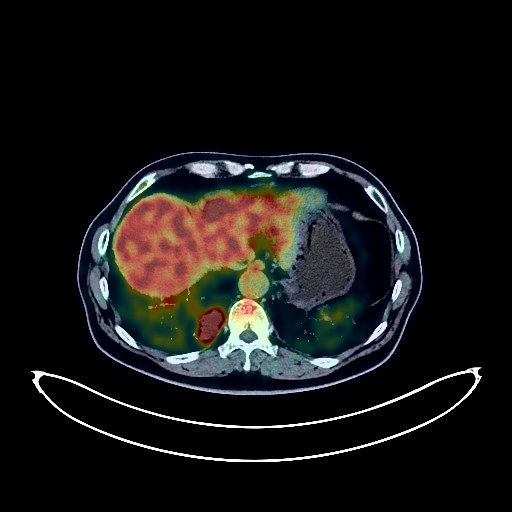
Lung Cancer PET/CT (case 983827-000133 from PETWB-REP)
0 views9 days agoWhole-body 18F-FDG PET/CT scan in a patient with Lung Cancer taken from the PETWB-REP dataset. The following English report (translated from original Chinese) is taken verbatim from the public dataset and has not been modified or otherwise checked for accuracy (see the end for citation). Impression a. Space-occupying lesion in the posterior basal segment of the right lower lobe, with increased FDG metabolism, suggestive of peripheral lung cancer. Multiple lymph node metastases in the right hilum, mediastinum, and right supraclavicular fossa. Liver metastases. T2 spinous process metastases. b. Interstitial pneumonia in both lungs. Slight pleural thickening bilaterally. Calcification of some arterial walls (including coronary arteries). A few ischemic lesions in the deep bilateral brain. Multiple liver cysts. Continuous increased FDG metabolism in parts of the colon and rectum, suggestive of inflammatory or physiological uptake; colonoscopy follow-up is recommended. Spinal degenerative changes. L4/5 and L5/S1 intervertebral disc bulges. This case is from PETWB-REP, a curated dataset of whole-body 18F-FDG PET/CT scans and corresponding radiology reports from 490 patients with a broad spectrum of malignancies. The data were retrospectively collected from patients who underwent clinically indicated whole-body 18F-FDG PET/CT scans at the Shanghai Universal Medical Imaging Diagnostic Center between 2021 and 2024. License: Creative Commons Attribution 4.0 International (CC BY 4.0) Citation: Xue, L., Feng, G., Wenbo, Z., Zhang, Y., Li, L., Wang, S., Peng, L., Peng, S., & Gao, X. (2026). PETWB-REP: A Multi-Cancer Whole-Body FDG PET/CT Dataset with Corresponding Radiology Reports [Data set]. Zenodo. https://doi.org/10.5281/zenodo.18670487
Whole BodyPET/CT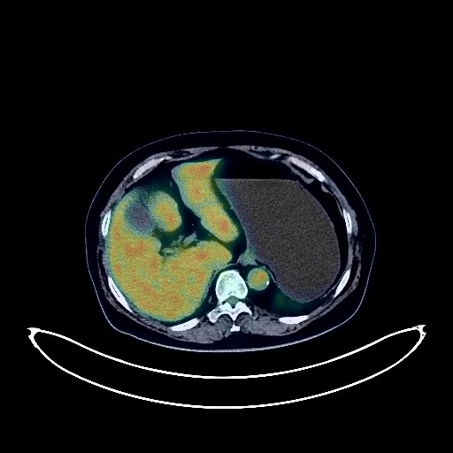
Cervical Cancer PET/CT (case 983827-000208 from PETWB-REP)
0 views9 days agoWhole-body 18F-FDG PET/CT scan in a patient with Cervical Cancer taken from the PETWB-REP dataset. The following English report (translated from original Chinese) is taken verbatim from the public dataset and has not been modified or otherwise checked for accuracy (see the end for citation). Impression Cervical mass with elevated FDG metabolism, suggestive of cervical cancer; please correlate with clinicopathology. Reactive hyperplasia of bilateral iliac vessels and bilateral inguinal lymph nodes. a. Mixed ground-glass nodule in the anterior segment of the left upper lobe with elevated FDG metabolism, highly suggestive of inflammation; anti-inflammatory treatment followed by CT scan recommended to rule out lung cancer. b. Several ground-glass nodules in both lungs, FDG metabolism normal, suggestive of inflammation or atypical adenomatous hyperplasia; annual CT scan recommended. c. Chronic inflammatory micronodules (solid) in both lungs. A few post-inflammatory lesions in both lungs. Calcification of some arterial walls (including coronary arteries). Postoperative changes after thyroid cancer surgery; no signs of tumor recurrence in the surgical area. Reactive hyperplasia of bilateral cervical lymph nodes. Fatty liver; small cyst in the right lobe of the liver. Chronic inflammatory changes in the gastric antrum; please follow up with endoscopy. Osteoporosis, degenerative changes in the spine, L4/5 and L5/S1 disc bulges. Old fracture of the right fourth anterior rib. Age-related brain abnormalities, deep lacunar infarcts. Chronic inflammation of the right ethmoid sinus. This case is from PETWB-REP, a curated dataset of whole-body 18F-FDG PET/CT scans and corresponding radiology reports from 490 patients with a broad spectrum of malignancies. The data were retrospectively collected from patients who underwent clinically indicated whole-body 18F-FDG PET/CT scans at the Shanghai Universal Medical Imaging Diagnostic Center between 2021 and 2024. License: Creative Commons Attribution 4.0 International (CC BY 4.0) Citation: Xue, L., Feng, G., Wenbo, Z., Zhang, Y., Li, L., Wang, S., Peng, L., Peng, S., & Gao, X. (2026). PETWB-REP: A Multi-Cancer Whole-Body FDG PET/CT Dataset with Corresponding Radiology Reports [Data set]. Zenodo. https://doi.org/10.5281/zenodo.18670487
Whole BodyPET/CT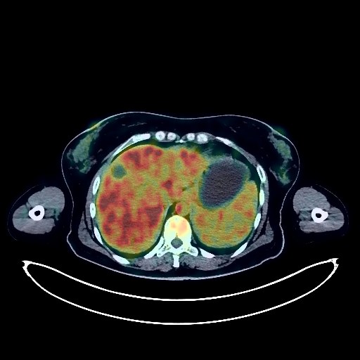
Glioma PET/CT (case 983827-000213 from PETWB-REP)
0 views9 days agoWhole-body 18F-FDG PET/CT scan in a patient with Glioma taken from the PETWB-REP dataset. The following English report (translated from original Chinese) is taken verbatim from the public dataset and has not been modified or otherwise checked for accuracy (see the end for citation). Impression a. Cystic mass with septa in the left temporal lobe, FDG metabolism absent; combined with contrast-enhanced MRI from another hospital, glioma is suspected. b. Left temporal pole arachnoid cyst. Combined with MRI from another hospital, hepatic Grinson's sheath edema is suspected. Multiple hepatic cysts. Reactive hyperplasia of hilar lymph nodes. Splenomegaly. Anemia. Bilateral breast hyperplasia. Uterine fibroids. Partial vertebral osteophyte formation. Low-density thyroid nodule, FDG uptake normal, nodular goiter is suspected; follow-up ultrasound is recommended. This case is from PETWB-REP, a curated dataset of whole-body 18F-FDG PET/CT scans and corresponding radiology reports from 490 patients with a broad spectrum of malignancies. The data were retrospectively collected from patients who underwent clinically indicated whole-body 18F-FDG PET/CT scans at the Shanghai Universal Medical Imaging Diagnostic Center between 2021 and 2024. License: Creative Commons Attribution 4.0 International (CC BY 4.0) Citation: Xue, L., Feng, G., Wenbo, Z., Zhang, Y., Li, L., Wang, S., Peng, L., Peng, S., & Gao, X. (2026). PETWB-REP: A Multi-Cancer Whole-Body FDG PET/CT Dataset with Corresponding Radiology Reports [Data set]. Zenodo. https://doi.org/10.5281/zenodo.18670487
Whole BodyPET/CT
Lung Cancer PET/CT (case 983827-000132 from PETWB-REP)
0 views9 days agoWhole-body 18F-FDG PET/CT scan in a patient with Lung Cancer taken from the PETWB-REP dataset. The following English report (translated from original Chinese) is taken verbatim from the public dataset and has not been modified or otherwise checked for accuracy (see the end for citation). Impression a. A mass in the posterior segment of the right upper lobe with significantly increased FDG metabolism in the solid portion, suggestive of lung cancer with minor obstructive pneumonia. Please confirm with pathology. b. Possible right hilar lymph node metastasis, highly likely reactive mediastinal lymph node hyperplasia. Please follow up with CT. c. Chronic inflammatory solid micronodules in both lungs, a few inflammatory ground-glass opacities in the left lower lobe. Bilateral emphysema with bullae, bilateral pulmonary fibrosis. a. Thickening of the gastric angle and antrum with ulceration and increased FDG metabolism suggest possible gastritis, but gastric cancer cannot be ruled out. Further gastroscopy is recommended. b. Physiological or inflammatory uptake in the lower esophagus, cardia, fundus, and part of the intestine. Duodenal diverticulum. c. Enlarged lymph nodes on the lesser curvature of the stomach, with no increase in FDG metabolism, suggesting possible reactive lymph node hyperplasia. Please follow up with CT to rule out other possibilities. Benign prostatic hyperplasia with calcification, uneven FDG metabolism; please follow up with MRI. Gallstones. Thickened perirenal septum bilaterally, left renal cyst. Slightly enlarged heart. Partial arteriosclerosis (including coronary arteries). Degenerative changes in the spine, L1 vertebral hemangioma, L4/5 intervertebral disc bulge. Bilateral shoulder joint and right acromioclavicular joint periarthritis. Left trapezius muscle lipoma. Physiological uptake of some muscles in the right abdomen. No obvious abnormalities seen on cranial scintigraphy. Minor chronic inflammation of the right maxillary sinus. This case is from PETWB-REP, a curated dataset of whole-body 18F-FDG PET/CT scans and corresponding radiology reports from 490 patients with a broad spectrum of malignancies. The data were retrospectively collected from patients who underwent clinically indicated whole-body 18F-FDG PET/CT scans at the Shanghai Universal Medical Imaging Diagnostic Center between 2021 and 2024. License: Creative Commons Attribution 4.0 International (CC BY 4.0) Citation: Xue, L., Feng, G., Wenbo, Z., Zhang, Y., Li, L., Wang, S., Peng, L., Peng, S., & Gao, X. (2026). PETWB-REP: A Multi-Cancer Whole-Body FDG PET/CT Dataset with Corresponding Radiology Reports [Data set]. Zenodo. https://doi.org/10.5281/zenodo.18670487
Whole BodyPET/CT
Lung Cancer PET/CT (case 983827-000023 from PETWB-REP)
0 views9 days agoWhole-body 18F-FDG PET/CT scan in a patient with Lung Cancer taken from the PETWB-REP dataset. The following English report (translated from original Chinese) is taken verbatim from the public dataset and has not been modified or otherwise checked for accuracy (see the end for citation). Impression "Right lung cancer after radiotherapy and chemotherapy," compared with the previous PET/CT scan (2022-02-08) taken at our center: a. The lesion in the right hilar region has significantly decreased in size compared to the previous scan, and FDG metabolism has decreased, suggesting that some tumor activity has been suppressed after treatment. b. Multiple lymph nodes in the right hilum, mediastinum, right internal mammary chain, and right supraclavicular region have decreased in size compared to the previous scan, and FDG metabolism has decreased, suggesting that some tumor activity has been suppressed; the lymph nodes in the hepatogastric space are roughly the same size as before; among them, one lymph node in the right visceral subpleural region is a newly developed lesion. c. Multiple bone metastases throughout the body, some lesions have slightly shrunk in size compared to the previous scan, and FDG metabolism has slightly decreased. d. Small nodules in the anterior abdominal wall and right abdominal wall space, above and behind the right kidney, in the space behind the diaphragm crus, and in the retroperitoneum, are roughly the same size as before, and FDG metabolism has slightly increased. e. Nodule in the upper segment of the right anterior lobe of the liver is similar in size as before, and FDG metabolism has decreased. a. Several nodules in both lungs; some nodules are smaller and some have disappeared compared to previous scans. FDG metabolism is normal. Close follow-up with CT is recommended. b. Minor chronic inflammation and sequelae in both lungs. Slight local thickening of the pleura bilaterally. Partial arteriosclerosis (including coronary arteries). Post-bladder tumor surgery, no obvious filling defect changes were observed. Please follow up with cystoscopy. Large cystic lesions in the right lower abdomen and pelvis, slightly smaller than before, likely benign. Please follow up with clinical findings. Increased FDG metabolism in the middle and lower esophagus and part of the gastric wall, considered physiological uptake or chronic inflammatory changes. Please follow up with gastroscopy. Multiple cysts in the left kidney. Spinal degeneration. L2-S1 intervertebral disc bulge. Minor lacunar infarcts in the deep bilateral brain, mild age-related brain changes. Please follow up with MRI. Minor chronic inflammation in both ethmoid sinuses. Decreased lens density bilaterally. Please follow up with clinical findings. Multiple nodules in both lobes of the thyroid gland, with no abnormalities in FDG metabolism, are considered likely to be benign nodules, similar to the previous findings. Please follow up with ultrasound. This case is from PETWB-REP, a curated dataset of whole-body 18F-FDG PET/CT scans and corresponding radiology reports from 490 patients with a broad spectrum of malignancies. The data were retrospectively collected from patients who underwent clinically indicated whole-body 18F-FDG PET/CT scans at the Shanghai Universal Medical Imaging Diagnostic Center between 2021 and 2024. License: Creative Commons Attribution 4.0 International (CC BY 4.0) Citation: Xue, L., Feng, G., Wenbo, Z., Zhang, Y., Li, L., Wang, S., Peng, L., Peng, S., & Gao, X. (2026). PETWB-REP: A Multi-Cancer Whole-Body FDG PET/CT Dataset with Corresponding Radiology Reports [Data set]. Zenodo. https://doi.org/10.5281/zenodo.18670487
Whole BodyPET/CT