Loading...
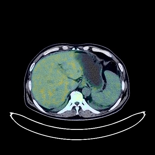
Pancreatic Cancer PET/CT (case 983827-000215 from PETWB-REP)
0 views9 days agoWhole-body 18F-FDG PET/CT scan in a patient with Pancreatic Cancer taken from the PETWB-REP dataset. The following English report (translated from original Chinese) is taken verbatim from the public dataset and has not been modified or otherwise checked for accuracy (see the end for citation). Impression A mass in the pancreatic tail with elevated FDG metabolism suggests pancreatic cancer invading the adjacent spleen and splenic flexure of the transverse colon. Please correlate with clinicopathology. Multiple lymph node metastases in the left mesenteric cavity and a small amount of effusion in the abdominopelvic cavity are also present. Thickened appendix with cystic dilatation and thickened intestinal wall with elevated FDG metabolism suggest possible cystadenoma, but cystadenoma should be ruled out. Please correlate with clinical findings and colonoscopy. Chronic inflammatory micronodules in the upper lobes of both lungs. Scattered post-inflammatory lesions in both lungs. Bilateral pleural thickening with a small amount of pleural effusion. Calcification of some arterial walls (including coronary arteries). Liver cyst. Splenic infarction. Possible gallbladder adenomyosis; residual contrast agent in the gallbladder and bladder. Benign prostatic hyperplasia with calcification. Chronic inflammatory changes in part of the gastric wall; please follow up with gastroscopy. Degenerative changes in the spine, L4/5 and L5/S1 intervertebral disc bulge, L5/S1 disc pneumoconiosis and degeneration. Low-density nodule in the right lobe of the thyroid gland, FDG metabolism normal, suggestive of nodular goiter, please confirm with ultrasound examination. Age-related brain, deep lacunar infarcts, please confirm with MRI examination. This case is from PETWB-REP, a curated dataset of whole-body 18F-FDG PET/CT scans and corresponding radiology reports from 490 patients with a broad spectrum of malignancies. The data were retrospectively collected from patients who underwent clinically indicated whole-body 18F-FDG PET/CT scans at the Shanghai Universal Medical Imaging Diagnostic Center between 2021 and 2024. License: Creative Commons Attribution 4.0 International (CC BY 4.0) Citation: Xue, L., Feng, G., Wenbo, Z., Zhang, Y., Li, L., Wang, S., Peng, L., Peng, S., & Gao, X. (2026). PETWB-REP: A Multi-Cancer Whole-Body FDG PET/CT Dataset with Corresponding Radiology Reports [Data set]. Zenodo. https://doi.org/10.5281/zenodo.18670487
Whole BodyPET/CT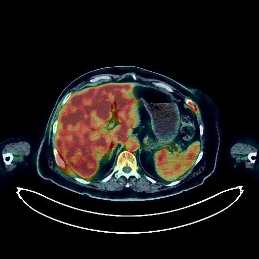
Breast Cancer PET/CT (case 983827-000158 from PETWB-REP)
0 views9 days agoWhole-body 18F-FDG PET/CT scan in a patient with Breast Cancer taken from the PETWB-REP dataset. The following English report (translated from original Chinese) is taken verbatim from the public dataset and has not been modified or otherwise checked for accuracy (see the end for citation). Impression a. Postoperative changes after right breast cancer surgery, no signs of tumor recurrence. b. Multiple bone metastases throughout the body (see description for details). c. Left temporal lobe metastasis; possible sellar region lesion. Further enhanced MRI is recommended for all of the above. d. Multiple lymph nodes in the hilum, mediastinum, and bilateral supraclavicular fossa show increased FDG metabolism, suggesting likely reactive hyperplasia; partial metastasis is pending. Follow-up is recommended. Chronic inflammatory nodules in both lungs. Inflammation in the lower lobe of the right lung, calcifications in both lungs, scattered post-inflammatory remnants in both lungs. Tracheal diverticulum. Calcification of some arterial walls (including coronary arteries). Fatty liver, calcifications in the right lobe of the liver, liver cysts. Angiomyolipoma of the left kidney. Intrauterine device (IUD) insertion, possible uterine fibroids. Chronic inflammatory changes or physiological uptake in parts of the intestine. Thyroid enlargement with multiple nodules, FDG metabolism normal, suggestive of nodular goiter. Osteoporosis, degenerative changes in the spine, L3/4, L4/5, L5/S1 intervertebral disc bulging. Bilateral frozen shoulder. Chronic inflammation of the right frontal sinus, bilateral ethmoid sinuses, and bilateral maxillary sinuses, submucosal cyst of the sphenoid sinus. Nasal mucosal thickening, chronic inflammation of the right lateral wall of the nasopharynx. This case is from PETWB-REP, a curated dataset of whole-body 18F-FDG PET/CT scans and corresponding radiology reports from 490 patients with a broad spectrum of malignancies. The data were retrospectively collected from patients who underwent clinically indicated whole-body 18F-FDG PET/CT scans at the Shanghai Universal Medical Imaging Diagnostic Center between 2021 and 2024. License: Creative Commons Attribution 4.0 International (CC BY 4.0) Citation: Xue, L., Feng, G., Wenbo, Z., Zhang, Y., Li, L., Wang, S., Peng, L., Peng, S., & Gao, X. (2026). PETWB-REP: A Multi-Cancer Whole-Body FDG PET/CT Dataset with Corresponding Radiology Reports [Data set]. Zenodo. https://doi.org/10.5281/zenodo.18670487
Whole BodyPET/CT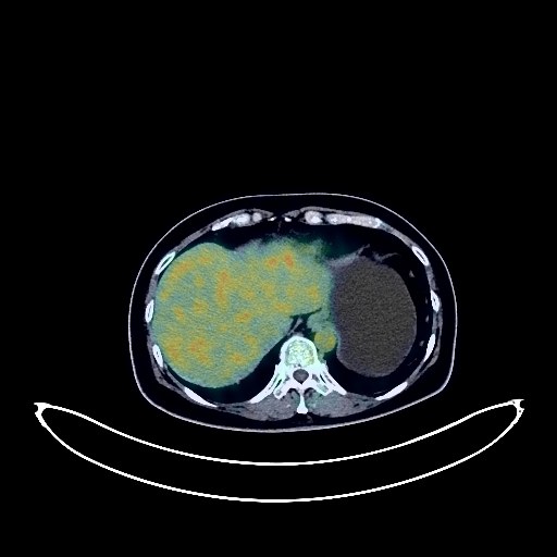
Renal Cancer PET/CT (case 983827-000120 from PETWB-REP)
0 views9 days agoWhole-body 18F-FDG PET/CT scan in a patient with Renal Cancer taken from the PETWB-REP dataset. The following English report (translated from original Chinese) is taken verbatim from the public dataset and has not been modified or otherwise checked for accuracy (see the end for citation). Impression Right renal mass with elevated FDG metabolism, suggestive of malignancy, most likely renal cell carcinoma; please correlate with clinicopathology. Reactive hyperplasia of retroperitoneal lymph nodes is possible, but metastasis should be ruled out; follow-up is recommended. Chronic inflammatory micronodules in the left lower lobe. Atelectasis in the right middle lobe, with a few post-inflammatory lesions in both lungs. Calcification of some arterial walls (including coronary arteries). Fatty liver, chronic cholecystitis, gallstones. Residual contrast agent in the urinary tract, small cyst in the left kidney. Degenerative changes in the spine, L5/S1 disc bulge with pneumothorax, L5/S1 endplate inflammation. No obvious abnormalities on cranial scintigraphy. Chronic inflammation of both ethmoid sinuses. Chronic inflammatory lymph nodes in the right deep cervical space. This case is from PETWB-REP, a curated dataset of whole-body 18F-FDG PET/CT scans and corresponding radiology reports from 490 patients with a broad spectrum of malignancies. The data were retrospectively collected from patients who underwent clinically indicated whole-body 18F-FDG PET/CT scans at the Shanghai Universal Medical Imaging Diagnostic Center between 2021 and 2024. License: Creative Commons Attribution 4.0 International (CC BY 4.0) Citation: Xue, L., Feng, G., Wenbo, Z., Zhang, Y., Li, L., Wang, S., Peng, L., Peng, S., & Gao, X. (2026). PETWB-REP: A Multi-Cancer Whole-Body FDG PET/CT Dataset with Corresponding Radiology Reports [Data set]. Zenodo. https://doi.org/10.5281/zenodo.18670487
Whole BodyPET/CT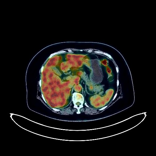
Ovarian Cancer PET/CT (case 983827-000211 from PETWB-REP)
0 views9 days agoWhole-body 18F-FDG PET/CT scan in a patient with Ovarian Cancer taken from the PETWB-REP dataset. The following English report (translated from original Chinese) is taken verbatim from the public dataset and has not been modified or otherwise checked for accuracy (see the end for citation). Impression a. Bilateral adnexal region lesions with increased FDG metabolism, suggestive of ovarian cancer involving the uterus; please correlate with clinicopathology. Multiple peritoneal seeding metastases. Liver metastases to be ruled out; enhanced MRI analysis recommended. b. Multiple reactive hyperplasia of retroperitoneal and bilateral inguinal lymph nodes. a. Pure ground-glass nodule in the anterior segment of the left upper lobe, normal FDG metabolism, suggestive of atypical adenomatous hyperplasia or inflammatory nodule; annual HRCT follow-up recommended. b. A few post-inflammatory lesions in both lungs. Fatty liver, small cyst in the left lateral lobe of the liver. Small kidney stone in the left kidney, multiple microlithiasis in the right kidney. Left adrenal hyperplasia. Increased FDG uptake in some colonic strips, suggestive of inflammation or physiological uptake. Focal increased FDG uptake in the anal region, suggestive of hemorrhoids. Slightly decreased thyroid density with calcification in the left lobe and increased FDG uptake suggest thyroiditis; ultrasound and thyroid function tests are recommended for follow-up. Spinal osteophyte formation. L4/5 disc bulge, L5/S1 disc herniation. Mild age-related brain changes. This case is from PETWB-REP, a curated dataset of whole-body 18F-FDG PET/CT scans and corresponding radiology reports from 490 patients with a broad spectrum of malignancies. The data were retrospectively collected from patients who underwent clinically indicated whole-body 18F-FDG PET/CT scans at the Shanghai Universal Medical Imaging Diagnostic Center between 2021 and 2024. License: Creative Commons Attribution 4.0 International (CC BY 4.0) Citation: Xue, L., Feng, G., Wenbo, Z., Zhang, Y., Li, L., Wang, S., Peng, L., Peng, S., & Gao, X. (2026). PETWB-REP: A Multi-Cancer Whole-Body FDG PET/CT Dataset with Corresponding Radiology Reports [Data set]. Zenodo. https://doi.org/10.5281/zenodo.18670487
Whole BodyPET/CT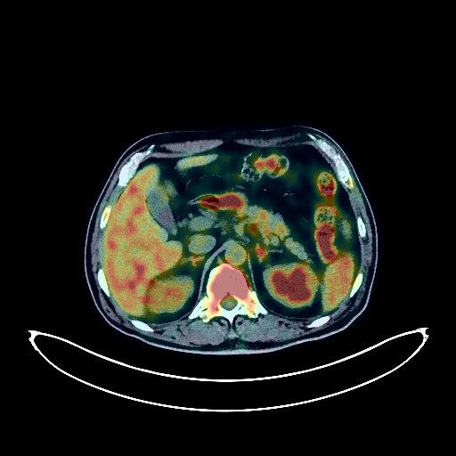
Esophageal Cancer PET/CT (case 983827-000028 from PETWB-REP)
0 views9 days agoWhole-body 18F-FDG PET/CT scan in a patient with Esophageal Cancer taken from the PETWB-REP dataset. The following English report (translated from original Chinese) is taken verbatim from the public dataset and has not been modified or otherwise checked for accuracy (see the end for citation). Impression a. Post-esophageal cancer surgery, increased FDG metabolism at the anastomosis site, likely due to inflammatory or physiological uptake; please follow up with gastroscopy to rule out other possibilities. b. Metastasis to the right paratracheal lymph nodes in the upper mediastinum. Metastatic tumor in the left lower pleural region. c. Possible retroperitoneal lymph node metastasis. Reactive hyperplasia of bilateral deep cervical, submandibular, and supraclavicular lymph nodes. Right renal mass; further enhanced MRI is recommended. Left renal cyst (including complex cysts). Prostatic calcification. Infection in the upper and middle lobes of the right lung and the lower lobe of the left lung. Scattered post-inflammatory lesions in both lungs. Calcification of some arterial walls (including coronary arteries). Physiological or inflammatory uptake of the gastric wall and parts of the intestine; please follow up with endoscopy. Partial vertebral osteophyte formation. L4/5 and L5/S1 intervertebral disc bulge. Reactive hyperplasia of the entire bone marrow. Old fracture of the left 8th rib. A few ischemic lesions deep in the brain. Age-related brain changes. Minor chronic inflammation of the right maxillary sinus. Nasal septum deviation. Inflammatory uptake in the right upper alveolar bone, palate, and base of the tongue. This case is from PETWB-REP, a curated dataset of whole-body 18F-FDG PET/CT scans and corresponding radiology reports from 490 patients with a broad spectrum of malignancies. The data were retrospectively collected from patients who underwent clinically indicated whole-body 18F-FDG PET/CT scans at the Shanghai Universal Medical Imaging Diagnostic Center between 2021 and 2024. License: Creative Commons Attribution 4.0 International (CC BY 4.0) Citation: Xue, L., Feng, G., Wenbo, Z., Zhang, Y., Li, L., Wang, S., Peng, L., Peng, S., & Gao, X. (2026). PETWB-REP: A Multi-Cancer Whole-Body FDG PET/CT Dataset with Corresponding Radiology Reports [Data set]. Zenodo. https://doi.org/10.5281/zenodo.18670487
Whole BodyPET/CT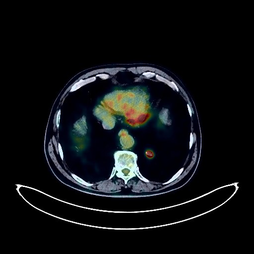
Bladder Cancer PET/CT (case 983827-000026 from PETWB-REP)
0 views9 days agoWhole-body 18F-FDG PET/CT scan in a patient with Bladder Cancer taken from the PETWB-REP dataset. The following English report (translated from original Chinese) is taken verbatim from the public dataset and has not been modified or otherwise checked for accuracy (see the end for citation). Impression a. Soft tissue lesions in the right kidney and bladder with increased FDG metabolism, all suggestive of malignancy, most likely renal pelvis cancer and bladder cancer. The bladder lesion involves the distal left ureter, with mild dilation and hydronephrosis of the left renal pelvis and ureter. b. Multiple metastatic lesions in the perirenal space. Retroperitoneal lymph node metastasis. c. Multiple metastatic tumors in both lungs. d. Metastatic tumor in the left iliac bone. Bilateral renal cysts. Prostatic calcification. A few ischemic lesions in the deep brain, age-related brain changes, MRI follow-up recommended. Left maxillary sinusitis. Emphysema and some interstitial changes in both lungs. Chronic inflammation and sequelae in both lungs. Reactive hyperplasia of mediastinal lymph nodes. Calcification of some arterial walls (including coronary arteries). Mild anterior slippage of the L4 vertebral body. Spinal degeneration. L4/5 and L5/S1 intervertebral disc bulge. This case is from PETWB-REP, a curated dataset of whole-body 18F-FDG PET/CT scans and corresponding radiology reports from 490 patients with a broad spectrum of malignancies. The data were retrospectively collected from patients who underwent clinically indicated whole-body 18F-FDG PET/CT scans at the Shanghai Universal Medical Imaging Diagnostic Center between 2021 and 2024. License: Creative Commons Attribution 4.0 International (CC BY 4.0) Citation: Xue, L., Feng, G., Wenbo, Z., Zhang, Y., Li, L., Wang, S., Peng, L., Peng, S., & Gao, X. (2026). PETWB-REP: A Multi-Cancer Whole-Body FDG PET/CT Dataset with Corresponding Radiology Reports [Data set]. Zenodo. https://doi.org/10.5281/zenodo.18670487
Whole BodyPET/CT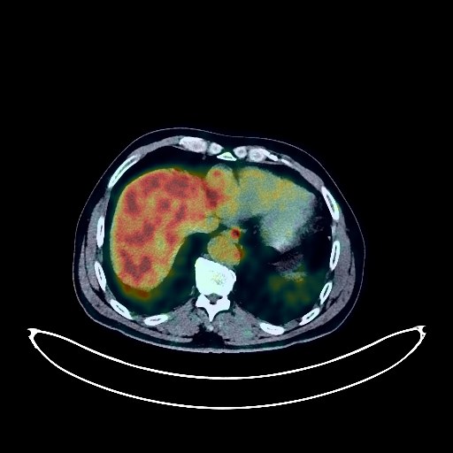
Rectal Cancer PET/CT (case 983827-000070 from PETWB-REP)
0 views9 days agoWhole-body 18F-FDG PET/CT scan in a patient with Rectal Cancer taken from the PETWB-REP dataset. The following English report (translated from original Chinese) is taken verbatim from the public dataset and has not been modified or otherwise checked for accuracy (see the end for citation). Impression a. Post-rectal cancer treatment: Slightly thickened anastomotic wall with increased FDG metabolism; thickening of the perirectal fascia and presacral fascia with mildly increased FDG metabolism. These are all likely post-treatment changes. Please follow up with colonoscopy and enhanced MRI to rule out recurrence. b. Post-operative changes in the anal region: Continuous increased FDG metabolism in parts of the intestine, suggesting physiological uptake or chronic inflammation. Please follow up with endoscopy. A mass in the lower segment of the right anterior lobe of the liver with increased FDG metabolism, suggesting a neoplastic lesion, possibly metastatic or primary. Please analyze comprehensively based on clinical findings and enhanced MRI images. Mild fatty liver. Slight dilation of the intrahepatic bile duct in the left medial lobe of the liver. Gallstones are possible; please use ultrasound. A slightly low-density nodule in the right lobe of the thyroid gland with increased FDG metabolism, suggesting a possible adenoma. Ultrasound follow-up is recommended to rule out malignancy; biopsy may be necessary. Reactive hyperplasia of bilateral cervical lymph nodes. Chronic inflammation and remnants in both lungs. Changes characteristic of chronic gastritis; please follow up with gastroscopy. Prostatic calcifications. Possible left renal cyst or small vessel leiomyolipomas. Scoliosis with degenerative changes. L2 and L5 vertebral instability. Right acromioclavicular arthritis. Chronic nasopharyngeal inflammation is highly likely; please consult a specialist. Minor inflammation of the bilateral ethmoid sinuses and right maxillary sinus. Inflammation of the left maxillary and mandibular alveolar regions. Right frontal lobe softening lesion, bilateral deep lacunar infarcts, age-related brain changes. This case is from PETWB-REP, a curated dataset of whole-body 18F-FDG PET/CT scans and corresponding radiology reports from 490 patients with a broad spectrum of malignancies. The data were retrospectively collected from patients who underwent clinically indicated whole-body 18F-FDG PET/CT scans at the Shanghai Universal Medical Imaging Diagnostic Center between 2021 and 2024. License: Creative Commons Attribution 4.0 International (CC BY 4.0) Citation: Xue, L., Feng, G., Wenbo, Z., Zhang, Y., Li, L., Wang, S., Peng, L., Peng, S., & Gao, X. (2026). PETWB-REP: A Multi-Cancer Whole-Body FDG PET/CT Dataset with Corresponding Radiology Reports [Data set]. Zenodo. https://doi.org/10.5281/zenodo.18670487
Whole BodyPET/CT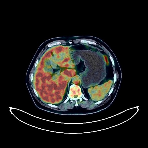
Lung Cancer PET/CT (case 983827-000182 from PETWB-REP)
0 views9 days agoWhole-body 18F-FDG PET/CT scan in a patient with Lung Cancer taken from the PETWB-REP dataset. The following English report (translated from original Chinese) is taken verbatim from the public dataset and has not been modified or otherwise checked for accuracy (see the end for citation). Impression a. Right middle lobe lung mass with elevated FDG metabolism, suggestive of lung cancer; please correlate with clinicopathology. b. Several small, solid, chronic inflammatory nodules in both lungs are highly probable; annual CT scan recommended. Minor chronic inflammation and old lesions in both lungs. Some arterial wall calcification. c. Left adrenal hyperplasia is highly probable; follow-up CT scan recommended to rule out metastasis. Prostatic calcification. Cervical, thoracic, and lumbar spondylosis. L4/5 and L5/S1 intervertebral disc bulge. Post-cervical spine surgery changes. Minor ischemic lesions in the deep bilateral brain, age-related encephalopathy. Chronic inflammation of bilateral palatine tonsils and oropharynx. Submucosal cyst of the left maxillary sinus. Minor chronic inflammation of bilateral ethmoid sinuses and the right maxillary sinus. Benign osteopathy of the 6th rib on the right side. This case is from PETWB-REP, a curated dataset of whole-body 18F-FDG PET/CT scans and corresponding radiology reports from 490 patients with a broad spectrum of malignancies. The data were retrospectively collected from patients who underwent clinically indicated whole-body 18F-FDG PET/CT scans at the Shanghai Universal Medical Imaging Diagnostic Center between 2021 and 2024. License: Creative Commons Attribution 4.0 International (CC BY 4.0) Citation: Xue, L., Feng, G., Wenbo, Z., Zhang, Y., Li, L., Wang, S., Peng, L., Peng, S., & Gao, X. (2026). PETWB-REP: A Multi-Cancer Whole-Body FDG PET/CT Dataset with Corresponding Radiology Reports [Data set]. Zenodo. https://doi.org/10.5281/zenodo.18670487
Whole BodyPET/CT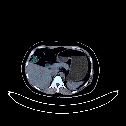
Cervical Cancer PET/CT (case 983827-000217 from PETWB-REP)
0 views9 days agoWhole-body 18F-FDG PET/CT scan in a patient with Cervical Cancer taken from the PETWB-REP dataset. The following English report (translated from original Chinese) is taken verbatim from the public dataset and has not been modified or otherwise checked for accuracy (see the end for citation). Impression Cervical mass with elevated FDG metabolism, consistent with cervical cancer, involving the lower segment of the uterus and upper segment of the vagina; left parailiac lymph node metastasis. Chronic inflammatory micronodules in the lower lobes of both lungs. Anemia. Mild fatty liver. Accessory spleen. Uterine fibroids. Chronic inflammatory changes or physiological uptake in some intestinal segments; please follow up with endoscopy. Bilateral sacroiliac joint condensing osteitis. Elevated FDG metabolism throughout the bone marrow cavity, suggestive of reactive bone marrow hyperplasia. No obvious abnormalities were found on cranial scintigraphy. This case is from PETWB-REP, a curated dataset of whole-body 18F-FDG PET/CT scans and corresponding radiology reports from 490 patients with a broad spectrum of malignancies. The data were retrospectively collected from patients who underwent clinically indicated whole-body 18F-FDG PET/CT scans at the Shanghai Universal Medical Imaging Diagnostic Center between 2021 and 2024. License: Creative Commons Attribution 4.0 International (CC BY 4.0) Citation: Xue, L., Feng, G., Wenbo, Z., Zhang, Y., Li, L., Wang, S., Peng, L., Peng, S., & Gao, X. (2026). PETWB-REP: A Multi-Cancer Whole-Body FDG PET/CT Dataset with Corresponding Radiology Reports [Data set]. Zenodo. https://doi.org/10.5281/zenodo.18670487
Whole BodyPET/CT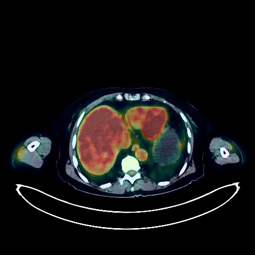
Lung Cancer PET/CT (case 983827-000197 from PETWB-REP)
0 views9 days agoWhole-body 18F-FDG PET/CT scan in a patient with Lung Cancer taken from the PETWB-REP dataset. The following English report (translated from original Chinese) is taken verbatim from the public dataset and has not been modified or otherwise checked for accuracy (see the end for citation). Impression a. A mass in the posterior segment of the right lower lobe with increased FDG metabolism, suggestive of lung cancer. b. A small solid nodule in the right middle lobe with mildly increased FDG metabolism, suggestive of possible metastasis; follow-up with CT scan is recommended. c. Scattered chronic inflammation and sequelae in both lungs. Reactive hyperplasia of hilar and mediastinal lymph nodes. Mediastinal calcification. Calcification of some arterial walls (including coronary arteries). Nodular goiter is highly probable; local malignancy in the right lobe is a possibility, further ultrasound examination is recommended. Punctate calcifications in the liver, calcification of the right lobe capsule. Bilateral renal cysts. Partial vertebral osteophyte formation. L4/5 and L5/S1 intervertebral disc bulges. Sacral canal cysts. Arachnoid cyst in the right frontal region. No obvious abnormalities were found on intracranial FDG imaging. This case is from PETWB-REP, a curated dataset of whole-body 18F-FDG PET/CT scans and corresponding radiology reports from 490 patients with a broad spectrum of malignancies. The data were retrospectively collected from patients who underwent clinically indicated whole-body 18F-FDG PET/CT scans at the Shanghai Universal Medical Imaging Diagnostic Center between 2021 and 2024. License: Creative Commons Attribution 4.0 International (CC BY 4.0) Citation: Xue, L., Feng, G., Wenbo, Z., Zhang, Y., Li, L., Wang, S., Peng, L., Peng, S., & Gao, X. (2026). PETWB-REP: A Multi-Cancer Whole-Body FDG PET/CT Dataset with Corresponding Radiology Reports [Data set]. Zenodo. https://doi.org/10.5281/zenodo.18670487
Whole BodyPET/CT