Loading...
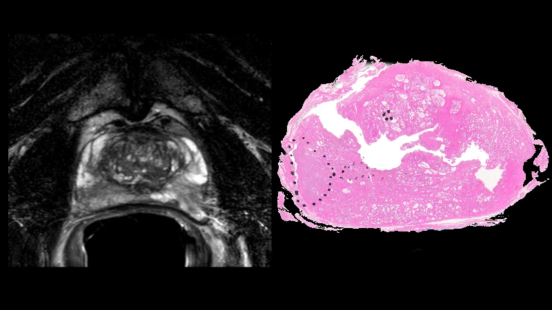
Prostate MRI - Case 0007
66 views6 months agoThis is a confirmed case of prostate cancer from the PROSTATE-MRI dataset. This collection of prostate MRIs was obtained with an endorectal and phased array surface coil at 3T (Philips Achieva). Each patient had biopsy confirmation of cancer and underwent a robotic-assisted radical prostatectomy. A mold was generated from each MRI, and the prostatectomy specimen was first placed in the mold, then cut in the same plane as the MRI. The data was generated at the National Cancer Institute, Bethesda, Maryland, USA between 2008-2010.Note: The prostatectomy color images are very high resolution. Zoom in to see fine detail if interested.License: CC BY 3.0Citation: Choyke P, Turkbey B, Pinto P, Merino M, Wood B. (2016). Data From PROSTATE-MRI. The Cancer Imaging Archive. http://doi.org/10.7937/K9/TCIA.2016.6046GUDv
PelvisMRIProstate MRI - Case 0006
119 views6 months agoThis is a confirmed case of prostate cancer from the PROSTATE-MRI dataset. This collection of prostate MRIs was obtained with an endorectal and phased array surface coil at 3T (Philips Achieva). Each patient had biopsy confirmation of cancer and underwent a robotic-assisted radical prostatectomy. A mold was generated from each MRI, and the prostatectomy specimen was first placed in the mold, then cut in the same plane as the MRI. The data was generated at the National Cancer Institute, Bethesda, Maryland, USA between 2008-2010.Note: The prostatectomy color images are very high resolution. Zoom in to see fine detail if interested.License: CC BY 3.0Citation: Choyke P, Turkbey B, Pinto P, Merino M, Wood B. (2016). Data From PROSTATE-MRI. The Cancer Imaging Archive. http://doi.org/10.7937/K9/TCIA.2016.6046GUDv
PelvisMRIProstate MRI - Case 0005
95 views6 months agoThis is a confirmed case of prostate cancer from the PROSTATE-MRI dataset. This collection of prostate MRIs was obtained with an endorectal and phased array surface coil at 3T (Philips Achieva). Each patient had biopsy confirmation of cancer and underwent a robotic-assisted radical prostatectomy. A mold was generated from each MRI, and the prostatectomy specimen was first placed in the mold, then cut in the same plane as the MRI. The data was generated at the National Cancer Institute, Bethesda, Maryland, USA between 2008-2010.Note: The prostatectomy color images are very high resolution. Zoom in to see fine detail if interested.License: CC BY 3.0Citation: Choyke P, Turkbey B, Pinto P, Merino M, Wood B. (2016). Data From PROSTATE-MRI. The Cancer Imaging Archive. http://doi.org/10.7937/K9/TCIA.2016.6046GUDv
PelvisMRIProstate MRI - Case 0004
113 views6 months agoThis is a confirmed case of prostate cancer from the PROSTATE-MRI dataset. This collection of prostate MRIs was obtained with an endorectal and phased array surface coil at 3T (Philips Achieva). Each patient had biopsy confirmation of cancer and underwent a robotic-assisted radical prostatectomy. A mold was generated from each MRI, and the prostatectomy specimen was first placed in the mold, then cut in the same plane as the MRI. The data was generated at the National Cancer Institute, Bethesda, Maryland, USA between 2008-2010.Note: The prostatectomy color images are very high resolution. Zoom in to see fine detail if interested.License: CC BY 3.0Citation: Choyke P, Turkbey B, Pinto P, Merino M, Wood B. (2016). Data From PROSTATE-MRI. The Cancer Imaging Archive. http://doi.org/10.7937/K9/TCIA.2016.6046GUDv
PelvisMRIProstate MRI - Case 0003
84 views6 months agoThis is a confirmed case of prostate cancer from the PROSTATE-MRI dataset. This collection of prostate MRIs was obtained with an endorectal and phased array surface coil at 3T (Philips Achieva). Each patient had biopsy confirmation of cancer and underwent a robotic-assisted radical prostatectomy. A mold was generated from each MRI, and the prostatectomy specimen was first placed in the mold, then cut in the same plane as the MRI. The data was generated at the National Cancer Institute, Bethesda, Maryland, USA between 2008-2010.Note: The prostatectomy color images are very high resolution. Zoom in to see fine detail if interested.License: CC BY 3.0Citation: Choyke P, Turkbey B, Pinto P, Merino M, Wood B. (2016). Data From PROSTATE-MRI. The Cancer Imaging Archive. http://doi.org/10.7937/K9/TCIA.2016.6046GUDv
PelvisMRIProstate MRI - Case 0002
92 views6 months agoThis is a confirmed case of prostate cancer from the PROSTATE-MRI dataset. This collection of prostate MRIs was obtained with an endorectal and phased array surface coil at 3T (Philips Achieva). Each patient had biopsy confirmation of cancer and underwent a robotic-assisted radical prostatectomy. A mold was generated from each MRI, and the prostatectomy specimen was first placed in the mold, then cut in the same plane as the MRI. The data was generated at the National Cancer Institute, Bethesda, Maryland, USA between 2008-2010.Note: The prostatectomy color images are very high resolution. Zoom in to see fine detail if interested.License: CC BY 3.0Citation: Choyke P, Turkbey B, Pinto P, Merino M, Wood B. (2016). Data From PROSTATE-MRI. The Cancer Imaging Archive. http://doi.org/10.7937/K9/TCIA.2016.6046GUDv
PelvisMRIProstate MRI - Case 0001
142 views6 months agoThis is a confirmed case of prostate cancer from the PROSTATE-MRI dataset. This collection of prostate MRIs was obtained with an endorectal and phased array surface coil at 3T (Philips Achieva). Each patient had biopsy confirmation of cancer and underwent a robotic-assisted radical prostatectomy. A mold was generated from each MRI, and the prostatectomy specimen was first placed in the mold, then cut in the same plane as the MRI. The data was generated at the National Cancer Institute, Bethesda, Maryland, USA between 2008-2010.Note: The prostatectomy color images are very high resolution. Zoom in to see fine detail if interested.License: CC BY 3.0Citation: Choyke P, Turkbey B, Pinto P, Merino M, Wood B. (2016). Data From PROSTATE-MRI. The Cancer Imaging Archive. http://doi.org/10.7937/K9/TCIA.2016.6046GUDv
PelvisMRI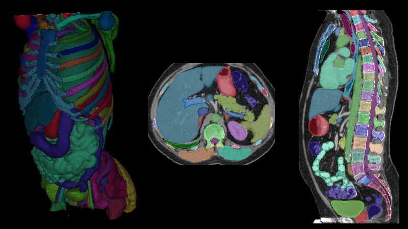
Body CT Anatomy Module from Total Segmentator Dataset (s1405)
46 views6 months agoCase and segmentations are taken directly from the Total Segmentator dataset, which is distributed under the CC Attribution 4.0 International license. If interested, you may display a default 3D view of the segmentations via the settings button in the bottom right corner of the DicomTube player.The following additional metadata are provided for this particular case in the dataset:Pathology: vascular Pathology Location: abdomenCitation:Jakob Wasserthal. (2023). Dataset with segmentations of 117 important anatomical structures in 1228 CT images (2.0.1) [Data set]. Zenodo. https://doi.org/10.5281/zenodo.10047292
APCTBody CT Anatomy Module from Total Segmentator Dataset (s1404)
15 views6 months agoCase and segmentations are taken directly from the Total Segmentator dataset, which is distributed under the CC Attribution 4.0 International license. If interested, you may display a default 3D view of the segmentations via the settings button in the bottom right corner of the DicomTube player.The following additional metadata are provided for this particular case in the dataset:Pathology: no_pathology Pathology Location: no_locationCitation:Jakob Wasserthal. (2023). Dataset with segmentations of 117 important anatomical structures in 1228 CT images (2.0.1) [Data set]. Zenodo. https://doi.org/10.5281/zenodo.10047292
CAPCTBody CT Anatomy Module from Total Segmentator Dataset (s1403)
14 views6 months agoCase and segmentations are taken directly from the Total Segmentator dataset, which is distributed under the CC Attribution 4.0 International license. If interested, you may display a default 3D view of the segmentations via the settings button in the bottom right corner of the DicomTube player.The following additional metadata are provided for this particular case in the dataset:Pathology: vascular Pathology Location: abdomenCitation:Jakob Wasserthal. (2023). Dataset with segmentations of 117 important anatomical structures in 1228 CT images (2.0.1) [Data set]. Zenodo. https://doi.org/10.5281/zenodo.10047292
APCT