Loading...
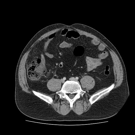
Appendicitis - Case from TR_Abdomen Rad_Emergency (20001)
82 views6 months agoDiagnosis: Appendicitis. This is an abdominal CT case from the TR_Abdomen Rad_Emergency dataset, a collection of anonymized emergency abdominal CT cases from the Republic of Türkiye Ministry of Health and made available publicly. Citation: Koç et al. Eur Radiol 34, 3588-3597 (2024). https://doi.org/10.1007/s00330-023-10391-y
Abdomen PelvisCT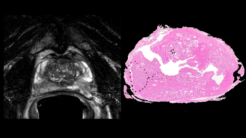
Prostate MRI - Case 0026
98 views6 months agoThis is a confirmed case of prostate cancer from the PROSTATE-MRI dataset. This collection of prostate MRIs was obtained with an endorectal and phased array surface coil at 3T (Philips Achieva). Each patient had biopsy confirmation of cancer and underwent a robotic-assisted radical prostatectomy. A mold was generated from each MRI, and the prostatectomy specimen was first placed in the mold, then cut in the same plane as the MRI. The data was generated at the National Cancer Institute, Bethesda, Maryland, USA between 2008-2010.Note: The prostatectomy color images are very high resolution. Zoom in to see fine detail if interested.License: CC BY 3.0Citation: Choyke P, Turkbey B, Pinto P, Merino M, Wood B. (2016). Data From PROSTATE-MRI. The Cancer Imaging Archive. http://doi.org/10.7937/K9/TCIA.2016.6046GUDv
PelvisMRIProstate MRI - Case 0025
89 views6 months agoThis is a confirmed case of prostate cancer from the PROSTATE-MRI dataset. This collection of prostate MRIs was obtained with an endorectal and phased array surface coil at 3T (Philips Achieva). Each patient had biopsy confirmation of cancer and underwent a robotic-assisted radical prostatectomy. A mold was generated from each MRI, and the prostatectomy specimen was first placed in the mold, then cut in the same plane as the MRI. The data was generated at the National Cancer Institute, Bethesda, Maryland, USA between 2008-2010.Note: The prostatectomy color images are very high resolution. Zoom in to see fine detail if interested.License: CC BY 3.0Citation: Choyke P, Turkbey B, Pinto P, Merino M, Wood B. (2016). Data From PROSTATE-MRI. The Cancer Imaging Archive. http://doi.org/10.7937/K9/TCIA.2016.6046GUDv
PelvisMRIProstate MRI - Case 0024
113 views6 months agoThis is a confirmed case of prostate cancer from the PROSTATE-MRI dataset. This collection of prostate MRIs was obtained with an endorectal and phased array surface coil at 3T (Philips Achieva). Each patient had biopsy confirmation of cancer and underwent a robotic-assisted radical prostatectomy. A mold was generated from each MRI, and the prostatectomy specimen was first placed in the mold, then cut in the same plane as the MRI. The data was generated at the National Cancer Institute, Bethesda, Maryland, USA between 2008-2010.Note: The prostatectomy color images are very high resolution. Zoom in to see fine detail if interested.License: CC BY 3.0Citation: Choyke P, Turkbey B, Pinto P, Merino M, Wood B. (2016). Data From PROSTATE-MRI. The Cancer Imaging Archive. http://doi.org/10.7937/K9/TCIA.2016.6046GUDv
PelvisMRIProstate MRI - Case 0023
78 views6 months agoThis is a confirmed case of prostate cancer from the PROSTATE-MRI dataset. This collection of prostate MRIs was obtained with an endorectal and phased array surface coil at 3T (Philips Achieva). Each patient had biopsy confirmation of cancer and underwent a robotic-assisted radical prostatectomy. A mold was generated from each MRI, and the prostatectomy specimen was first placed in the mold, then cut in the same plane as the MRI. The data was generated at the National Cancer Institute, Bethesda, Maryland, USA between 2008-2010.Note: The prostatectomy color images are very high resolution. Zoom in to see fine detail if interested.License: CC BY 3.0Citation: Choyke P, Turkbey B, Pinto P, Merino M, Wood B. (2016). Data From PROSTATE-MRI. The Cancer Imaging Archive. http://doi.org/10.7937/K9/TCIA.2016.6046GUDv
PelvisMRIProstate MRI - Case 0022
73 views6 months agoThis is a confirmed case of prostate cancer from the PROSTATE-MRI dataset. This collection of prostate MRIs was obtained with an endorectal and phased array surface coil at 3T (Philips Achieva). Each patient had biopsy confirmation of cancer and underwent a robotic-assisted radical prostatectomy. A mold was generated from each MRI, and the prostatectomy specimen was first placed in the mold, then cut in the same plane as the MRI. The data was generated at the National Cancer Institute, Bethesda, Maryland, USA between 2008-2010.Note: The prostatectomy color images are very high resolution. Zoom in to see fine detail if interested.License: CC BY 3.0Citation: Choyke P, Turkbey B, Pinto P, Merino M, Wood B. (2016). Data From PROSTATE-MRI. The Cancer Imaging Archive. http://doi.org/10.7937/K9/TCIA.2016.6046GUDv
PelvisMRIProstate MRI - Case 0021
84 views6 months agoThis is a confirmed case of prostate cancer from the PROSTATE-MRI dataset. This collection of prostate MRIs was obtained with an endorectal and phased array surface coil at 3T (Philips Achieva). Each patient had biopsy confirmation of cancer and underwent a robotic-assisted radical prostatectomy. A mold was generated from each MRI, and the prostatectomy specimen was first placed in the mold, then cut in the same plane as the MRI. The data was generated at the National Cancer Institute, Bethesda, Maryland, USA between 2008-2010.Note: The prostatectomy color images are very high resolution. Zoom in to see fine detail if interested.License: CC BY 3.0Citation: Choyke P, Turkbey B, Pinto P, Merino M, Wood B. (2016). Data From PROSTATE-MRI. The Cancer Imaging Archive. http://doi.org/10.7937/K9/TCIA.2016.6046GUDv
PelvisMRIProstate MRI - Case 0020
93 views6 months agoThis is a confirmed case of prostate cancer from the PROSTATE-MRI dataset. This collection of prostate MRIs was obtained with an endorectal and phased array surface coil at 3T (Philips Achieva). Each patient had biopsy confirmation of cancer and underwent a robotic-assisted radical prostatectomy. A mold was generated from each MRI, and the prostatectomy specimen was first placed in the mold, then cut in the same plane as the MRI. The data was generated at the National Cancer Institute, Bethesda, Maryland, USA between 2008-2010.Note: The prostatectomy color images are very high resolution. Zoom in to see fine detail if interested.License: CC BY 3.0Citation: Choyke P, Turkbey B, Pinto P, Merino M, Wood B. (2016). Data From PROSTATE-MRI. The Cancer Imaging Archive. http://doi.org/10.7937/K9/TCIA.2016.6046GUDv
PelvisMRIProstate MRI - Case 0019
69 views6 months agoThis is a confirmed case of prostate cancer from the PROSTATE-MRI dataset. This collection of prostate MRIs was obtained with an endorectal and phased array surface coil at 3T (Philips Achieva). Each patient had biopsy confirmation of cancer and underwent a robotic-assisted radical prostatectomy. A mold was generated from each MRI, and the prostatectomy specimen was first placed in the mold, then cut in the same plane as the MRI. The data was generated at the National Cancer Institute, Bethesda, Maryland, USA between 2008-2010.Note: The prostatectomy color images are very high resolution. Zoom in to see fine detail if interested.License: CC BY 3.0Citation: Choyke P, Turkbey B, Pinto P, Merino M, Wood B. (2016). Data From PROSTATE-MRI. The Cancer Imaging Archive. http://doi.org/10.7937/K9/TCIA.2016.6046GUDv
PelvisMRIProstate MRI - Case 0018
113 views6 months agoThis is a confirmed case of prostate cancer from the PROSTATE-MRI dataset. This collection of prostate MRIs was obtained with an endorectal and phased array surface coil at 3T (Philips Achieva). Each patient had biopsy confirmation of cancer and underwent a robotic-assisted radical prostatectomy. A mold was generated from each MRI, and the prostatectomy specimen was first placed in the mold, then cut in the same plane as the MRI. The data was generated at the National Cancer Institute, Bethesda, Maryland, USA between 2008-2010.Note: The prostatectomy color images are very high resolution. Zoom in to see fine detail if interested.License: CC BY 3.0Citation: Choyke P, Turkbey B, Pinto P, Merino M, Wood B. (2016). Data From PROSTATE-MRI. The Cancer Imaging Archive. http://doi.org/10.7937/K9/TCIA.2016.6046GUDv
PelvisMRI