Loading...
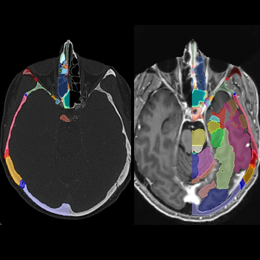
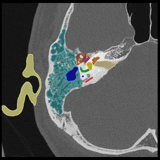
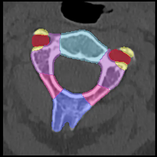
Comprehensive Head CT + MRI Anatomy: Brain, Skullbase and Sinonasal Cavity
3,461 views6 months agoHighly detailed annotated anatomy of the skull, skullbase, brain, and sinonasal cavity on registered CT and MRI images from the same subject. There are multiple display presets available which you can access from the settings button in the bottom right of the DicomTube player.The brain cortical segmentation is predominantly auto generated using Freesurfer software and the Desikan-Killiany Atlas. For further details visit the Freesurfer website or associated publication. There are, however, a number of manual edits and custom post processing also performed on the cortical segmentations to make sure they respect the anatomy better.
HeadMRI, CT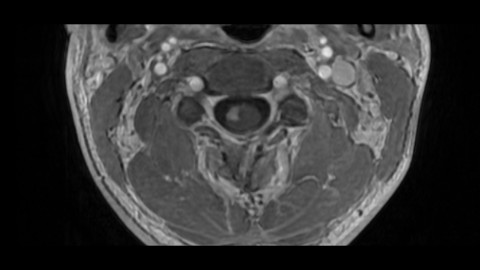
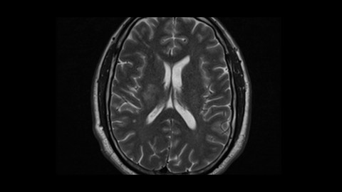
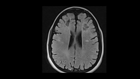
Acute myelitis, first onset Multiple Sclerosis
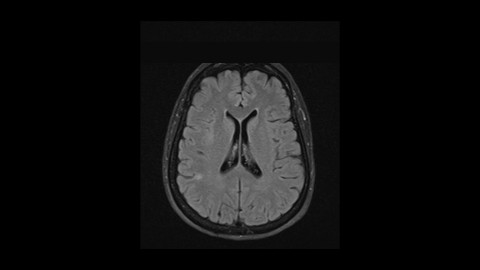
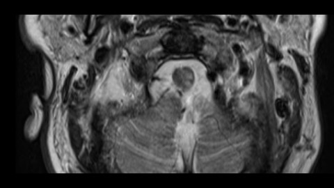
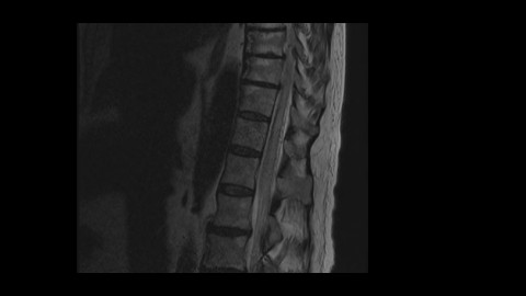
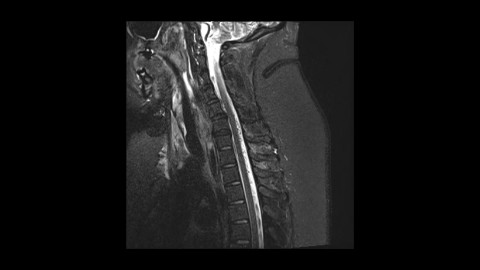
437 views7 months agoThere is an avidly enhancing short segment cord lesion in the lateral and anterior cord (@Key Finding 1, @Key Finding 2). This involves the lateral spinothalamic tract carrying pain and temperature sensation. These nervers decussate immediately in the cord (@Key Finding 6) and so a right sided cord lesion would carry sensory information from the LEFT side of the body. This patient was having left sided burning sensation from the shoulder down.With focal cord lesions like these, multiple sclerosis should be a major consideration and brain MRI is prudent. Here, both periventricular (@Key Finding 3) as well as infratentorial (@Key Finding 4 and @Key Finding 5) lesions are present and so the patient meets imaging criteria for dissemination in space (3 of the 4 MS spaces in this case) and dissemination in time (enhancing + non-enhancing) needed for MS. Notice how much easier the infratentorial lesion is to see on PD images but it is clearly present on the FLAIR images as well. Always look at traditional T2 images (and PD if you have them) for the posterior fossa.
Brain, SpineMRI